A sinus of Valsalva aneurysm (SOVA) is an uncommon cardiac anomaly occurring in less than 1% of patients undergoing open cardiac surgical procedures [1]. Approximately 65-85% of SOVAs originate from the right sinus of Valsalva, while SOVAs originating from noncoronary (10-30%) and left sinuses (< 5%) are less common [2]. It may be congenital or acquired in origin and may rupture into any of the cardiac chambers to form an aorto-cardiac fistula. When intra-cardiac rupture occurs, early surgical intervention is indicated as rupture may lead to serious hemodynamic instability, acute heart failure and, if left untreated, has a mean survival period of 1-2 years [3]. Although there have been successful reports of transcatheter closure [4, 5], no dedicated closure devices exist and open surgical repair remains the mainstay of treatment. We present a case of a chronic ruptured non-coronary SOVA illustrating the classical “windsock” appearance of the aorta to right atrial fistula tract and correlate the non-invasiveimaging appearances with those seen at the time of open surgical repair.
A 50-year-old female presented with a 2-week history of sudden onset shortness of breath, impaired exercise tolerance and orthopnoea. Past medical history included rheumatoid arthritis. Her vital signs were: heart rate 102 beats per minute, respiratory rate 28 breaths per minute, blood pressure 112/52mmHg, oxygen saturation 98% on room air and NYHA class III. On cardiovascular examination she was noted to have clinical signs of biventricular heart failure with a sinus tachycardia, elevated jugular venous pulse, a continuous murmur, bilateral pulmonary crepitations and pitting edema in the lower limbs. Transesophageal echocardiogram (TEE) confirmed a ruptured non-coronary SOVA with a torrential aortic to right atrial shunt and right heart volume overload. The fistula tract was seen as a “windsock” originating in the non-coronary sinus and projecting into the right atrium (Figure 1).
Figure 1.

Short Axis view of TEE confirmed a ruptured non-coronary SOVA with torrential aortic to right atrial shunt.
AV = Aortic Valve; FT = Fistula Tract; RA = Right Atrium.
There were no other cardiac abnormalities identified on TEE. Right and left heart catheterization studies showed normal coronary anatomy, elevated right heart pressures [mean RA: 31 mmHg, RV: 62/24 mmHg, PA: 55/31 (mean = 43 mmHg] and a large left to right shunt (Qp:Qs 3.2:1) (Figure 2).
Figure 2.

Left heart catheterization study showing a large aortic to right atrial shunt.
Asc = Ascending Aorta; FT = Fistula Tract; RA = Right Atrium.
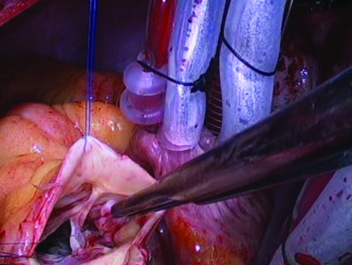
The patient was operated on with urgent status (during current hospital stay) after a short intensive period of diuretic therapy. At operation there was a 2cm defect in the non coronary sinus with a fistula tract of thinned aortic wall protruding from the base of the non coronary sinus into the right atrium with the appearances of a “windsock” (Figure 3).
Figure 3.
Intra operative image of the fistula tract protruding from the base of the non-coronary sinus with a “windsock” appearance. Fistula tract is identified by the forceps.
The “windsock” was delivered back into the aorta from its right atrial position and its apex was seen to have multiple perforations resulting in the fistula. The tissue that made up the “windsock” was excised and a bovine pericardial patch was used to repair the defect in the non-coronary sinus. After weaning from cardiopulmonary bypass, intra-operative TEE revealed a satisfactory repair with no residual fistula connection and normal aortic and tricuspid valve function. The patient had an unremarkable recovery and was discharged home on the 6th post operative day. At 6-week follow up she was symptom free and transthoracic echocardiogram revealed normal cardiac function with no detectable shunt.
This case serves to highlight the importance of early identification and treatment of a ruptured SOVA as it can bring rapid and permanent symptomatic relief. Surgery can be performed with low morbidity and mortality with 10-year survival rates of 63-93% [3, 6]. In the presence of an otherwise normal aortic valve and root, the SOVA can be treated locally with either pericardial patch closure of the defect or replacement of the sinus with a Dacron patch with preservation of the native aortic valve. When a diagnosis of ruptured SOVA is made, we advocate early surgical repair as if left untreated progressive heart failure and premature death may occur.
Footnotes
Source of Support Nil.
Disclosures None declared.
Cite as: Perera NK, Galvin SD, Farouque O, Matalanis G. Aortic to right atrial fistula secondary to chronic ruptured sinus of Valsalva aneurysm. Heart, Lung and Vessels. 2015; 7(3): 260-262.
References
- Takach T J, Reul G J, Duncan J M, Cooley D A, Livesay J J, Ott D A. et al. Sinus of Valsalva aneurysm or fistula: management and outcome. The Annals of thoracic surgery. 1999;68:1573–1577. doi: 10.1016/s0003-4975(99)01045-0. [DOI] [PubMed] [Google Scholar]
- Meier J H, Seward J B, Miller FA F A Jr., Oh J K, Enriquez-Sarano M. Aneurysms in the left ventricular outflow tract: clinical presentation, causes, and echocardiographic features. J Am Soc Echocardiogr. 1998;11:729–745. doi: 10.1053/je.1998.v11.a91047. [DOI] [PubMed] [Google Scholar]
- Sarikaya S, Adademir T, Elibol A, Buyukbayrak F, Onk A, Kirali K. Surgery for ruptured sinus of Valsalva aneurysm: 25-year experience with 55 patients. Eur J Cardiothorac Surg. 43:591–596. doi: 10.1093/ejcts/ezs450. [DOI] [PubMed] [Google Scholar]
- Chen F, Li S H, Qin Y W, Li P, Liu S X, Dong J. et al. Transcatheter closure of giant ruptured sinus of valsalva aneurysm. Circulation. 2013;128:1–3. doi: 10.1161/CIRCULATIONAHA.112.113365. [DOI] [PubMed] [Google Scholar]
- Kuriakose E M, Bhatla P, McElhinney D B. Comparison of Reported Outcomes With Percutaneous Versus Surgical Closure of Ruptured Sinus of Valsalva Aneurysm. Am J Cardiol. 2015;115:392–398. doi: 10.1016/j.amjcard.2014.11.013. [DOI] [PubMed] [Google Scholar]
- Moustafa S, Mookadam F, Cooper L, Adam G, Zehr K, Stulak J. et al. Sinus of Valsalva aneurysms--47 years of a single center experience and systematic overview of published reports. The American journal of cardiology. 2007;99:1159–1164. doi: 10.1016/j.amjcard.2006.11.047. [DOI] [PubMed] [Google Scholar]


