Abstract
Introduction Bone conduction implants are indicated for patients with conductive and mixed hearing loss, as well as for patients with single-sided deafness (SSD). The transcutaneous technology avoids several complications of the percutaneous bone conduction implants including skin reaction, skin growth over the abutment, and wound infection. The Bonebridge (MED-EL, Austria) prosthesis is a semi-implantable hearing system: the BCI (Bone Conduction Implant) is the implantable part that contains the Bone Conduction-Floating Mass Transducer (BC-FMT), which applies the vibrations directly to the bone; the external component is the audio processor Amadé BB (MED-EL, Austria), which digitally processes the sound and sends the information through the coil to the internal part. Bonebridge may be implanted through three different approaches: the transmastoid, the retrosigmoid, or the middle fossa approach.
Objective This systematic review aims to describe the world́s first active bone conduction implant system, Bonebridge, as well as describe the surgical techniques in the three possible approaches, showing results from implant centers in the world in terms of functional gain, speech reception thresholds and word recognition scores.
Data Synthesis The authors searched the MEDLINE database using the key term Bonebridge. They selected only five publications to include in this systematic review. The review analyzes 20 patients that received Bonebridge implants with different approaches and pathologies.
Conclusion Bonebridge is a solution for patients with conductive/mixed hearing loss and SSD with different surgical approaches, depending on their anatomy. The system imparts fewer complications than percutaneous bone conduction implants and shows proven benefits in speech discrimination and functional gain.
Keywords: bone conduction hearing, conductive hearing loss, mixed hearing loss, single sided deafness, bonebridge, bone conduction implants
Introduction
Bone conduction implants are indicated for patients with conductive and mixed hearing loss that do not benefit from conventional hearing aids or for those that cannot use them for anatomy abnormalities or medical conditions. These implants are also indicated for patients with single-sided deafness (SSD), which consists of one ear with profound hearing loss and the contralateral with normal hearing. Bonebridgés (MED-EL, Austria) transcutaneous technology avoids several complications of the percutaneous bone conduction implants including skin reaction, growth of skin over the abutment, implant extrusion, and wound infection. Audiological criteria for the Bonebridge include patients with conductive or mixed hearing loss with bone conduction thresholds up to 45 dB, as well as those with single-sided deafness (Contralateral ear thresholds between 0–20dB). The Bonebridge implant is a semi-implantable hearing system with two parts: the inner one (Bone Conduction Implant -BCI) contains a magnet that holds the external audio processor in place (Amadé BB) and transmits the signal through the coil to make this system transcutaneous. The Bone Conduction Implant (BCI) may be implanted through different approaches: the mastoid approach, retrosigmoid approach, or middle fossa approach.
Review of Literature
As this is a recently used prosthesis, this review begins describing the prosthesis and the surgical technique applied.
Implant Description
The Bonebridge (BB) from MED-EL Company (Innsbruck, Austria) is the world's first active, intact skin bone conduction implant system, intended for individuals with conductive or mixed hearing loss and Single-Sided Deafness (SSD). It is a semi-implantable hearing system consisting of an implantable part, the BCI (Bone Conduction Implant), and the external audio processor, Amadé (Fig. 1).
Fig. 1.
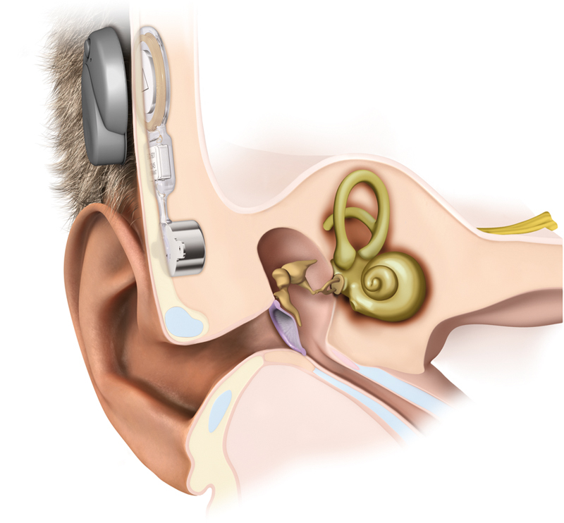
Bonebridge in mastoid.
The Amadé contains two microphones, a digital signal processor and a battery. The BCI is the implantable part of the Bonebridge and consists of a magnet surrounded by the receiver coil, the electronics (demodulator), a bendable transition and the Bone Conduction-Floating Mass Transducer (BC-FMT), which vibrates in a controlled manner according to user need. The information processed in the Amadé travels transcutaneously to the BCI and the BC-FMT generates vibrations. The transmitted vibrations stimulate the auditory system and are interpreted by the patient as sound.
The BC-FMT is ∼8.7mm in height, 15.8mm in diameter and weighs ∼10g. Two screws located laterally of the BC-FMT with a 23.8mm distance between them are responsible for the transmission of vibration to the bone. Given that the BC-FMT is secured to bone by screws, the osseointegration process is not required and may be activated within two or three weeks after it is implanted. The Bonebridge supports MRI up to 1.5 Tesla due to its patented design magnets (Fig. 2).
Fig. 2.
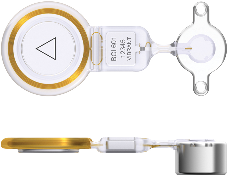
Bone Conduction Implant (BCI).
Audiological Criteria
In 2012, the Bonebridge received its first approval for patients over 18 years and, in 2014, was approved for children over 5 years (CE Mark approval).
Audiological criteria for patient selection is divided into two:
-
Conductive or mixed hearing loss by audiometric testing with bone conduction thresholds better than or equal to 45dB HL at 500Hz, 1KHz, 2KHz, and 3KHz (Fig. 3).
Contraindication, in the case of mixed and conductive hearing loss, is the presence of retrocochlear or central disorders.
Single-sided deafness (SSD), that is, severe to profound sensorineural deafness in one ear while the other ear has normal hearing, with air conduction equal or better than 20dB HL measured at 500Hz, 1KHz, 2KHz, and 3KHz (Fig. 4).
Fig. 3.
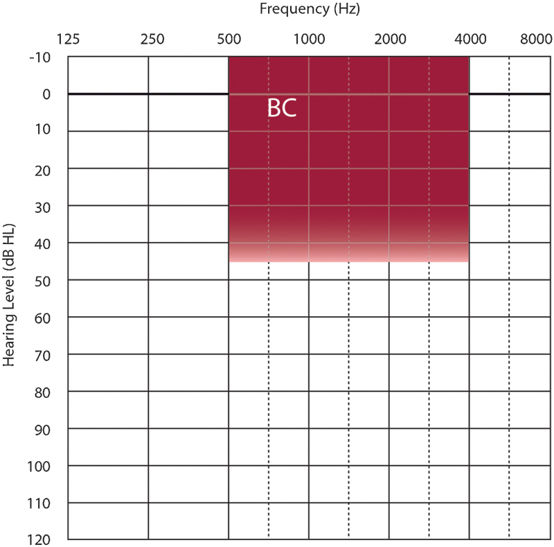
Audiological criteria for conductive and mixed hearing loss.
Fig. 4.
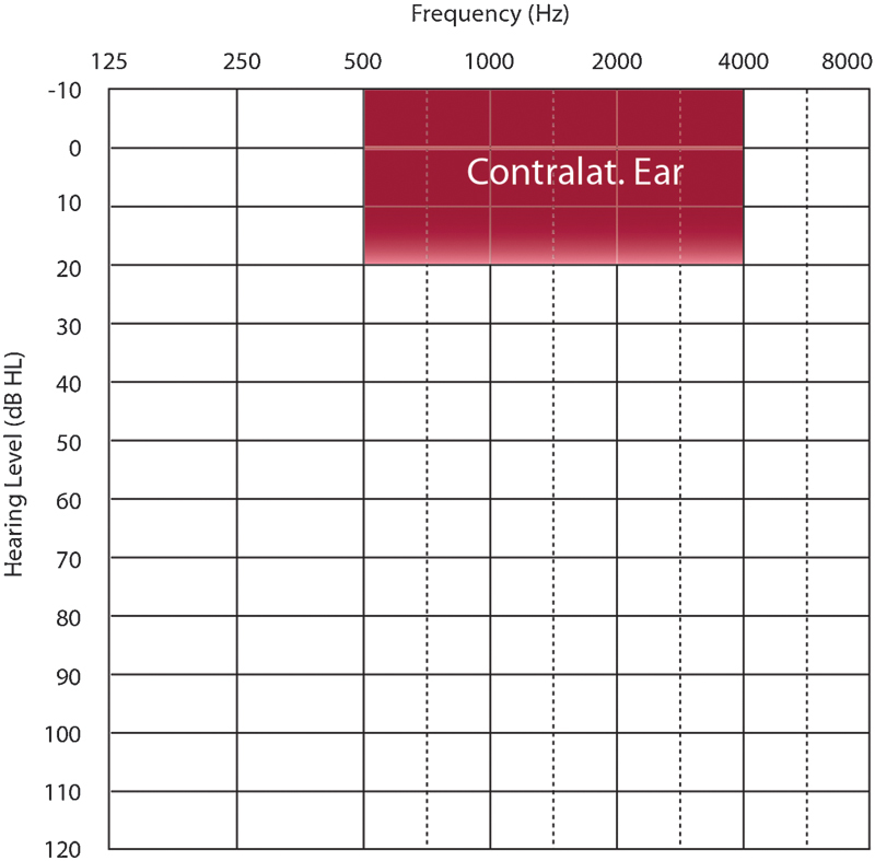
Audiological criteria for Single-Sided Deafness (SSD).
Radiological Planning Prior to Surgery
Imaging tools are useful for previous planning for the placement of the device. There are two programs available for this purpose. The first one is the 3D Slicer (http://www.slicer.org). It can plan the location of BC-FMT in the mastoid bone. This allows one to determine in advance the location and possible structures involved in the implantation.
The authors currently use the BB FastView program (http://www1.ceit.es/cg/BBfastView/BBFastView.zip) developed by the Center for Technical Studies and Research at the University of Navarra, Spain. This program allows the correct placement of the BC-FMT according to each patient́s anatomy using CT images in DICOM format simulated in the coronal, sagital, axial, and even three-dimensional reconstructions.1
Prior radiological programming allows specialists to know exactly where to place the BCI, aside from planning the use of alternative routes and prevent possible complications.
Surgery
Bonebridge surgery is relatively simple and quick. There are three main approaches and techniques for placement.2 3 The most common is via the mastoid; the second most common is the retrosigmoid and, finally, the middle fossa.
Transmastoid Approach
The transmastoid approach is the ideal approach for cases of otosclerosis, middle ear surgeries with poor functional outcomes (tympanoplasties and ossiculoplasties), and congenital aural atresia. This approach depends on the mastoid size, as explained by the preoperative radiological measurement.
The approach begins with a retroauricular classical incision (no more than 5cm), after which two flaps are performed: one from skin and subcutaneous layers and another muscular. On the mastoid cortex, we mark the size with the BC-FMT demo (bone conduction floating mass transducer). A bed is drilled to make a cylindrical cavity that can accommodate the entire BC-FMT, considering that the holes for the screws should rest perfectly on the surface of the cortical bone. The drilling needs to be precise and the angles between the sidewall and the bottom of the cavity must be at right angles. It is best to avoid both the dura and the sigmoid sinus and, despite the fact that the size of the mastoid is not large enough, exposing these structures does not mean a larger problem. If the patient́s anatomy does not allow enough space to place the BCI, there is no issue in partially pressing the meninges, or even partially depressing the sigmoid sinus. It is recommendable to cover the surface of these noble structures with a resorbable material (gel foam, for example) and carry on with the surgery.
Next, two small holes are performed with a special burr with a stop, just deep enough to perforate the cortical and facilitate the placement of the screw. After this step, the physician may proceed with placing the Bonebridge, starting with the magnet part and the electronics package that sits in a pocket beneath the periosteum. Then, the BC-FMT should be placed in a way that allows a 30° angle in the vertical plane and 90° in the horizontal plane, leaving the coil in the best position for subsequent use of the external part (Amadé). Once in position, the self-tapping screws are secured. Adequate fixation may be measured with a tool called torque wrench, whereby the ideal force would be ∼20 Ncm. If any of the screws spin loosely or improperly or break, there is a rescue screw in the surgical kit that is thicker than the regular, to save the situation. Subsequently, we perform the closure flaps and place compression bandage.2 3
Retrosigmoid Approach
The Retrosigmoid Approach is indicated for patients whose anatomy makes it difficult to provide an adequate or sufficient mastoid space or in cases of low middle fossa or very anterior sigmoid sinus. It is also the ideal choice for patients who have undergone previous mastoidectomy for chronic otitis media or cholesteatoma, with the canal wall down technique.4 5
In this case, the incision is 4 or 5 cm, one or two centimeters behind the sigmoid sinus area and running parallel to it. The two flaps and subsequent steps are performed in the same way as the mastoid approach. At this stage, it is common to find the mastoid emissary vein, which should be cauterized or blocked with bone wax to prevent uncomfortable bleeding. Next, the bed is performed.6 7
Similarly, the dura is commonly found in this procedure and should be treated in the same manner as previously described. It is important to avoid injury to the dura and to cover it to prevent damage. Implantation is performed with screw fixation. The convexity of the skull often renders it difficult to provide ideal support for both screws. In these cases, there are spacers called lifts that range from 1 to 4 mm. Lifts are little rings designed to avoid dead space between the wings of the BC-FMT and the cortex of the skull, achieving a perfect fit. These supplements are also very useful in young children in the mastoid approach, when the depth of the bone is not sufficient (Figs. 5, 6, 7).
Fig. 5.
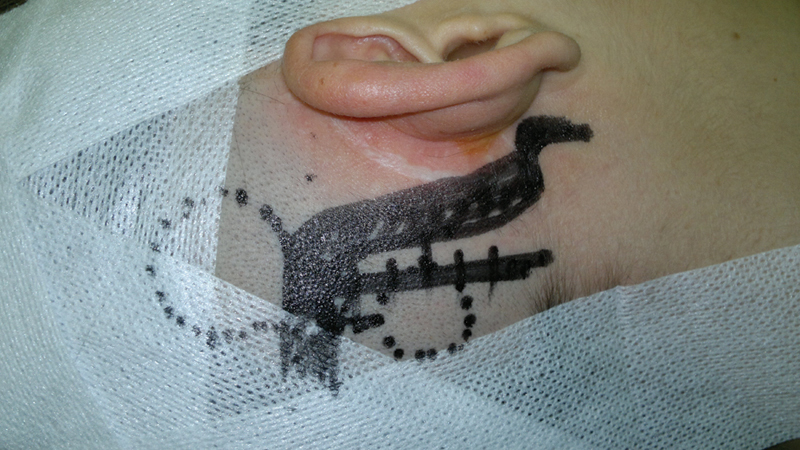
Previous planification showing the position of BB in retrosigmoidal approach.
Fig. 6.
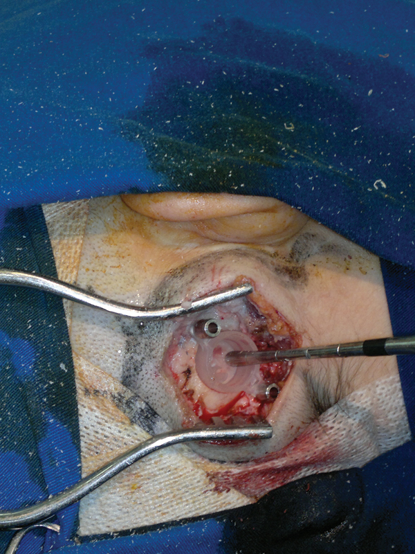
Retrosigmoid approach.
Fig. 7.
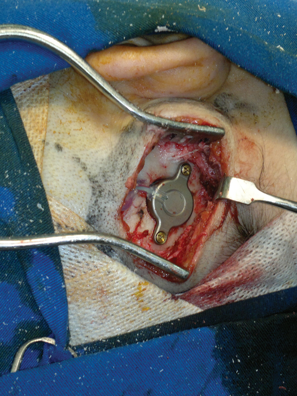
BB placement in a retrosigmoidal approach.
Middle Fossa Approach
Exceptionally, the two aforementioned approaches may not be the preferred due to anatomical reasons, leaving the option of the middle fossa approach, as described by Dr. Sumit Agrawal from the Western University (Ontario, Canada). The incision is made above the position of the ear of the parietal bone and is usually smaller (around 3cm). At the height at which the bed is made, the entire prosthesis is placed close to the horizontal line, so that the area of the magnet is posterior to the ear. Here we always expose dura and depress it to place the BC-FMT. In any of the techniques described, the compression of the dura by the BC-FMT does not cause any risk or sequel. This approach is faster as it allows the use of a 15mm drill head (neuro drill), which significantly shortens the operating time. Also, the use of spacers is common in this approach.
Complications
The complications that may arise from these procedures are similar to those of other implantable prosthesis, including a cochlear implant. These may be major complications, requiring re-operation or hospitalization, or minor ones, which are often solved with the patient in the office. However, it is important to remember that a refined technique and good preoperative planning avoids most complications.
With BB cases, there are no reports of severe complications. The flap necrosis or infection is similar to that of the cochlear implant surgery. We recommend performing the double flap with good vascularization and minimal incisions, as this minimizes the risk of flap complications.
Regarding the problem of the magnet on the skin, BB is the only active and transcutaneous prosthesis, whereas other bone conduction prosthesis are active but percutaneous. For this reason, BB has the lowest weight (8gr versus 15–23gr) and the lowest external profile (9mm versus 16, or more), a feature that reduces the chances of injury to the skin and improves the quantity of hours of use. In cases of infection or partial necrosis, we strongly recommend using pediculate flaps to cover the zone.
Injury to the meninges or sigmoid sinus is necessarily a result of abrupt maneuvers and, therefore is generally avoidable. The management of these lesions is the same as in any other ear surgery and involves repair and meningeal gap with the use of suture or synthetic materials.
With respect to the possibility that the BC-FMT may touch or press the meninges and cause any damage or problems, there has been no report to date that establishes a higher prevalence of headaches in the implanted population compared to the general population.
Data Synthesis
We searched the MEDLINE database from the date of BB approbation (May 2012) up to July 30, 2014, using Bonebridge as the search term. The initial search included 19 studies. We included only five publications with descriptions of the surgical techniques and results in this systematic review (Table 1). The studies involved 20 patients that underwent Bonebridge implants with different approaches and pathologies.
Table 1. Details of the systematic Review.
Several groups around the world have reported different audiological results (functional gain, speech reception thresholds, and word recognition scores).2 3 4 6 8 Based on our experience, 90% of patients with conductive hearing loss got the total closure of air-bone gap, which means a gain of 30 to 60dB. In cases of congenital atresia, this is achieved in almost all patients, being less prevalent in patients with other etiologies, such as otosclerosis or chronic otitis media.
Fig. 8 shows the free-field functional gain in a total of 20 patients with mixed and conductive hearing loss from five different implant centers. Functional gains range between 24dB and 43dB.
Fig. 8.
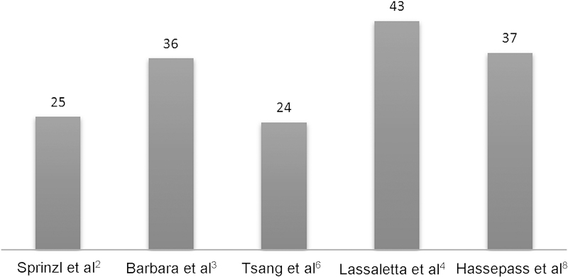
Functional gain as reported by five different implant centers. The graph includes a total 20 subjects with mixed and conductive hearing loss.
Discussion
This new kind of bone conduction implant allows treatment conductive and mixed hearing losses, usually in patients who showed bad results with other available treatment solutions.
Regarding congenital aural atresia, Siegert, in a recent work, proposes three surgical steps, including treatment of microtia with plastic reconstruction. In this publication, the author reports that 76% of patients present hearing with thresholds near 30 dB.9
In a review conducted at Oklahoma University, only 50% of patients reach thresholds near 30 dB.10 Yellow et al. published results in atresioplasty surgery with pure tone average thresholds and air-bone gaps (ABG) of 37.5 and 29.4 dB, respectively.11
Regarding osseointegrated hearing aids, it is worth noting that some reports claim 85.1% of patients with external atresia.12 Despite the functional outcome being mostly positive, this surgery has an important number of major complications. Hobson et al., in a series of over 600 implants, reported a complication rate of 23.9%, while presenting a surgical revision rate of 12.1%.13 Another review mentioned postoperative complications that included severe skin infections in 8% of patients, while 18% of the cases presented problems with osseointegration, and an additional 8% of patients presented skin growing over the external connector.14 Finally, it is important to note that the support (Abutment) used is percutaneous, which many patients reject for aesthetic reasons and constant maintenance needs.
Most authors acknowledge that the Bonebridge can overcome some of the issues with bone-anchored hearing devices. Tsang et al. state “unlike traditional percutaneous bone anchored hearing aid, the use of the Bonebridge system eliminates postoperative pin tract infections, which greatly enhances the patient's quality of life as it obviates the need for daily implant wound care.”6
Chronic otitis media is generally associated with some degree of hearing loss. Barbara et al. state: “regards the comparison with the other transcutaneous devices whose efficacy is directly related to the magnetic attraction between the magnet of the external unit and the metallic implanted plaque that, in some cases, may cause problems as regards the interposed skin. In the [Bonebridge], the magnet only acts as a coupling tool for the external unit with the implanted BC-FMT and its strength does not affect the efficacy of the system, making it unlikely that any skin problems will arise.”3
All these publications show many problems with conventional surgery and with bone-anchored hearing aids, especially in patients suffering from CAA, otosclerosis and failed ossiculoplasty. Therefore, the Bonebridge as the first active bone conduction implant could be a good alternative.
Conclusion
The Bonebridge is a novel solution for patients with conductive/mixed hearing loss and SSD, and surgical approaches vary depending on their anatomy. The BB device presents fewer complications than percutaneous bone-anchored hearing aids and bring proven benefits to patients, in addition to speech discrimination and functional gain. Finally, the Bonebridge decreases post-surgical complications due to intact skin.
Footnotes
Conflicts of Interest Andrea Bravo Sarasty is employed by the device manufacturer. The other authors declare no conflicts of interest.
References
- 1.Güldner C, Heinrichs J, Weiß R. et al. Visualisation of the Bonebridge by means of CT and CBCT. Eur J Med Res. 2013;18(1):30. doi: 10.1186/2047-783X-18-30. [DOI] [PMC free article] [PubMed] [Google Scholar]
- 2.Sprinzl G, Lenarz T, Ernst A. et al. First European multicenter results with a new transcutaneous bone conduction hearing implant system: short-term safety and efficacy. Otol Neurotol. 2013;34(6):1076–1083. doi: 10.1097/MAO.0b013e31828bb541. [DOI] [PubMed] [Google Scholar]
- 3.Barbara M, Perotti M, Gioia B, Volpini L, Monini S. Transcutaneous bone-conduction hearing device: audiological and surgical aspects in a first series of patients with mixed hearing loss. Acta Otolaryngol. 2013;133(10):1058–1064. doi: 10.3109/00016489.2013.799293. [DOI] [PubMed] [Google Scholar]
- 4.Lassaletta L, Sanchez-Cuadrado I, Munoz E, Gavilan J. Retrosigmoid implantation of an active bone conduction stimulator in a patient with chronic otitis media. Auris Nasus Larynx. 2014;41(1):84–87. doi: 10.1016/j.anl.2013.04.004. [DOI] [PubMed] [Google Scholar]
- 5.Canis M, Ihler F, Blum J, Matthias C. CT-assisted navigation for retrosigmoidal implantation of the Bonebridge. HNO. 2013;61(12):1038–1044. doi: 10.1007/s00106-012-2652-5. [DOI] [PubMed] [Google Scholar]
- 6.Tsang W S, Yu J K, Bhatia K S, Wong T K, Tong M C. The Bonebridge semi-implantable bone conduction hearing device: experience in an Asian patient. J Laryngol Otol. 2013;127(12):1214–1221. doi: 10.1017/S0022215113002144. [DOI] [PubMed] [Google Scholar]
- 7.Clarós P Diouf M S Clarós A Setting up a “Bonebridge” Rev Laryngol Otol Rhinol (Bord) 20121334-5217–220. French [PubMed] [Google Scholar]
- 8.Hassepass F, Bulla S, Aschendorff A. et al. The bonebridge as a transcutaneous bone conduction hearing system: preliminary surgical and audiological results in children and adolescents. Eur Arch Otorhinolaryngol. 2015;272(9):2235–2241. doi: 10.1007/s00405-014-3137-9. [DOI] [PubMed] [Google Scholar]
- 9.Siegert R. Combined reconstruction of congenital auricular atresia and severe microtia. Adv Otorhinolaryngol. 2010;68:95–107. doi: 10.1159/000314565. [DOI] [PubMed] [Google Scholar]
- 10.Digoy G P, Cueva R A. Congenital aural atresia: review of short- and long-term surgical results. Otol Neurotol. 2007;28(1):54–60. doi: 10.1097/01.mao.0000227897.73032.95. [DOI] [PubMed] [Google Scholar]
- 11.Yellon R F. Combined atresiaplasty and tragal reconstruction for microtia and congenital aural atresia: thesis for the American Laryngological, Rhinological, and Otological Society. Laryngoscope. 2009;119(2):245–254. doi: 10.1002/lary.20023. [DOI] [PubMed] [Google Scholar]
- 12.Ricci G, Della Volpe A, Faralli M. et al. Results and complications of the Baha system (bone-anchored hearing aid) Eur Arch Otorhinolaryngol. 2010;267(10):1539–1545. doi: 10.1007/s00405-010-1293-0. [DOI] [PubMed] [Google Scholar]
- 13.Hobson J C, Roper A J, Andrew R, Rothera M P, Hill P, Green K M. Complications of bone-anchored hearing aid implantation. J Laryngol Otol. 2010;124(2):132–136. doi: 10.1017/S0022215109991708. [DOI] [PubMed] [Google Scholar]
- 14.Badran K, Arya A K, Bunstone D, Mackinnon N. Long-term complications of bone-anchored hearing aids: a 14-year experience. J Laryngol Otol. 2009;123(2):170–176. doi: 10.1017/S0022215108002521. [DOI] [PubMed] [Google Scholar]


