Dear Editor,
Although various underlying conditions such as increased intracranial pressure, inflammation, ischemia, or compression can induce optic disc edema (ODE), all forms of disc swelling ultimately result from blockage of axoplasmic transport [1]. In contrast to these types of ODE, which can generally be managed by systemic treatment of the underlying etiology, we describe a case of ODE treated with intravitreal bevacizumab injection in a patient with polyneuropathy, organomegaly, endocrinopathy, monoclonal gammopathy, and skin changes (POEMS) syndrome, a rare multisystemic disorder associated with a high serum vascular endothelial growth factor (VEGF) level [2].
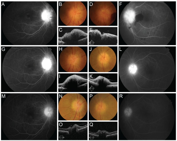
A 58-year-old man with POEMS syndrome was referred to the ophthalmology department with an 1-month history of bilateral blurred vision and an 1-week history of a round blind spot in the left eye. He also had an 8-year history of POEMS syndrome, which had been in remission for the past 7 years, following treatment with chemotherapy combined with steroids for the first 1 year. He was admitted because of generalized edema and pulmonary hypertension, which began 1 week before. He underwent 3-week systemic treatment with high-dose dexamethasone, which was started one day before the first ophthalmologic examination. He did not have any other past medical problems. Best-corrected visual acuity was 20 / 30 in the right eye and 20 / 50 in the left eye. Fundus examination and optical coherence tomography revealed bilateral ODE and subretinal fluid in the left eye, and fluorescein angiography (FA) demonstrated prominent leakage from the optic disc in both eyes (Fig. 1A-1F). Neither intraretinal microvascular abnormalities nor retinal neovascularization were observed (Fig. 1A and 1F).
Fig. 1. (A-F) Initial evaluation with color photos (B,D) and optical coherence tomography (C,E) revealed optic disc edema (ODE) in the right eye (B,C) and in the left eye (D,E). Fluorescein angiography demonstrated prominent leakage in the right eye (A) and in the left eye (F). (G-L) Two months after completion of three monthly injections of intravitreal bevacizumab in the left eye, ODE and leakage were decreased in the left eye (J-L) and remained unchanged in the right eye (G-I). Eight months after completion of three monthly injections of intravitreal bevacizumab in the left eye, ODE and disc leakage were decreased in the right eye (M-O) and no definite ODE or leakage was observed in the left eye (P-R).
After obtaining informed consent, three monthly injections of intravitreal bevacizumab (1.25 mg) were administered to the left eye. Two months after the final injection, both eyes were evaluated with color photos, optical coherence tomography and FA (Fig. 1G-1L). ODE and subretinal fluid showed gradual response and best-corrected visual acuity was improved to 20 / 20 in the left eye (Fig. 1J and 1K). FA also demonstrated decreased disc leakage in the left eye (Fig. 1L). In contrast, right ODE remained unchanged on both optical coherence tomography and FA (Fig. 1G-1I). Eight months after the final injection, ODE and disc leakage were decreased in both eyes and general condition was improved (Fig. 1M-1R). No definite ODE or leakage was observed in the left eye (Fig. 1P-1R).
POEMS syndrome is associated with manifestations of increased vascular permeability such as peripheral edema, ascites, pleural effusion, and ODE [3]. Serum VEGF levels of patients with POEMS have been shown to be significantly higher than those of normal controls and other monoclonal gammopathy patients [2]. As VEGF is a potent vascular permeabilizing cytokine, increased serum VEGF in POEMS is thought to play an important role in inducing extravascular volume overload. The responsiveness of ODE to anti-VEGF treatment in our case supports that ODE in POEMS syndrome should also be considered a symptom of extravascular volume overload, similar to peripheral edema, ascites and pleural effusion.
It is interesting that ODE and subretinal fluid secondary to optic disc leakage were the only structural ocular abnormalities in POEMS syndrome [3]. No peripheral retinal vessels responded to the increase in serum VEGF. This phenomenon can be explained by the differential actions of VEGF and the anatomy of the optic nerve head (ONH). As VEGF receptors VEGFR1 and VEGFR2 show differential apicobasal distribution, only abluminal VEGF can increase permeability in contrast to luminal VEGF that facilitates cytoprotection instead [4]. Moreover, only the ONH has the border tissue of Elschnig in the prelaminar region, which lacks barrier function and leaks material freely [5]. In POEMS patients, plasma with a high concentration of VEGF would leak into the choroidal interstitial fluid through fenestrated choriocapillaris. VEGF would thus diffuse into the ONH through the border tissue of Elschnig and increases the permeability of capillaries of the ONH by affecting vascular endothelial cells abluminally, while the permeability of the retinal capillaries is not affected by luminal VEGF. The FA findings of our study also showed leakage limited to around the ONH.
Our case suggests that intravitreal anti-VEGF injection such as bevacizumab may resolve ODE and lead to visual improvement in POEMS patients.
Footnotes
Conflict of Interest: No potential conflict of interest relevant to this article was reported.
References
- 1.Van Stavern GP. Optic disc edema. Semin Neurol. 2007;27:233–243. doi: 10.1055/s-2007-979684. [DOI] [PubMed] [Google Scholar]
- 2.Watanabe O, Arimura K, Kitajima I, et al. Greatly raised vascular endothelial growth factor (VEGF) in POEMS syndrome. Lancet. 1996;347:702. doi: 10.1016/s0140-6736(96)91261-1. [DOI] [PubMed] [Google Scholar]
- 3.Kaushik M, Pulido JS, Abreu R, et al. Ocular findings in patients with polyneuropathy, organomegaly, endocrinopathy, monoclonal gammopathy, and skin changes syndrome. Ophthalmology. 2011;118:778–782. doi: 10.1016/j.ophtha.2010.08.013. [DOI] [PubMed] [Google Scholar]
- 4.Hudson N, Powner MB, Sarker MH, et al. Differential apicobasal VEGF signaling at vascular blood-neural barriers. Dev Cell. 2014;30:541–552. doi: 10.1016/j.devcel.2014.06.027. [DOI] [PMC free article] [PubMed] [Google Scholar]
- 5.Cohen AI. Is there a potential defect in the blood-retinal barrier at the choroidal level of the optic nerve canal? Invest Ophthalmol. 1973;12:513–519. [PubMed] [Google Scholar]


