

Abstract
Background and Purpose
Movement dysfunction in the trunk and lower extremity (e.g., apparent hamstring tightness) may produce pain, as well as decrease range of motion, function, and performance in athletes. Novel treatments not frequently studied in the literature, such as Total Motion Release® (TMR®) and instrument‐assisted soft ‐tissue mobilization (IASTM), have anecdotal claims of immediate, gross gains of mobility that far exceed conventionally reported results. The purpose of this case report was to examine the efficacy of TMR® in treating an apparent tissue tightness/extensibility dysfunction and to determine if IASTM would improve outcomes if TMR® techniques failed to produce maintained improvement.
Case Description
A 27‐year old former competitive speed walker presented with a chronic history of bilateral pain and posterior leg tightness. The patient met the criteria for diagnosis of a bilateral tissue extensibility dysfunction in the posterior lower extremity and was treated with TMR® and IASTM (Técnica Gavilán®; Tracy, California, United States).
Outcomes
After the first week of treatment, the patient increased her sit and reach by 5cm and her active straight leg raise (ASLR) by an average of 31.5 ° bilaterally. Following the second week of treatment, the patient experienced an additional increase in sit and reach and ASLR. At discharge, the patient displayed negative 90/90 Active Knee Extension, Tripod, and Slump tests bilaterally, normalized ASLR and a resolution of her complaints. Follow‐up examinations completed at one month and three months post‐discharge indicated maintenance of the outcomes without any additional interventions.
Discussion
The subject in this case report demonstrated the potential use of TMR® in classifying apparent hamstring tightness and provided evidence to support the use of TMR® and IASTM in addressing mobility deficits associated with hamstring inflexibility/tightness. Based on these findings, clinicians should consider the use of TMR® to improve classification and treatment of patients with a chief complaint of hamstring “tightness.”
Level of Evidence
Level 4; single case report.
Keywords: Apparent hamstring tightness, tissue extensibility dysfunction, total motion release
BACKGROUND AND PURPOSE
Tissue extensibility dysfunction (TED), joint mobility dysfunction (JMD), and/or stability motor control dysfunction (SMCD) in the trunk and lower extremity have been theorized to produce pain, as well as decrease range of motion (ROM), function, and performance in athletes.1–3 A variety of potential causes, including muscular inflexibility,1 musculoskeletal injury,4,5 loss of normal neurodynamics,6 or neuromuscular protection patterns1 may result in a patient presenting with decreased mobility at the trunk and hip. A clinician should be able to efficiently identify the underlying cause of the patient's reduced mobility, dysfunction, and/or pain to best match the appropriate intervention to presentation of the individual patient.
Several clinicians (e.g., Mulligan7, Dalonzo‐Baker8) suggest that rapid and immediate changes in many clinical signs and symptoms may occur when “apparent hamstring inflexibility/tightness” is found during clinical examination. The reported anecdotal expectations of these clinicians, however, far exceed what can be expected if “true” muscular tissue tightness is present.7,8 Thus, the classification of “apparent hamstring inflexibility/tightness” is used when a patient presents with positive findings using traditional evaluative measures (e.g., 90/90 Active Knee Extension Test, etc.), but does not have “true” tissue length/extensibility as the root cause of the patient's dysfunction. “True” tissue tightness would only be considered when a length deficit resulting from an inability of tissue to appropriately elongate/extend10 is present and this condition should require a tissue remodeling intervention for resolution. Given the range of possible causes of conditions that present as apparent muscular tightness/extensibility dysfunction, it is not surprising that intervention and injury surveillance research has produced such disparate and contradictory results.10–14 Although the underlying mechanisms resulting in apparent hamstring tightness/extensibility dysfunction are not well understood, the possibility2,7,8 of immediate, gross gains of mobility that far exceed conventionally reported results using interventions designed to increase tissue length/extensibility12–15 requires investigation.
Total Motion Release®
Total Motion Release® (TMR®) is a technique based on the assessment of asymmetry within the body.16 Tom Dalonzo‐Baker, the founder of TMR®, has suggested that whenever apparent hamstring tightness exists, forward trunk flexion with rotation should be tested both with the feet together and the feet apart (Figure 1‐3).17 The TMR® forward flexion trunk twist (FFTT) technique for hamstring tightness may be performed in either a standing or long‐sitting position.8 The technique requires the patient to forward bend with feet together and then rotate the trunk to the right and to the left while identifying the “good side” (e.g., side with more motion, smoother motion, less pain). The patient grades each side using a 0‐100 scale, where 0 = “no problems at all” and 100 = “the worst”.8 Once the “good side” is identified, the patient is then asked to do 10 rotation repetitions to the good side to end range of motion or to hold the rotation at the terminal range of motion for 20‐30 seconds. During the repetitions, the patient is asked to release what he/she feels is preventing further motion (e.g., bending one knee, extending the trunk to finish the rotation) whenever resistance is experienced during the movement. The patient completes this movement/exercise in sets (e.g., two sets of 10 repetitions, two sets of 20 second isometric holds), while re‐checking their primary complaint after completing the exercise sets. Typically, the patient will continue to perform the TMR® movement/exercise until completing sets of the movement no longer improves the primary complaint (e.g., performing a “toe‐touch”).
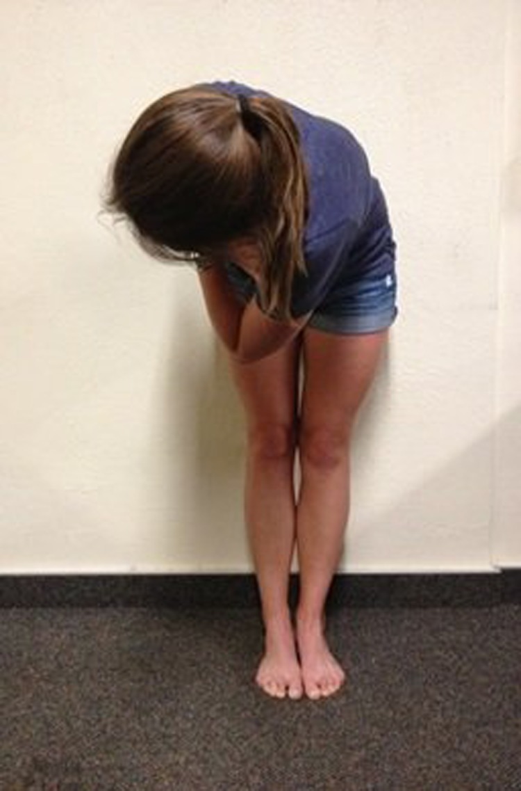
Figure 1.

Total Motion Release® Hamstring Technique feet together starting position.
Figure 3.

Total Motion Release® Hamstring Technique testing trunk rotation to the right with feet apart.
Figure 2.
Total Motion Release® Hamstring Technique testing trunk rotation to the right with feet together.
After completing the initial treatment, the patient may be asked to test a more advanced motion in the TMR® system. The next motion test for the apparent hamstring technique requires the patient to place the feet in a straddle position, several feet apart (e.g., shoulder width). The patient determines the “good side” by rotating the trunk to the left and right over each leg. After identifying the “best” out of the four motions, the patient performs the same treatment process (i.e., rotation until a barrier is felt and then the restricting motion segment is released to continue the motion).
Following the completion of treatment, the patient should reassess his/her movement within the TMR® system (e.g., forward trunk flexion with rotation) and against the primary complaint (e.g., toe‐touch). The expectation is that a dramatic increase in motion with a decrease in symptoms will occur; the change should far exceed what would be expected according to tissue remodeling theory typically employed with traditional interventions (e.g., static stretching). It is important to note that if neither of the TMR® motion tests demonstrate appreciable differences between sides on the 0‐100 scale, then the patient would likely benefit from other interventions in lieu of the TMR® hamstring tightness techniques.8 If effective, the TMR® technique could not only serve as an effective intervention, but may also work as an effective screening tool used to identify patients who need an intervention that would work through a different mechanism of action (e.g. static stretching).
Instrument Assisted Soft Tissue Mobilization
Instrument Assisted Soft Tissue Mobilization (IASTM) is a non‐invasive technique that may be utilized in combination with TMR® when it appears tissue remodeling may be necessary to restore full function. Researchers have suggested that the use of IASTM initiates a healing cascade, reduces soft tissue adhesion, mechanically mobilizes fascial tissues, and improves tissue healing.18–21 Most IASTM techniques are performed statically, while the Técnica Gavilán® (Tracy, California, United States) method incorporates IASTM with movement and loading (e.g., stretching, muscular contraction) of the tissue being treated.22 In theory, using the Técnica Gavilán® method is beneficial to the healing process because it combines the benefits of mechanical load with therapeutic exercise during IASTM treatments.3,22
The purpose of this case report was to examine the efficacy of TMR® in treating an apparent tissue tightness/extensibility dysfunction and to determine if IASTM would improve outcomes if TMR® techniques failed to produce maintained improvement. The case report was guided by three clinical research questions: 1) If a patient presents with apparent TED, will movement impairments be present in loaded and unloaded positions? 2) In this patient, did the TMR® FFTT hamstring tightness technique decrease the level of disablement as measured by the Numeric Rating Scale (NRS), goniometric active ROM measurements, and special tests (i.e., standing flexion, sit and reach, 90/90 Active Knee Extension test, Tripod test, and Slump test)? 3) In this patient, would the use of IASTM promote treatment effectiveness if the clinician perceives the effects of the basic TMR® FFTT hamstring technique had plateaued?
CASE DESCRIPTION: SUBJECT HISTORY AND SYSTEMS REVIEW
The subject, a 27‐year old former competitive speed walker, presented with a chronic history (over five years) of bilateral pain and posterior leg tightness, which included discomfort during activities of daily living (ADLs). She presented without any previous history of trauma or injury to her lower extremities, but had a previous lumbar spine pathology that had been resolved without recurrence for 10 years. The subject experienced tingling and pain through her posterior thigh during hamstring stretching activities, but these sensations did not progress below the knee. She also reported sensations of tightness and discomfort during ADLs which placed her posterior leg on stretch, but otherwise did not have any other complaints in the spine or lower extremity. She reported receiving various traditional treatments (e.g., thermal modalities, stretching) over the previous five years without improvement in her symptoms. The history portion of the exam did not reveal any red flag symptoms and the subject reported being otherwise healthy. The study was approved by the University of Idaho Institutional Review Board and the subject provided written consent for participation in the case study.
CLINICAL IMPRESSION #1
“True” hamstring tightness is commonly addressed using treatments focused on lengthening the soft tissue (e.g., muscle, fascia, etc.). As the subject had not reported any previous improvement following traditional warm‐up and stretching interventions, the cause of the subject's chief complaint was hypothesized to be a result of apparent (i.e., non‐tissue length related) hamstring tightness/extensibility dysfunction. Further evaluation needed to be performed to identify whether the subject could be classified with true versus apparent hamstring tightness based on traditional evaluation techniques.
EXAMINATION
Clinical evaluation did not reveal obvious deformity, erythema, edema, ecchymosis, and gait or postural abnormalities. Physical examination revealed palpable tension in her posterior thigh/leg, but she did not report any other point tenderness in her spine, sacroiliac joint, or lower extremities. Active ROM assessment revealed decreased active straight leg raise (SLR) (61°on left leg, 56° on right leg) bilaterally that produced pain rated 5/10 on the NRS. She reported the pain felt like a “burning” sensation and traveled from her proximal thigh to just proximal to her knee. Passive SLR assessment produced a firm end‐feel with equal ROM deficiency when compared to active ROM and the subject reported experiencing the same pain (i.e., severity and patterning) during testing. All other hip (i.e., abduction, adduction, extension, internal rotation, external rotation, and bent knee hip flexion) and ankle (i.e., dorsiflexion, plantarflexion, inversion, and eversion) ranges of motion were within normal limits actively and passively. The subject's manual muscle tests (including hip flexion, hip extension, hip internal rotation, hip external rotation, hip adduction, hip adduction, knee flexion, knee extension) were normal (5/5) bilaterally. Orthopedic special tests were negative for fracture, disc pathology, facet, and hip pathology; however the 90/90 Active Knee Extension (AKE), Tripod, and Slump Tests were positive bilaterally.
The subject also displayed a flexion limitation in a weight‐bearing (i.e., standing multi‐segmental flexion) position and experienced pain with that movement in her posterior thigh (i.e., approximately mid‐thigh to just proximal to popliteal fossa). She also demonstrated a flexion limitation in a non‐weight‐bearing (i.e., seated multi‐segmental flexion) position with a sacral angle of less than 70° (Table 1) and experienced pain with that movement in her posterior thigh. The subject had normal, pain‐free motion when pulling her knees to her chest (i.e., passive hip and lumbar flexion). When asked to perform the TMR® trunk twist to each side, the subject reported that it was much easier to twist to her right (thus, defined as the good side). Within the TMR® grading system, the subject rated her trunk twist to the right a 0/100 and the trunk twist to the left a 70/100. To further rule out other lumbar spine involvement, a second clinician, who had completed the McKenzie Mechanical Diagnosis and Therapy (MDT) diploma program, completed an MDT exam and cleared the lumbar spine for possible involvement. The lower quarter screen was also unremarkable. The subject reported a Patient Specific Functional Scale (PSFS) score of 4 out of 10.
Table 1.
Range of Motion Measurement Techniques and Procedures.
| Measurement* | Measurement Device | Patient Position | Patient Instructions | Test Performance |
|---|---|---|---|---|
| Sit & Reach23 | Sit & Reach Box | Long‐sit position | Keep your feet together and your knees extended and slowly bend forward as far as you can and hold the furthest position for 2 seconds. Please keep your hands overlapped and maintain contact with the measuring portion of the box. | Record the distance based on the most distal point reached and held with the fingertips. |
| Finger‐Floor Distance Test24 | Ruler | Standing | Keep your feet together and your knees extended and slowly bend forward as far as you can as you try to touch the floor with your fingers/hands | Measure the distance between the tip of the patient's middle finger and the floor |
| 90/90 (AKE) Test24 | Plastic Universal Goniometer | Supine, with 90° of hip and knee flexion; 90° of hip flexion maintained during procedure | Extend knee until tension is felt in posterior thigh | Measure the knee flexion angle using the lateral femoral epicondyle as the fulcrum, the lateral midline of the fibula as the distal arm, and the lateral midline of the femur as the stationary arm |
| ASLR Flexion24 | Plastic Universal Goniometer | Supine with both hips at 0° of rotation, adduction, and abduction | Extend your knee, plantarflex your foot, and lift your leg until tension is felt in posterior thigh | Measure the amount of hip flexion using the greater trochanter as the fulcrum, the lateral midline of the femur as the distal arm, and the lateral midline of the pelvis as the proximal arm |
| Tripod (Sign) Test | Plastic Universal Goniometer | Short‐seated with knees flexed to 90°at the edge of the plinth25 | None | Passive extension of the knee until the trunk extends or tension is felt in posterior thigh.25 Measure the knee flexion angle using the lateral femoral epicondyle as the fulcrum, the lateral midline of the fibula as the distal arm, and the lateral midline of the femur as the stationary arm.** |
| Sacral Angle1 | Gravity Dependent Inclinometer | Long‐sit position | Extend your knees/legs and slowly bend forward and touch your toes. | Place inclinometer over median sacral crests withdistal inclinometer on sacral base with; measure angle when motion is complete |
The same devices (e.g., goniometer) were used by the same clinician for all measurements.
Measurement of the knee flexion angle is not traditionally part of the Tripod (Sign) Test, but was used as to make the test objectively measurable.
CLINICAL IMPRESSION #2
The working clinical diagnosis was an apparent lower extremity TED based on the ROM measurements, special tests, and TMR® trunk twist assessments. As the subject's complaints were consistent with the examination results and traditional treatments had failed to improve her symptoms previously, treatment was focused on her movement asymmetry during the TMR® trunk twist assessment. It was theorized that utilizing TMR® to address the trunk twist asymmetry could assist in resolving any underlying core SMCD and neural tension issues that may have been contributing to her dysfunction. Follow‐up time points were chosen, one and three months post‐discharge.
INTERVENTION
After initial examination, a treatment protocol using only TMR® was performed by the same clinician during the first week of treatment. The TMR® technique utilized has been suggested for use to address apparent hamstring flexibility dysfunction.17 The intervention began with a five‐minute warm‐up on a stationary bicycle at a set resistance followed by the completion of all measurements (Table 1). The TMR® FFTT technique utilized involved forward flexion trunk rotations (1 set of 10 repetitions to end‐range without an isometric hold) to the subject's side with less restriction with her feet next to each other and bent‐over trunk rotations (1 set of 10 repetitions to end‐range without an isometric hold) to the side with less restriction with her feet shoulder width apart. On each repetition, when resistance during the movement was felt, the subject was asked to release the body part/segment she felt was preventing further motion (e.g., bending one knee, extending the trunk) to finish the rotation (Figure 4). All of her measurements were then reassessed. The subject was instructed to avoid any additional treatment (e.g., stretching, thermal modalities), but was otherwise not restricted from any activity. The TMR® treatment was applied three times on non‐consecutive days during the first week as a means for addressing movement asymmetries/dysfunction and required less than two minutes to complete. The subject was not given a home exercise program to complete between clinic visits and agreed to not perform any other interventions (e.g., stretching) while receiving treatment.
Figure 4.

Example of the “knee release” when moving into end‐range trunk rotation while performing treatment using the Total Motion Release® Hamstring Technique.
The second week of treatment included a continuation of the TMR® technique, followed by the application of IASTM with movement to the hamstring (two minutes) and triceps surae (one minute) muscle groups on each leg. When treating the hamstring muscle groups, the patient was placed in a supine position so that she could perform active‐assistive hip flexion/extension through a pain‐free ROM while the clinician applied the instruments. When treating the triceps surae muscle groups, the patient was placed in a prone position so that she could actively move her ankle through pain‐free dorsiflexion/plantarflexion while the clinician applied the instruments. The IASTM application was added as an intervention to address potential true TED symptoms that appeared to be limiting maintained improvements from the third TMR® treatment (i.e., Day 5 post‐measurement to Day 8 pre‐measurement). During this week, the subject began with a five‐minute warm‐up on a stationary bicycle at a set resistance followed by the completion of all measurements. The TMR® technique was then applied and all measurements were taken again. The IASTM technique was then applied and the measurements were completed a final time. All IASTM treatments were provided by a clinician who was certified by Técnica Gavilán® (Tracy, CA). The treatment protocol was also applied three times on non‐consecutive days during the second week. Over the course of her care, all treatments and measurements were provided/collected by the same clinician.
OUTCOMES
After the first week of treatment (TMR® only), the subject increased her sit and reach by 5cm and her SLR hip flexion by an average 31.5 ° bilaterally and a resolution of her complaints with ADLs as reported on the PSFS (Table 2). Following the second week of treatment (TMR® and IASTM), the subject experienced an additional increase in sit and reach (5cm) and SLR hip flexion (7.5 ° bilaterally) (Table 3). Additionally, the subject displayed negative 90/90 AKE, Tripod, and Slump tests bilaterally at discharge. Following the sixth treatment, 12 days after initial exam, the subject was discharged with normal ROM, a TMR trunk twist score of 0/100 in both directions, and a resolution of her complaints (i.e., PSFS score of 10/10). Over the six treatments, the subject reported a five‐point decrease in pain on the NRS, representing a change that exceeds the established standards for a minimal clinically important difference (MCID) of two points.26,27 Follow‐up examinations were completed at one and three months post‐discharge and indicated maintenance of the final measurements and a continued resolution of symptoms without any additional interventions (Table 3).
Table 2.
Results of TMR® Treatment During the First Week of Treatment.
| Day 1 | Day 3 | Day 5 | Day 8 | ||||
|---|---|---|---|---|---|---|---|
| Outcome | Pre‐TMR | Post‐TMR | Pre‐TMR | Post‐TMR | Pre‐TMR | Post‐TMR | Pre‐TX |
| Sit & Reach | 26.2cm | 31.1cm | 29.8cm | 33.3cm | 30.6cm | 35.3cm | 31.1cm |
| Finger‐Floor Distance Test | ‐5.3cm | IPs and MCPs on floor | 0cm (Fingertips to floor) | IPs and MCPs on floor | DIPs on floor | MCPs on floor, distal metacarpals on floor | DIPs on floor |
| 90/90 (AKE) Test | 32°(L), 28°(R) | 52°(L), 58°(R) | 49°(L), 50°(R) | 68°(L), 64°(R) | 69°(L), 62°(R) | 79°(L), 74°(R) | 69°(L), 64°(R) |
| ASLR | 61°(L), 56°(R) | 85°(L), 82°(R) | 84°(L), 78°(R) | 87°(L), 88°(R) | 88°(L), 89°(R) | 94°(L), 92°(R) | 94°(L), 86°(R) |
| Tripod (Sign) Test | 66°(L), 62°(R) | 81°(L), 71°(R) | 74°(L), 71°(R) | 79°(L), 83°(R) | 83°(L), 82°(R) | 89°(L), 87°(R) | 84°(L), 82°(R) |
| Slump Test | “+” bilaterally w/DF (3/10 on left and 5/10 on right) | “‐“ on L, R “+” with DF, but 1/10 | “+” with DF bilaterally; 1/10 on L and 3/10 on R | “‐“ on L, R “+” at end of DF, but 1/10 | “+” with DF bilaterally; 1/10 on L and 3/10 on R | “‐“ on L, R “+” at end of DF, but 1/10 | “+” with DF bilaterally; 2/10 on L and 4/10 on R |
| PSFS Score | 4 | 10 | 10 | 10 | 10 | 10 | 10 |
AKE = Active Knee Extension, ASLR = Active Straight Leg Raise, PSFS = Patient Specific Functional Scale, TMR = Total Motion Release, TX = Treatment, IPs = Interphalangeal Joints, MCPs = Metacarpophalangeal Joints, DIPs – Distal Interphalangeal Joints, L = Left, R = Right, DF = Dorsiflexion, “+” = positive, “‐“ = negative.
Table 3.
Results of TMR® and IASTM Treatment During 2nd Week and at Follow‐up Exams.
| Day 8 | Day 10 | Day 12 | Day 15 | Day 43 | Day 94 | |||||||
|---|---|---|---|---|---|---|---|---|---|---|---|---|
| Outcome | Pre‐TX | Post‐TMR | Post‐IASTM | Pre‐TX | Post‐TMR | Post‐IASTM | Pre‐TX | Post‐TMR | Post‐IASTM | Final | Final | Final |
| Sit & Reach | 31.1cm | 35.1cm | 36.8cm | 33cm | 35.7cm | 37.9cm | 35.1cm | 37.7cm | 38.4cm | 37.3cm | 37.4cm | 36.2cm |
| Finger‐Floor Distance Test | DIPs on floor | MCPs & distal metacarpals on floor | Distal carpals on floor | MCPs on floor, distal metacarpals on floor | Distal carpals on floor | Palms flat on floor & position maintained | Palms flat on floor | Palms flat on floor & position maintained | Palms flat on floor & “feels” like she can go further | Palms flat on floor & position maintained | Palms flat on floor & position maintained | Palms flat on floor & position maintained |
| 90/90 (AKE) Test | 69°(L), 64°(R) | 77°(L), 74°(R) | 86°(L), 82°(R) | 82°(L), 78°(R) | 86°(L), 80°(R) | 90°(L), 86°(R) | 89°(L), 83°(R) | 89°(L), 84°(R) | 90°(L), 90°(R) | 90°(L), 90°(R) | 90°(L), 90°(R) | 90°(L), 90°(R) |
| ASLR | 94°(L), 86°(R) | 94°(L), 89°(R) | 100°(L, 94°(R) | 99°(L), 94°(R) | 101°(L), 99°(R) | 106°(L), 103°(R) | 103°(L), 101°(R) | 104°(L), 101°(R) | 106°(L), 105°(R) | 106°(L), 104°(R) | 106°(L), 103°(R) | 103°(L), 102°(R) |
| Tripod (Sign) Test | 84°(L), 82°(R) | 84°(L), 85°(R) | 90°(L), 89°(R) | 89°(L), 86°(R) | 90°(L), 89°(R) | 90°(L), 90°(R) | 90°(L), 88°(R) | 90°(L), 90°(R) | 90°(L), 90°(R) | 90°(L), 90°(R) | 90°(L), 90°(R) | 90°(L), 90°(R) |
| Slump Test | “+” with DF bilaterally; 2/10 on L & 4/10 on R | “‐“ on L, R “+” at end of DF, but 1/10 | “‐“ on L, R “+” at end of DF, but 1/10 | “‐“ on L, R “+” at end of DF, but 1/10 | “‐“ on L, R “+” at end of DF, but 1/10 | Negative bilaterally | “‐“ on L, R “+” at end of DF, but 1/10 | “‐“ on L, R “+” at end of DF, but 1/10 | Negative bilaterally | “‐“ on L, R “+” at end of DF, but 1/10 with overpressure | “‐“ on L, R “+” at end of DF, but 1/10 with overpressure | “‐“ on L, R “+” at end of DF, but 1/10 with overpressure |
| PSFS Score | 10 | ‐ | 10 | 10 | ‐ | 10 | 10 | ‐ | 10 | 10 | 10 | 10 |
AKE = Active Knee Extension, ASLR = Active Straight Leg Raise, PSFS = Patient Specific Functional Scale, TMR = Total Motion Release, IASTM = Instrument Assisted Soft Tissue Mobilization, TX = Treatment, IPs = Interphalangeal Joints, MCPs = Metacarpophalangeal Joints, DIPs – Distal Interphalangeal Joints, L = Left, R = Right, DF = Dorsiflexion, “+” = positive, “‐“ = negative.
DISCUSSION
This case report was performed to investigate the effectiveness of TMR® and IASTM to treat apparent chronic hamstring tightness and pain that was identified with traditional hamstring flexibility measures. The interventions were effective at addressing subject dysfunction as indicated by the normalized ROM, resolution of pain (clinically significant changes), and the subject being asymptomatic at discharge exam in a total of six treatments over two weeks. Total Motion Release® was selected as an intervention because it is advocated to rapidly change apparent hamstring tightness and the subject presented with an asymmetrical movement pattern during TMR® testing. The IASTM intervention was selected because it is indicated for patients classified with a TED.3
Tissue extensibility dysfunction, especially in the hamstring muscle group, is a common complaint in physically active individuals.1,28–30 Various research studies have been conducted to determine the effect of different stretching protocols on improving hamstring flexibility. For example, a recent investigation using the American College of Sports Medicine stretching guidelines demonstrated an average increase of 16.6 ° of hip flexion in subjects, over a 12 week static stretching program.31 In a male athletic population, static stretching produced an increase of 12 ° in knee extension after a six‐week static stretching program.12 Similarly, use of a six‐week proprioceptive neuromuscular facilitation (PNF) stretching protocol demonstrated an increase of 19 ° in knee extension in subjects.32 Flexibility improvements associated with stretching protocols are found for both static and PNF stretching, but the findings also suggest the improvements required repetitive application of the stretching protocol performed over a long period of time. Few studies, if any, establish criteria for determining if participants should be included in a stretching protocol by determining if the apparent ROM deficiencies truly need to be addressed through tissue lengthening interventions, which may be necessary given certain findings.
For example, there is evidence in the literature to support the theory that joint33,34 and soft‐tissue3 mobilization with movement can improve ROM measures for patients with apparent hamstring tightness. The traction SLR is a joint mobilization with movement indicated when patients present with a limited SLR and posterior thigh pain that does not extend below the knee.7,33,35 In a case report, researchers found a 21 ° increase in hip flexion SLR and an elimination of pain following two treatments of the traction SLR.36 Other researchers have compared the traction SLR to stretching and found that it was significantly more effective than both static and PNF stretching.34 Similarly, the use of IASTM with movement using Técnica Gavilán® in three patients classified with a TED resulted in an average ROM increase of over 44° in SLR measures in an average of 7.33 treatments.3 Another option to address reduced hamstring ROM is the use of neurodynamic sliding of the sciatic nerve. In patients with reduced hamstring ROM on a passive SLR, neurodynamic sciatic sliding has resulted in improved ROM compared to static stretching, placebo, and control groups.11,37 Following a single treatment session, subjects receiving neurodynamic sciatic sliding intervention improved by 9.86o while subjects in the static stretching group improved by 5.5o and the placebo group improved by 0.03o on a passive SLR measurement.11 Similar results have been demonstrated following a one week course (three treatment sessions) of neurodynamics with subjects in the sciatic sliding intervention group improving by 9.4o compared to 0.2o in the control group.37
Although there are currently no published studies about the TMR® treatment for apparent hamstring tightness, researchers have provided evidence supporting the underlying theories of neural coupling and cross education. Neural coupling is the brain‐based modulation and coordination of sensation and movement that occurs within the body.37,39 Researchers40,41 demonstrated that movement of the upper limbs promotes neuromuscular activation of the lower limbs, possibly providing support to the theory that trunk and upper body rotation during a TMR® treatment may have an impact on trunk and lower extremity movement. Completing the “release” maneuver (e.g., bending knee, extending trunk) increases total body movement and allows for maximal motion during the activity, which may optimize neuromuscular activity through the trunk and lower extremity. Other researchers16 who addressed ROM deficits in the upper extremity found significant and clinically meaningful increases in shoulder mobility following a TMR® treatment compared to a traditional warm‐up protocol. Additionally, performing a motion on the good side may have a cross education effect, resulting in bilateral improvements, as has been demonstrated in the literature following stretching,42 strengthening,43 and surgical procedures.44
In this case report, the use of TMR® alone improved “loaded” (i.e., standing hip flexion) and “unloaded” (i.e., sit and reach) functional measurements, while also producing an increase of 30 ° (56 ° to 86 °) on the right side and of 33 ° (61 ° to 94 °) on the left side during active SLR, with an increase of 36 ° (28 ° to 64 °) increase in right knee extension and a 37 ° increase in left knee extension (32 ° to 69 °) measured using the Active 90/90 Knee Extension Test in three treatments of two minutes over five days. Additionally, the patient's complaints during ADLs were resolved after the first TMR® treatment as reported on the PSFS. The combination of TMR® with IASTM (three additional treatments over five days) continued to produce patient improvement in both the “loaded” and “unloaded” functional measurements, while also resulting in SLR measurements greater than 100 ° bilaterally. Additionally, the Active 90/90 AKE, Tripod, and Slump tests were no longer positive at discharge. The use of TMR® and IASTM produced greater results at a faster rate than is reported in the literature for common stretching protocols and the gains were maintained at three month follow‐up without further intervention. The findings of this case report illuminate the potential need to re‐examine the clinical paradigm that leads clinicians to recommending a stretching protocol for a patient.
Limitations and Future Research
As the results of a single case report cannot be generalized to other patients, additional research is needed to determine the effectiveness of this protocol in a greater number of subjects. In this case report, the clinician and patient were not blinded to the treatment or outcomes, which could bias the results. Within the TMR® system, there is potential that performing more than one set of the technique, performing the technique multiple times per day, or treating another area of the body presenting with greater asymmetry in movement may produce better outcomes. Designing a randomized control trial that uses a greater number of research participants, utilizes a control group, blinds researchers, and examines different TMR® interventions would be useful in determining the value of TMR® for improving movement asymmetries and ROM.
CONCLUSION
The combined treatment interventions of TMR® and IASTM resolved the subject's complaints in six visits and improved the patient's dysfunction in “loaded” and “unloaded” positions. Additionally, the results were maintained without further intervention at the one and three month follow‐up exams. The present case report is an example of the potential need to re‐examine the clinical paradigm that leads clinicians to recommending a traditional stretching protocol for a patient.
REFERENCES
- 1.Cook G. Movement: Functional Movement Systems: Screening, Assessment and Corrective Strategies. Aptos, CA: On Target Publications;2010. [Google Scholar]
- 2.Loutsch RA Baker RT May JM Nasypany AM. Reactive neuromuscular training results in immediate and long‐term improvements of measures of hamstring flexibility: A case report. Int J Sports Phys Ther. 2015;10(3):371‐377. [PMC free article] [PubMed] [Google Scholar]
- 3.Baker RT Nasypany A Seegmiller JG Baker JG. Instrument‐assisted soft tissue mobilization treatment for tissue extensibility dysfunction. Int J Ath Ther Train. 2013; 18(5):16‐21. [Google Scholar]
- 4.Jarvinen T Jarvinen T Kaariainen M Kalimo H Jarvinen M. Muscle injuries: Biology and treatment. Am J Sports Med. 2005;33(5):745‐766. [DOI] [PubMed] [Google Scholar]
- 5.Melham TJ Sevier TL Malnofski MJ Wilson JK Helfst RH. Chronic ankle pain and fibrosis successfully treated with a new noninvasive augmented soft tissue mobilization technique (ATSM): A case report. Med Sci Sports Exerc. 1998;30(6):801‐804. [DOI] [PubMed] [Google Scholar]
- 6.Butler D. The Sensitive Nervous System. Adelaide, Australia: Noigroup Publications;2000. [Google Scholar]
- 7.Mulligan BR. Manual Therapy. NAGS, SNAGS, MWMs, etc. Wellington, New Zealand: Plane View Services, Ltd; 2010. [Google Scholar]
- 8.Dalonzo‐Baker T. Total Motion Release Seminars. Total Motion Release; 2012. [Google Scholar]
- 9.Sherry MA Best TM. A comparison of 2 rehabilitation programs in the treatment of acute hamstring strains. J Orthop Sports Phys Ther. 2004;34(3):116‐125. [DOI] [PubMed] [Google Scholar]
- 10.Kendall FP McCreary EK Provance PG Rodgers MM Romani WA. Muscles, Testing and Function with Posture and Pain. 5th ed. Baltimore, MD: Lippincott Williams & Wilkins;2005.. [Google Scholar]
- 11.Castellote‐Caballero Y Valenza M C E, et al. Immediate Effects of Neurodynamic Sliding versus Muscle Stretching on Hamstring Flexibility in Subjects with Short Hamstring Syndrome. J Sports Med. 2014;2014:e127471. [DOI] [PMC free article] [PubMed] [Google Scholar]
- 12.Davis DS Ashby PE McCale KL McQuain JA Wine JM. The effectiveness of 3 stretching techniques on hamstring flexibility using consistent stretching parameters. J Strength Cond Res. 2005;19(1):27‐32. [DOI] [PubMed] [Google Scholar]
- 13.Lim KI Nam HC Jung KS. Effects on Hamstring Muscle Extensibility, Muscle Activity, and Balance of Different Stretching Techniques. J Phys Ther Sci. 2014;26:209‐213. [DOI] [PMC free article] [PubMed] [Google Scholar]
- 14.Puentedura EJ Huijbregts PA Celeste S, et al. Immediate Effects of Quantified Hamstring Stretching: Hold‐Relax Proprioceptive Neuromuscular Facilitation Versus Static Stretching. Phys Ther Sport. 2011;12:122‐126. [DOI] [PubMed] [Google Scholar]
- 15.Draper DO Castro JL Feland B Schulthies S Eggett D. Shortwave diathermy and prolonged stretching increase hamstring flexibility more than prolonged stretching alone. J Orthop Sports Phys Ther. 2004;34(1):13–20. [DOI] [PubMed] [Google Scholar]
- 16.Gamma S Baker RT Iorio S Nasypany A Seegmiller JG. Total motion release warm‐up improves dominant arm shoulder internal and external rotation in baseball players. Int J Sports Phys Ther. 2014;9(4):509‐517. [PMC free article] [PubMed] [Google Scholar]
- 17.Dalonzo‐Baker T. Getting rid of tight hamstrings quickly – the TMR way: Part 2. [Video]. Retrieved from https://www.youtube.com/watch?v=oCMnMpUlGHo. Last update 9 March 2012. Accessed 9 June 2015.
- 18.Sevier TL Gehlsen GM Wilson JK Stover SA Helfst RH. Traditional physical therapy vs. graston augmented soft tissue mobilization in the treatment of lateral epicondyilitis. Med Sci Sports Exerc. 1995;27(5):S52. [Google Scholar]
- 19.Davidson CJ Gavion LR Gehlsen GM, et al. Rat tendon morphologic and functional changes resulting from soft tissue mobilization. Med Sci Sports Exerc. 1997;29:313‐319. [DOI] [PubMed] [Google Scholar]
- 20.Gehlsen GM Ganion LR Helfst R. Fibroblast responses to variation in soft tissue mobilization pressure. Med Sci Sports Exerc. 1999;31(4):531‐535. [DOI] [PubMed] [Google Scholar]
- 21.Loghmani MT Stuart JW. Instrument‐Assisted cross fiber massage accelerates knee ligament healing. J Orthop Sports Phys Ther. 2009;39(7):506‐514. [DOI] [PubMed] [Google Scholar]
- 22.Lang G. Técnica Gavilán. Hands‐on Workshop presented at California Baptist University; Jan 2011; Riverside, CA.
- 23.Medicine AC. ACSM's Guidelines for Exercise Testing and Prescription. 9th ed. Baltimore, MD: Lippincott Williams & Wilkins;2014. [Google Scholar]
- 24.Norkin CC White DJ. Measurement of Joint Motion – A Guide to Goniometry. 4th ed. Philadelphia, PA: F.A. Davis Co;2009. [Google Scholar]
- 25.Magee David J. Orthopedic physical assessment. Elsevier Health Sciences, 2014. [Google Scholar]
- 26.Pool JJ Ostelo RW Hoving JL Bouter LM de Vet HC. Minimal clinically important change of the neck disability index and the numerical rating scale for patients with neck pain. Spine. 2007;32(26):3047‐3051. [DOI] [PubMed] [Google Scholar]
- 27.Farrar JT Young JP LaMoreaux L Werth JL Poole M. Clinical importance of changes in chronic pain intensity measured on an 11‐point numerical pain rating scale. Pain. 2001;94:149‐158. [DOI] [PubMed] [Google Scholar]
- 28.Chandler TJ Kibler WB Uhl TL Wooten B Kiser A Stone E. Flexibility comparisons of junior elite tennis players to other athletes. Am J Sports Med. 1990;18(2):134–136. [DOI] [PubMed] [Google Scholar]
- 29.Mangine RE Noyes FR Mullen MP Barber SD. A Physiological Profile of the Elite Soccer Athlete 1. J Orthop Sports Phys Ther. 1990:12(4):147–152. [DOI] [PubMed] [Google Scholar]
- 30.Weerasekara I. The Prevalence of Hamstring Tightness among the Male Athletes of University of Peradeniya in 2010, Sri Lanka. Int J Phys Med Rehab. 2013;01(01). [Google Scholar]
- 31.Sainz de Baranda PS Ayala F. Chronic flexibility improvement after 12 week of stretching program utilizing the ACSM recommendations: Hamstring flexibility. Sports Med. 2010;31:389‐396. [DOI] [PubMed] [Google Scholar]
- 32.Yuktasir B Kaya F. Investigation into the long‐term effects of static and PNF stretching exercises on range of motion and jump performance. J Bodyw Mov Ther. 2009;13(1):11–21. [DOI] [PubMed] [Google Scholar]
- 33.Hall T Hardt S Schäfer A Wallin L. Mulligan bent leg raise technique—a preliminary randomized trial of immediate effects after a single intervention. Man Ther. 2006;11(2):130–135. [DOI] [PubMed] [Google Scholar]
- 34.Hall T Cacho A McNee C Riches J Walsh J. Effects of the mulligan traction straight leg raise technique on range of movement. J Man Manip Ther. 2001;9(3):128. [Google Scholar]
- 35.Pratishtha K Jagga V. Effect of mulligan stretching techniques (TSLR AND BLR) on biceps femoris muscle and pelvic rotation by using surface EMG and bubble inclinometer respectively. J Exerc Sci Phys. 2012;8(1):39‐42. [Google Scholar]
- 36.Eusea J, Nasypany A, Baker R, Seegmiller J. The mulligan traction straight leg raise: A case report. Case study abstract presented at the American College of Sports Medicine's Annual Meeting: San Diego, CA; 2014.
- 37.Castellote‐Caballero Y Puentedura EJ Penas CF Valenza MC Martin LM Martos IC. Effects of a neurodynamic sliding technique on hamstring flexibility in healthy male soccer players: A pilot study. Physical Therapy in Sport. 2013;14(3):156‐162. [DOI] [PubMed] [Google Scholar]
- 38.Balter JE Zehr EP. Neural coupling between the arms and legs during rhythmic locomotor‐like cycling movement. J Neurophys. 2007;97(2):1809‐1818. [DOI] [PubMed] [Google Scholar]
- 39.Zehr EP Duysens J. Regulation of arm and leg movement during human locomotion. Neuroscientist. 2004;10(4):347‐361. [DOI] [PubMed] [Google Scholar]
- 40.Huang HJ Ferris DP. Neural coupling between upper and lower limbs during recumbent stepping. J Appl Physiol. 2004;97(4):1299‐1308. [DOI] [PubMed] [Google Scholar]
- 41.Huang HJ Ferris DP. Upper and lower limb muscle activation is bidirectionally and ipsilaterally coupled. Med Sci Sports Exerc. 2009;41(9):1778‐1789. [DOI] [PMC free article] [PubMed] [Google Scholar]
- 42.Kofotolis ND Kellis E. Cross‐training effects of a proprioceptive neuromuscular facilitation exercise programme on knee musculature. Phys Ther Sport. 2007;8:109‐116. [Google Scholar]
- 43.Farthing JP Borowsky R Chilibeck PD Binsted G Sarty GE. Neurophysiological adaptations associated with cross‐education of strength. Brain Topogr. 2007;20:77‐88. [DOI] [PubMed] [Google Scholar]
- 44.Alfredson H Spang C Forsgren S. Unilateral surgical treatment for patients with midportion Achilles tendinopathy may result in bilateral recovery. Br J Sports Med. 2014;48(19):1421‐1424. [DOI] [PubMed] [Google Scholar]