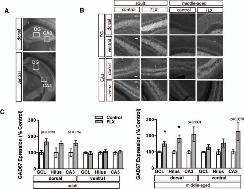
Figure 4. Fluoxetine treatment increases the expression of GAD67 in dorsal DG of middle-aged mice.
A. Representative GAD67 immunostained dorsal and ventral coronal hippocampal sections of adult control mice. Boxes indicate approximate locations of high magnification micrographs in B. B. Representative high magnification images of GAD67 immunostained dorsal and ventral DG and CA3 from adult and middle-aged mice. Fluoxetine treatment 18mg/kg/day for 28 days. C. GAD67 staining intensity was calculated from six hemisections per mouse (3 dorsal, 3 ventral). Mean intensity (±SEM) is expressed as percent of control staining for matched region in age-matched controls. Adult n=3 (VEH), 5 (FLX) mice per group, dGCL p=0.0539, vGCL p=0.8602, dHilus p=0.1065, vHilus p=0.5456, dCA3 p=0.0757, vCA3 p=0.7003; Middle-aged n=3 (VEH), 4 (FLX), dGCL p=0.0351, vGCL p=0.1423, dHilus p=0.0309, vHilus p=0.1464, dCA3 p=0.1001, vCA3 p=0.0878). *, P<0.05. Scale bar 50μM.

