

Abstract
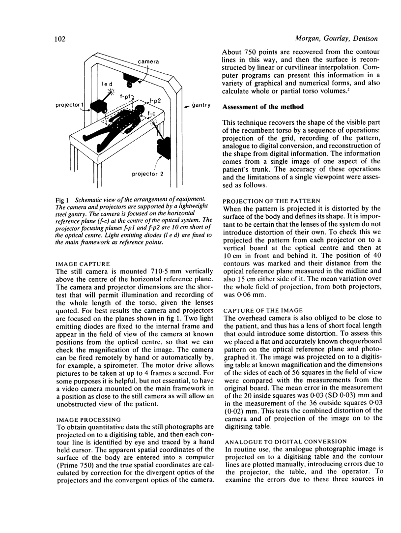
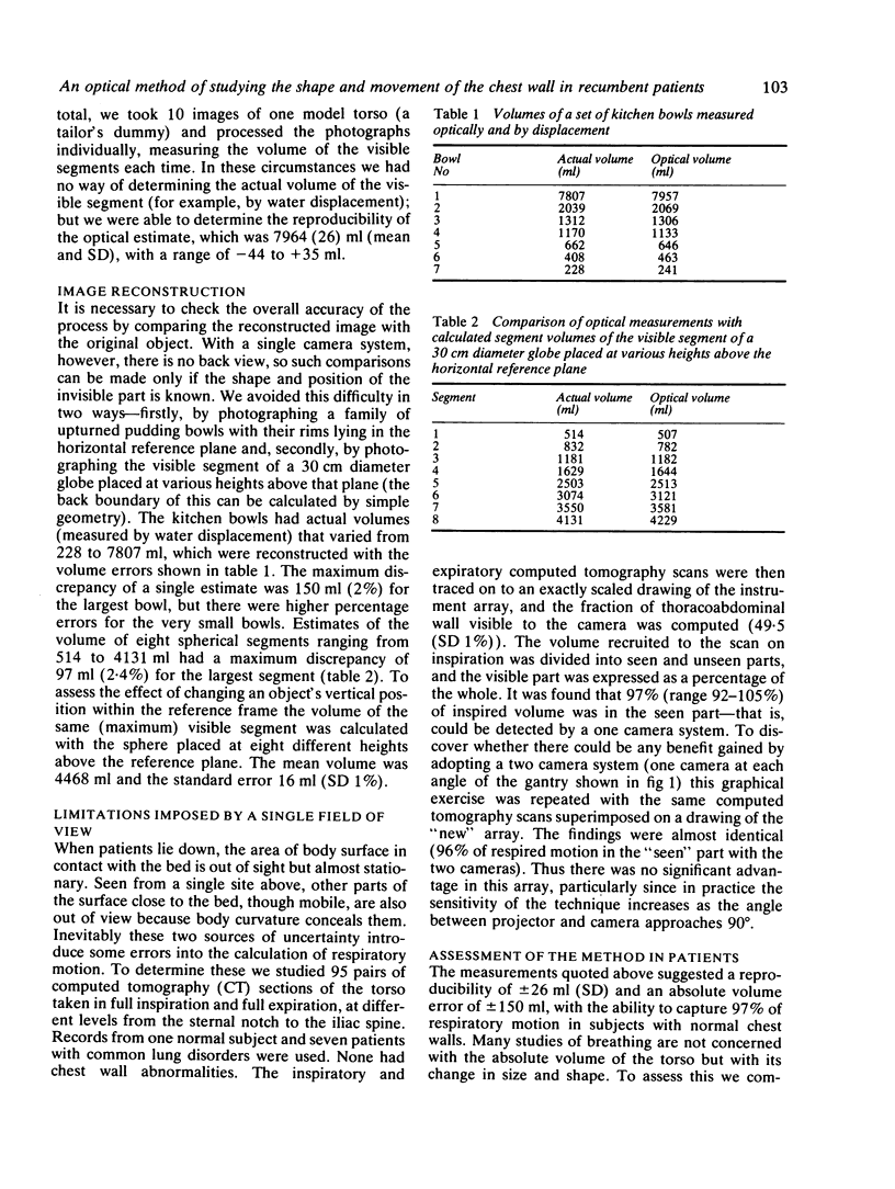
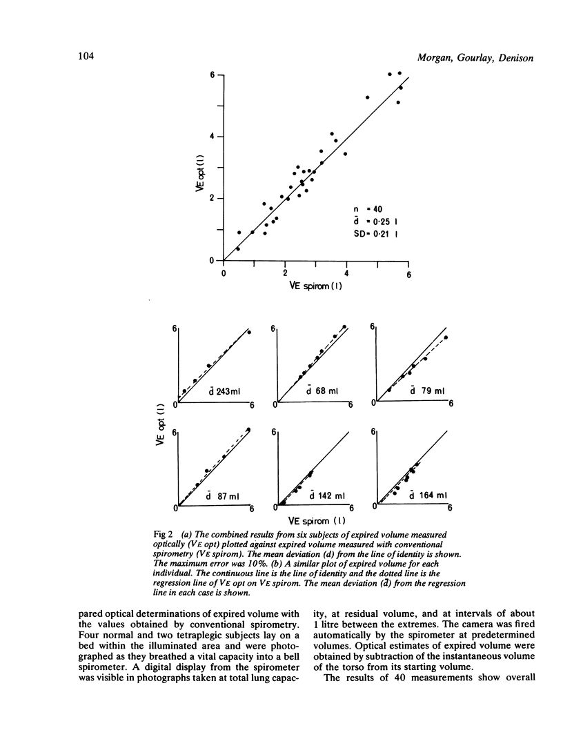
A method of optical mapping has been developed that allows the shape and motion of the chest wall to be studied in recumbent patients. It uses a single camera at a fixed viewpoint to determine the three dimensional coordinates of the visible surface of the body, and hence to measure the volume and shape of the visible segment. Measurements on different test objects (volumes 228-7807 ml) in different positions suggest a reproducibility of volume measurement of +/- 26 ml (SD) and a maximum volume error of 150 ml. Studies of computed tomography scans in inspiration and expiration show that it should be possible to capture 97% of respiratory motion by this method. The mean difference between optical and spirometric measurements of expired volume in six subjects was 0.25 (SD 0.2) 1. Within subject systematic errors of up to 10% were noted. The mean deviation about the regression line of optically measured expired volume on spirometric expired volume varied from 68 to 243 ml in the six subjects (mean 131 (SD 92) ml). To examine the potential of the optical technique, for rapid data acquisition it was used to construct a 24 point flow volume loop from a normal subject.
Full text
PDF
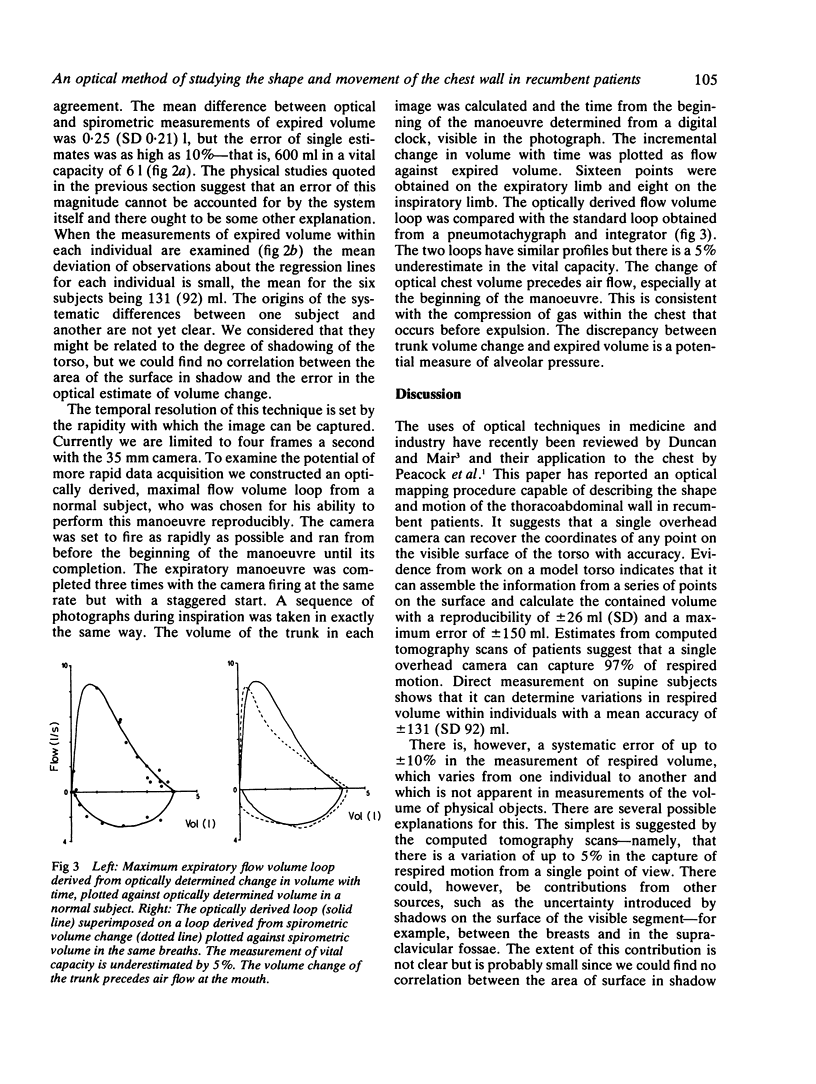
Selected References
These references are in PubMed. This may not be the complete list of references from this article.
- Peacock A. J., Morgan M. D., Gourlay S., Turton C., Denison D. M. Optical mapping of the thoracoabdominal wall. Thorax. 1984 Feb;39(2):93–100. doi: 10.1136/thx.39.2.93. [DOI] [PMC free article] [PubMed] [Google Scholar]