
Figure 1. Immunohistochemical features of multinodular adrenal glands.
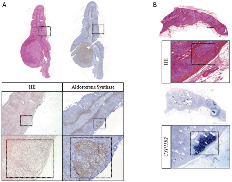
A. HES staining and aldosterone synthase IHC of an adrenal gland resected for lateralized PA showing an APA and secondary micronodules. Left. HES staining with the identification of secondary nodules in an adrenal carrying an APA. Right. Aldosterone synthase IHC positive in one secondary adrenal nodule. B. HES staining (upper panels) and CYP11B2 ISH (bottom panels) in an adrenal carrying an APA and one APCC region.