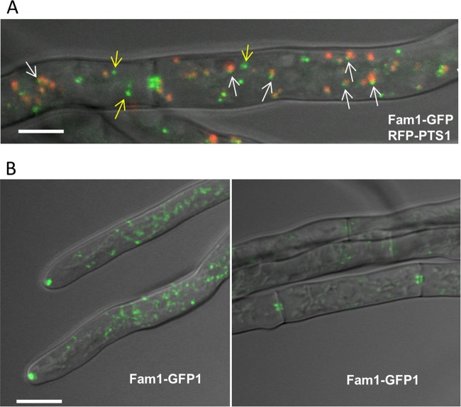
FIG 4 .
Subcellular localization of Fam1 in C. orbiculare. (A) Fam1-GFP and RFP-PTS1 were coexpressed in hyphae of the wild type and observed by confocal microscopy. RFP-PTS1 localized to the peroxisome matrix, while Fam1-GFP localized asymmetrically in small foci at the peroxisome periphery (white arrows) or in punctate structures distinct from peroxisomes (yellow arrows) in the overlay projection image of bright-field, GFP, and RFP channels. Bar, 2.5 µm. (B) Fam1-GFP accumulated in punctate structures at hyphal tips (left) and near septal pores (right). Overlay projection images of bright-field and GFP channels are shown. Bar, 5 µm.

