FIG 7 .
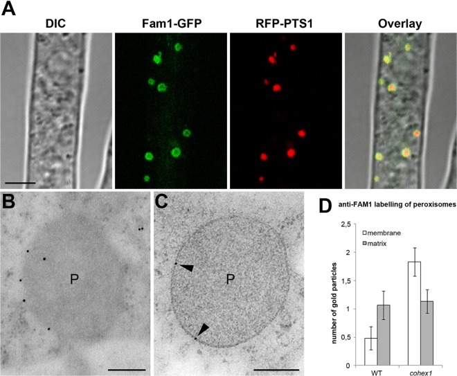
Subcellular localization of Fam1 in Woronin body-deficient cohex1 mutants. (A) In vegetative hyphae of the cohex1 mutant coexpressing Fam1-GFP and RFP-PTS1, Fam1-GFP localized to the periphery of peroxisomes. Confocal images show differential interference contrast (DIC), GFP or RFP fluorescence, merged GFP and RFP channels, and an overlay of all channels. Bar, 5 µm. (B and C) TEM and immunogold labeling with anti-Fam1 antibodies detect Fam1 on the peroxisome membrane (black arrowheads) of the cohex1 mutant. Bars, 200 nm. (D) Quantification of immunogold labeling by anti-Fam1 antibodies. The histogram shows mean numbers of gold particles located on the membrane or matrix of wild-type (WT) peroxisomes (n = 29) and peroxisomes of the cohex1 mutant (n = 30). Each error bar depicts 1 standard error.

