FIG 8 .
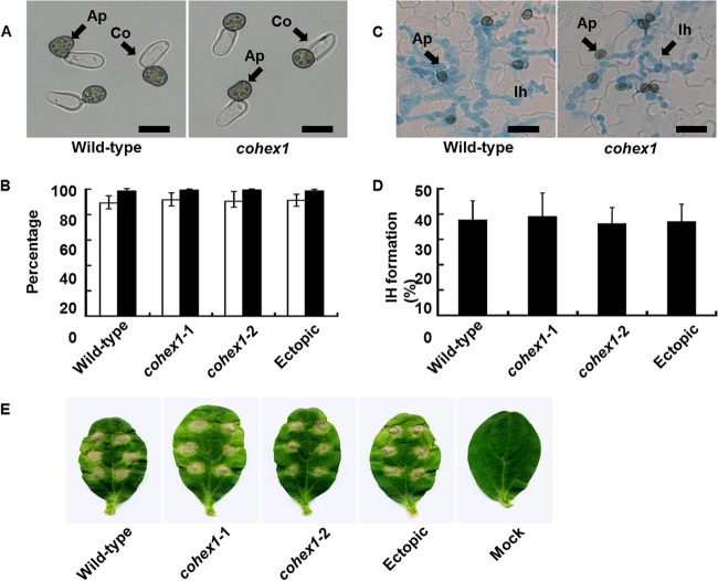
Appressorium formation and pathogenicity of cohex1 mutants. (A) Microscopic observation of appressorium formation in vitro. Conidial suspensions of the C. orbiculare wild-type strain and cohex1 mutants were incubated on glass slides for 24 h. Ap, Appressoria; Co, conidia. Bars = 10 µm. (B) Percentage of conidia forming appressoria. A total of at least 200 conidia were scored for each genotype, based on the examination of three replicates. The wild-type strain, cohex-1-1 and cohex-1-2 CoHEX1 disruption mutants, and CoHEX1 ectopic transformant were tested. Means ± standard deviations (error bars) were calculated from three independent experiments. (C) Microscopic observation of host infection. Conidial suspensions of the wild-type strain and cohex1 mutant were inoculated on the abaxial surface of cucumber cotyledons and viewed after 72 h. Ap, appressoria on the leaf surface; Ih, infection hyphae inside epidermal cells stained with aniline blue. Bars, 10 µm. (D) Percentage of appressoria penetrating to form visible infection hyphae. A total of at least 200 appressoria were scored for each sample, based on the examination of three replicates. The wild-type strain, cohex-1-1 and cohex-1-2 CoHEX1 disruption mutants, and CoHEX1 ectopic transformant were tested. Means plus standard deviations were calculated from three independent experiments. (E) Pathogenicity assay of cohex1 mutants on intact cucumber cotyledons. Droplets of conidial suspension were inoculated onto detached cucumber cotyledons and incubated for 7 days. The wild-type strain, cohex-1-1 and cohex-1-2 CoHEX1 disruption mutants, and CoHEX1 ectopic transformant, were tested. Distilled water was used as a control (mock).

