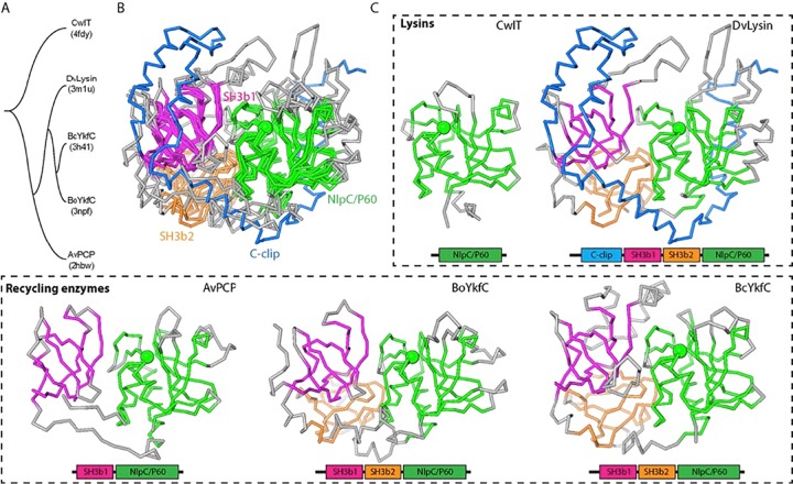
FIG 3 .
Structure comparisons of a prototypic NlpC/P60 domain and SH3b-NlpC/P60 fusion proteins (DvLysin, AvPCP, BoYkfC, and BcYkfC). (A) Tree representation of relationships based on structural similarity. (B) Superimposed structures. Equivalent residues are colored by domain (magenta, SH3b1; orange, SH3b2; green, NlpC/P60), while nonsuperimposed residues are shown in gray, except that the c-clip domain of DvLysin is in blue. The Cα atoms of catalytic cysteines are shown as green spheres. (C) Side-by-side comparison of the domain architecture of lysins and cell wall recycling enzymes.

