Abstract
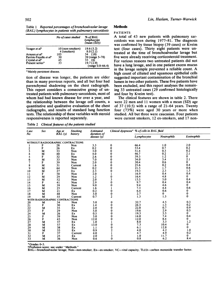
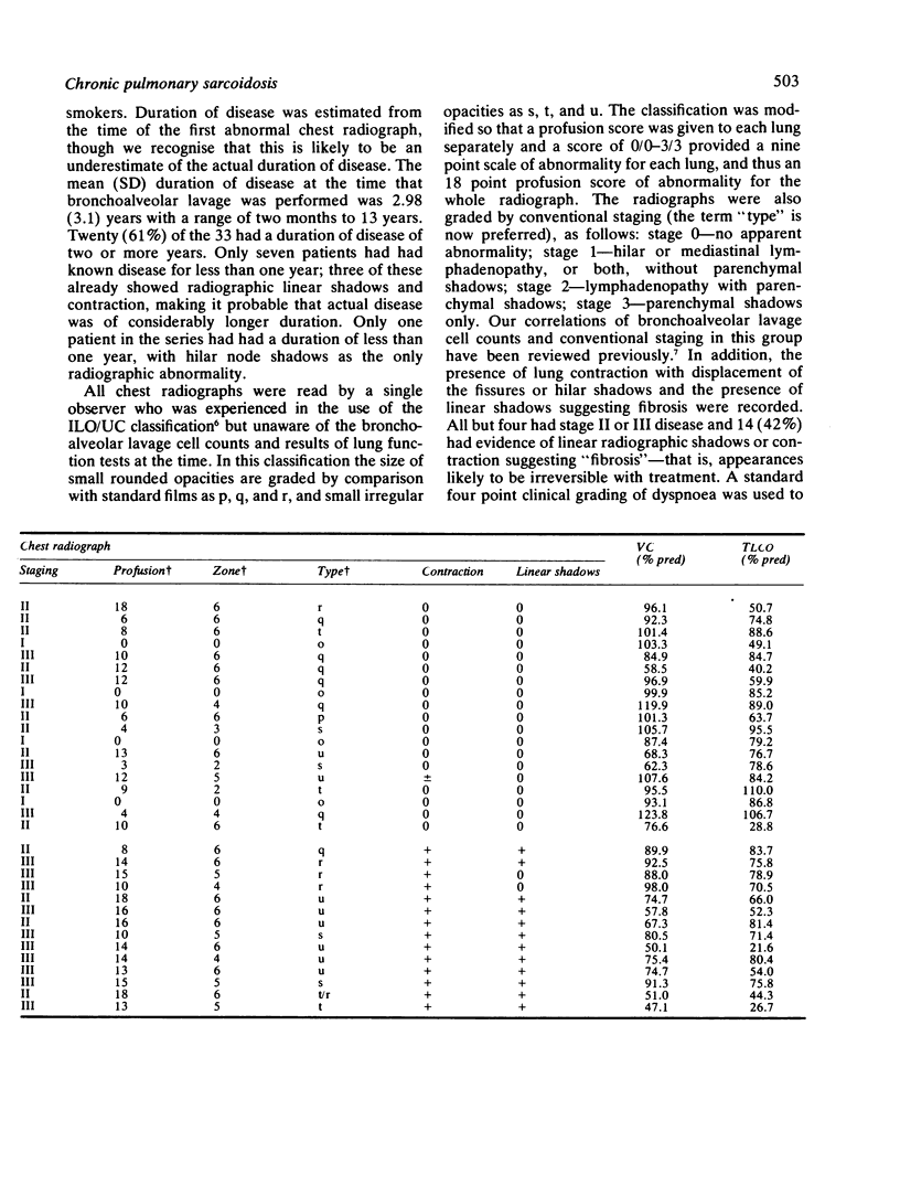
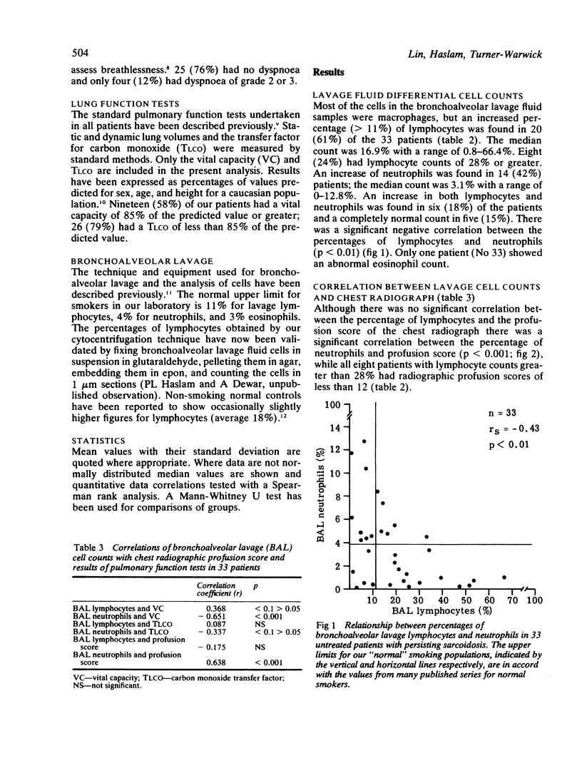
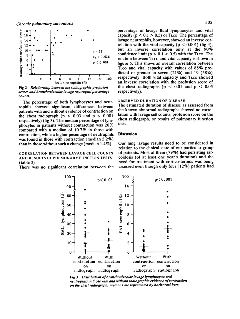
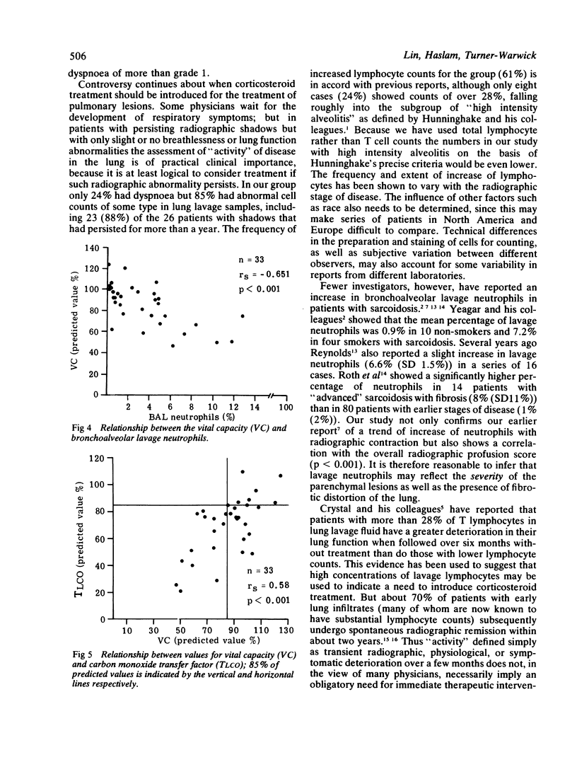
Thirty three consecutive untreated patients with pulmonary sarcoidosis, confirmed histologically or by Kveim test, were investigated to correlate cell counts in bronchoalveolar lavage fluid with clinical features, the chest radiograph, and results of lung function tests. A persistently abnormal radiograph had been observed for one year or more in 26 (79%) and for two years or more in 20 (61%), but only 24% had dyspnoea. Twenty (61%) of 33 patients showed an increased percentage of lymphocytes in bronchoalveolar lavage fluid, although only eight (24%) exceeded 28%. A moderate increase of neutrophils, up to 12%, was found in 14 (42%). Lymphocyte percentage counts were higher in the group of patients without evidence of radiographic contraction suggesting fibrosis, and this contrasted with higher percentage neutrophil counts in those with contraction. There was also a correlation between the percentages of neutrophils and increasing radiographic profusion scores (p less than 0.001), suggesting that neutrophils may reflect the severity of the parenchymal legions as well as fibrotic distortion, and an inverse correlation with vital capacity (p less than 0.001) and transfer factor (TLCO) (p less than 0.1 greater than 0.05). No significant correlation was found between the lymphocyte counts and radiographic profusion scores, vital capacity or TLCO; but it was noted that all eight patients with high lymphocyte counts (greater than 28%) had radiographic profusion scores less than 12. This study shows that, especially in sarcoidosis with more extensive radiographic shadows of long duration, bronchoalveolar lavage neutrophils may be important as well as lymphocytes in clinical assessment of "activity" of disease. These observations are important because they throw doubt on whether the lavage lymphocyte count alone can be used as an indicator of the need to start corticosteroid treatment.
Full text
PDF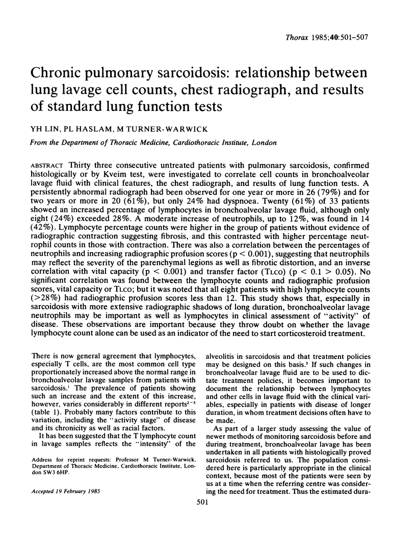
Selected References
These references are in PubMed. This may not be the complete list of references from this article.
- Haslam P. L., Turton C. W., Lukoszek A., Salsbury A. J., Dewar A., Collins J. V., Turner-Warwick M. Bronchoalveolar lavage fluid cell counts in cryptogenic fibrosing alveolitis and their relation to therapy. Thorax. 1980 May;35(5):328–339. doi: 10.1136/thx.35.5.328. [DOI] [PMC free article] [PubMed] [Google Scholar]
- Hunninghake G. W., Gadek J. E., Kawanami O., Ferrans V. J., Crystal R. G. Inflammatory and immune processes in the human lung in health and disease: evaluation by bronchoalveolar lavage. Am J Pathol. 1979 Oct;97(1):149–206. [PMC free article] [PubMed] [Google Scholar]
- NIH conference. Pulmonary sarcoidosis: a disease characterized and perpetuated by activated lung T-lymphocytes. Ann Intern Med. 1981 Jan;94(1):73–94. doi: 10.7326/0003-4819-94-1-73. [DOI] [PubMed] [Google Scholar]
- Reynolds H. Y., Newball H. H. Analysis of proteins and respiratory cells obtained from human lungs by bronchial lavage. J Lab Clin Med. 1974 Oct;84(4):559–573. [PubMed] [Google Scholar]
- Reynolds H. Y. The importance of lymphocytes in pulmonary health and disease. Lung. 1978;155(3):225–242. doi: 10.1007/BF02730695. [DOI] [PubMed] [Google Scholar]
- Roth C., Huchon G. J., Arnoux A., Stanislas-Leguern G., Marsac J. H., Chretien J. Bronchoalveolar cells in advanced pulmonary sarcoidosis. Am Rev Respir Dis. 1981 Jul;124(1):9–12. doi: 10.1164/arrd.1981.124.1.9. [DOI] [PubMed] [Google Scholar]
- Yeager H., Jr, Williams M. C., Beekman J. F., Bayly T. C., Beaman B. L. Sarcoidosis: analysis of cells obtained by bronchial lavage. Am Rev Respir Dis. 1977 Nov;116(5):951–954. doi: 10.1164/arrd.1977.116.5.951. [DOI] [PubMed] [Google Scholar]


