Figure 3.
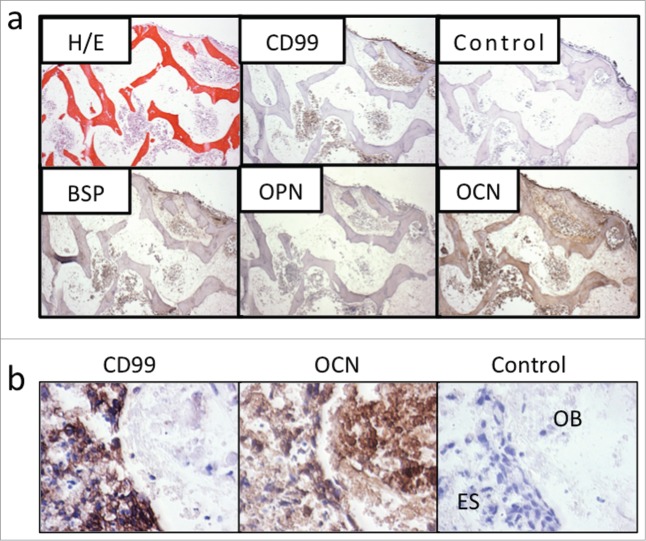
Tissue-engineered bone models express osteomimicry. (a) Tissue-engineered (TE) model of Ewing's sarcoma. Bone markers BSP, OPN and OCN are re-expressed in the TE-Ewing's sarcoma 3D model, as shown histologically (hematoxylin and eosin) and by immunostains (Ewing's sarcoma marker CD99; bone markers BSP, OPN and OCN) after 6 weeks of culture. Negative control without primary antibody is shown for comparison. Counterstaining was performed with hematoxylin QS (blue). (b) Marker expression after 4 weeks in co-culture. Data are shown for bone pellets in co-culture with Ewing's sarcoma aggregates. Immunohistochemical stainings of the aggregates model for Ewing's sarcoma marker CD99 and bone marker OCN at week 4 in co-culture with osteoblasts-derived from hMSC. Negative control without primary antibody is shown for comparison. OB = Osteoblasts; ES = Ewing's sarcoma cells. (Reproduced with permission from Villasante et al. Biomaterials 2014; 35: 5785-94).

