Abstract
Cutaneous larva migrans or creeping eruptions is a cutaneous dermatosis caused by hookworm larvae, Ancylostoma braziliense. A 2-month-old female child presented with a progressive rash over the left buttock of 4 days duration. Cutaneous examination showed an urticarial papule progressing to erythematous, tortuous, thread-like tract extending a few centimeters from papule over the left gluteal region. A clinical diagnosis of cutaneous larva migrans was considered. Treatment with albendazole led to complete resolution, confirming the diagnosis. This is to the best of our knowledge, the youngest age at which this condition is being reported.
Keywords: Cutaneous larva migrans, creeping eruptions, infancy
What was known?
Cutaneous larva migrans, although common in children is rare in infants.
Introduction
Cutaneous larva migrans (CLM) also known as creeping eruptions and epidermatitis linearis migrans is caused by hookworm, most commonly Ancylostoma braziliense and Ancylostoma caninum. This acquired dermatosis is caused by infective larval form of the dog or cat hookworm which may accidentally penetrate the intact skin and then wander through the epidermis. It is common in warmer tropical and subtropical countries.[1,2] Although condition is common in children, it is rare in infants. We describe the youngest infant with this condition.
Case Report
A 2-month-old female child presented with a progressive rash over the left buttock of 4 days duration. Child was irritable. Initially the lesion started as a solid, raised swelling, which gradually progressed in a serpiginous manner in the left gluteal region. Fever, cough, loose motion and systemic complaints were absent. Baby was exclusively breastfed and developmental milestones were normal. Vaccination status was up to date.
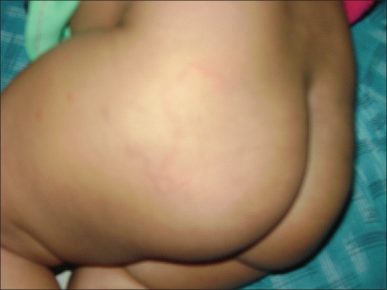
On examination the child was afebrile without pallor, clubbing, cyanosis, icterus and lymphadenopathy. Height, weight, head circumference and vitals were normal. Cutaneous examination showed an urticarial papule progressing to erythematous, tortuous, thread-like tract extending a few centimeters from the papule over the left gluteal region [Figure 1].
Figure 1.

Urticarial papule with serpiginous tract over left gluteal region
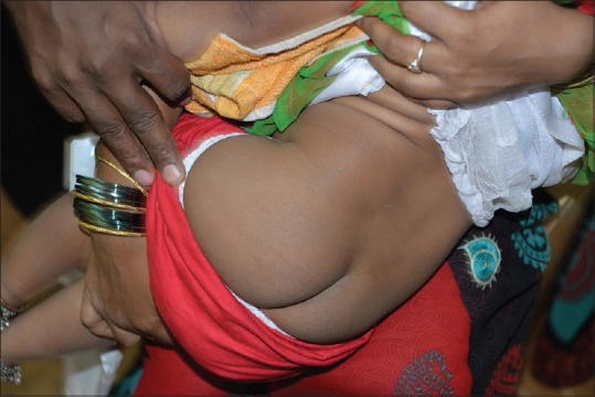
A clinical diagnosis of CLM was made. Complete hemogram was normal except for mild eosinophilia. Liver and renal function tests were normal. Venereal disease research laboratory test, human immunodeficiency virus and hepatitis surface antigen were negative. Parents refused biopsy. Baby was started on syrup albendazole 15 mg/kg body weight for three consecutive days. Complete resolution was seen at the end of first week [Figure 2].
Figure 2.

Complete resolution following treatment with albendazole
Discussion
Although CLM is globally distributed, it is endemic in Caribbean islands, Africa, south America, south east Asia and south eastern United States. It is a peculiar dermatosis caused by hookworm larvae such as A. braziliense, A. caninum, A. ceylonium, Uncinaria stenocephala and Bubostomum phlebotomus. Other rare causes are Strongyloides stercoralis, Dirofilaria spp, Gnathostoma spp and Loa Loa.[1]
It was first described in 1874. People frequently visiting beaches, working in crawl spaces under houses, children especially playing in sand boxes are at high risk.[1,3] A few reports of CLM in infants of crawling age are reported.[4,5,6] However, our patient was very young and unable to move. Prolonged contact with contaminated soil either at home or at the parent's work place was the probable route of spread, in our patient.
These nematodes normally do not parasitize human skin, but the infective larval form of the dog or cat hookworm may accidentally penetrate the intact skin and then wander through the epidermis.[3] Humans are the accidental hosts and are unable to complete their life cycle. The larvae secrete proteases and hyaluronidase, which facilitate the penetration and migration through epidermis.[2]
Clinically, tingling or pricking sensation may be noted, followed by an erythematous, linear or serpiginous raised lesion that advances at the rate 2 mm–3 cm per day. Most common sites of involvement are feet (interdigital spaces, dorsa of feet and medial aspects of sole), buttocks and hands. Rarely anterior abdominal wall, penile shaft, perianal area, sole, oral cavity or breasts may be involved. Multiple tracts may also be seen. Secondary impetization, eczematization, bullous lesion, folliculitis or visceral larva migrans including Loeffler's pneumonia may complicate. Differential diagnosis include larva currens, cercarial dermatitis, contact dermatitis, scabies and migratory myiasis.[3,7,8]
It is usually self-limiting, but can last up to 2 years. Diagnosis is usually based on history and clinical examination. Peripheral eosinophilia and increased serum IgE may be seen. Although a skin biopsy is of limited value, when taken ahead of the track, may show larva (PAS-positive) suprabasally, spongiosis, intra-epidermal vesiculation and dermal eosinophilic infiltrate. Epiluminescence microscopy, reflectance confocal microscopy and optical coherence tomography are other useful diagnostic tools.[3,7]
Treatment includes topical albendazole, thiabendazole and freezing. Systemic thiabendazole, albendazole, flubendazole and recently ivermectin are recommended.[1] Awareness, early recognition and treatment help in preventing complication. Avoiding contact with contaminated soil helps in prevention.
What is new?
Our case illustrates the youngest infant to be affected.
Footnotes
Source of support: Nil
Conflict of Interest: Nil.
References
- 1.Karthikeyan K, Thappa DM. Cutaneous larva migrans. Indian J Dermatol Venerol Leprol. 2002;68:252–8. [PubMed] [Google Scholar]
- 2.Balfour E, Zalka A, Lazova R. Cutaneous larva migrans with parts of the larva in the epidermis. Cutis. 2002;69:368–70. [PubMed] [Google Scholar]
- 3.Brenner MA, Patel MB. Cutaneous larva migrans: The creeping eruption. Cutis. 2003;72:111–5. [PubMed] [Google Scholar]
- 4.Masuria BL, Batra A, Kothiwala RK, Khuller R, Singhi MK. Creeping eruption. Indian J Dermatol Venereol Leprol. 1999;65:51. [PubMed] [Google Scholar]
- 5.Paul Y, Singh J. Cutaneous larva migrans in an infant. Indian Pediatr. 1994;31:1089–91. [PubMed] [Google Scholar]
- 6.Black MD, Grove DI, Butcher AR, Warren LJ. Cutaneous larva migrans in infants in the Adelaide hills. Australas J Dermatol. 2010;51:281–4. doi: 10.1111/j.1440-0960.2010.00696.x. [DOI] [PubMed] [Google Scholar]
- 7.Upendra Y, Mahajan VK, Mehta KS, Chauhan PS, Chander B. Cutaneous larva migrans. Indian J Dermatol Venereol Leprol. 2013;79:418–9. doi: 10.4103/0378-6323.110770. [DOI] [PubMed] [Google Scholar]
- 8.Mehta V, Shenoi SD. Extensive larva migrans. Indian J Dermatol Venerol Leprol. 2004;70:373–4. [PubMed] [Google Scholar]


