

Abstract
Parapharyngeal abscess (PPA)-like lesion is a very rare manifestation of Kawasaki disease (KD). Here we report a Chinese case of KD initially mimicking PPA, which is the first one reported in Asia.
A 3-year-old male patient presented with fever, drooling, and bilateral painful cervical lymphadenopathy for 3 days. Chest X-ray and echocardiogram were normal. With substantial elevation of white blood count and C-reactive protein, purulent cervical lymphadenitis was considered. Symptoms did not improve after treatment with vancomycin, and the patient further developed trismus and restricted neck movement. Neck CT revealed a 2 × 1.5 cm hypodense lesion in the right parapharyngeal space with peripheral enhancement. PPA was suspected and on the 3rd day following admission, the patient received surgical incision and drainage. One milliliter of serous fluid was drained without bacterial growth on cultures. Fever persisted after surgery. As the clinical course proceeded, additional major signs of KD gradually evolved, and on the 6th day following admission the patient completely fulfilled the diagnostic criteria for KD. Rapid clinical improvement was observed following treatment with high-dose immunoglobulin and aspirin. Due to the parapharyngeal operation, the patient was fed milk through a nasogastric tube for 15 days. His neck incision became infected but healed gradually following dressing change and antibiotic treatment. Currently he remains asymptomatic during regular follow-up and repeated echocardiograms are normal.
Both pediatricians and otolaryngologists can learn from this case that KD may initially manifest as PPA. Careful observation for major signs of KD during the clinical course can help to achieve a prompt and correct diagnosis. Thus, unnecessary surgery and cardiac complications of KD may be avoided.
INTRODUCTION
Kawasaki disease (KD), also known as mucocutaneous lymph node syndrome, predominantly affects infants and young children. Due to lack of specific diagnostic tests, the diagnosis of KD is mainly based on its principal clinical criteria.1 However, as an acute systemic vasculitis, KD may be involved in multiple organs and systems, causing unusual manifestations and thus making the diagnosis difficult.2 Here we report a case of KD which initially manifested as parapharyngeal abscess (PPA), leading to delayed diagnosis. Furthermore, this case included unnecessary surgery and thus the risk of surgical complications was increased.
CASE PRESENTATION
A previously healthy 3-year-old male patient was admitted into our pediatric department with fever (up to 39°), neck swelling, dysphagia, and drooling for 3 days. On physical examination, the patient was toxic and irritable, and showed hyperemic throat, enlarged tonsils and bilateral tender cervical lymphadenopathy. There was no obvious respiratory difficulty. Laboratory investigations revealed white blood count (WBC): 23,800/μL (neutrophils: 79.2%, lymphocytes: 11.6%), hemoglobin: 12.7 g/dL, platelet count: 256,000/μL and C-reactive protein (CRP) 210 mg/L. Chest X-ray and echocardiogram were normal. Blood and throat cultures were negative. Purulent cervical lymphadenitis was considered and vancomycin (40 mg/kg/day) was prescribed. On the following day, the painful cervical lymphadenopathy did not show any improvement and the patient developed trismus and restricted neck movement. Computed tomography (CT) of the neck revealed a 2 cm × 1.5 cm low-density lesion in the right parapharyngeal space (Figure 1: asterisk), with peripheral enhancement (Figure 1: arrow) as well as multiple lymph node enlargements in the posterior cervical space. PPA was suspected and the patient was transferred to the otorhinolaryngologic department for surgery. On the 3rd day following admission, the patient developed bilateral bulbar conjunctival injection and red lips. Due to the possibility of upper airway obstruction, incision and drainage of the PPA was performed. One milliliter of serous fluid was drained without bacterial growth on cultures. After surgery, his neck pain and trismus alleviated, however high fever persisted. During the 5th day of hospitalization, the patient manifested fissured red lips and strawberry tongue. On the 6th day, edema of the hands and feet and polymorphous generalized erythematous skin rash appeared. Laboratory investigations revealed WBC: 39,780/μL (neutrophils: 75%, lymphocytes: 18%), hemoglobin: 12.3 g/dL, platelet count: 395,000/μL, erythrocyte sedimentation rate: 132 mm/h, CRP: 171 mg/L, pro-albumin: 72 mg/L, and nonseptic pyuria. Despite antibiotic treatment with vancomycin and cefoperazone, the fever persisted. The patient was transferred back to the pediatric department from the otorhinolaryngologic department on the 8th day following admission. Upon re-evaluation of his clinical and laboratory findings, the diagnosis of KD was considered and intravenous immunoglobulin (IVIG) at 2 g/kg along with aspirin at 30 mg/kg/day was given. Fever and other symptoms subsided within 24 hours after administration. Desquamation of the fingers and toes developed on the 11th day following admission. No coronary artery abnormalities were found on repeated echocardiogram. Due to the parapharyngeal operation, the patient was fed milk through a nasogastric tube for 15 days. His neck incision became infected but healed gradually following dressing change and antibiotic treatment. Finally, the patient was discharged on the 20th day of hospitalization, on a low dose of aspirin. During the 2 months of follow-up, he remains asymptomatic and repeated echocardiograms were normal.
FIGURE 1.
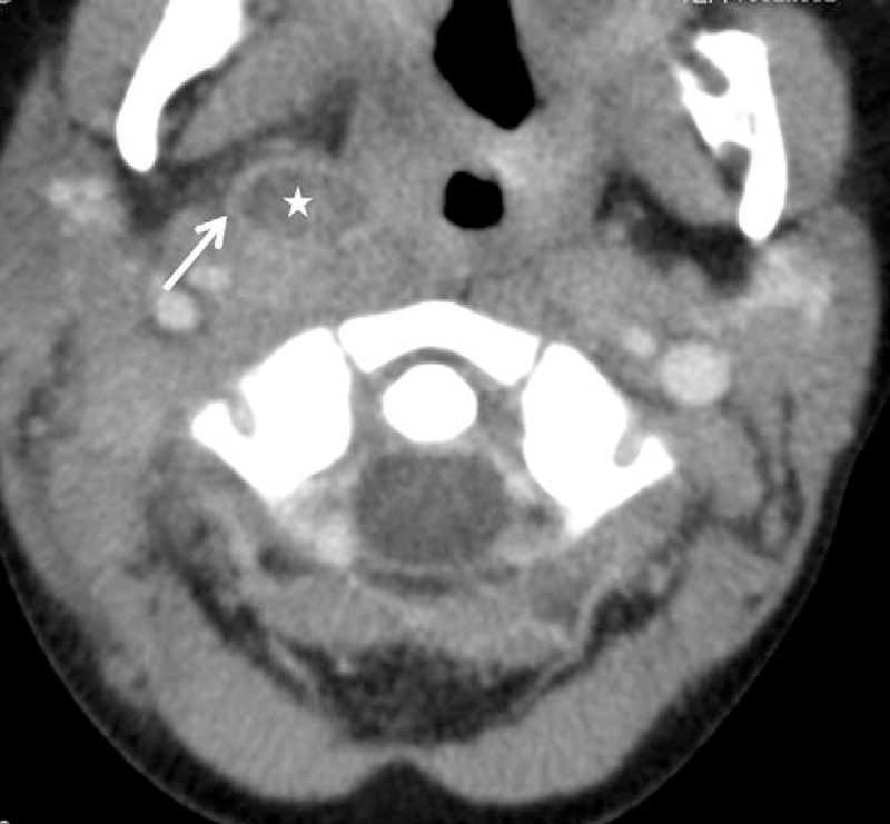
Neck CT scan showing a hypodense lesion (asterisk) in the right parapharyngeal space with peripheral enhancement (arrow).
DISCUSSION
Parapharyngeal abscess (PPA)-like lesion is a very rare manifestation of KD. To the best of our knowledge, there has only been one similar case reported, occurring in the USA.3 This case is the first one reported in Asia.
Besides PPA, KD can mimic other kinds of deep neck infections, such as peritonsillar abscess, peritonsillar cellulitis, or retropharyngeal abscess (RPA). The rarity of these presentations often leads to missed or delayed diagnosis and treatment.4–6 In this patient, although infection cannot be ruled out at the early stage, the fact that blood and throat cultures were negative and there was an excellent response within 24 hours after administration of intravenous immunoglobulin (IVIG) and aspirin makes the diagnosis of KD presenting as a parapharyngeal abscess-like lesion more plausible than an infection coexisting with KD.
In this case, the boy manifested like infectious PPA at the beginning: fever, dysphagia, neck rigidity, remarkably elevated WBC and CRP, and an abscess-like lesion on the CT. The major KD signs emerged gradually as the clinical course proceeded. Similarly, in most of the previously reported KD cases mimicking RPA, fever and deep neck infection like-symptoms were the only clinical findings at admission. Additional features of KD appeared gradually during hospitalization.6,7 After carefully comparing children with KD accompanied by retropharyngeal edema to, those with RPA, Nomura et al6 concluded that close attention to core manifestations but not laboratory examinations of KD helps clinicians to differentiate these 2 conditions. Obviously, our findings agree with Nomura's report.
In our opinion, both pediatricians and otolaryngologists should draw lessons from this case. Indeed, the patient had already developed bilateral bulbar conjunctival injection and red lips on the 3rd day following admission. From this case we need to realize that PPA-like lesion can be the initial presentation of KD. Therefore, when some signs of non-septic vasculitis emerge, pediatricians must carefully observe for other signs of KD. Otolaryngologists are excellent at managing deep neck infections, but not at diagnosing and treating KD. In this case, although the patient had already met the criteria of KD on the 6th day following admission, proper diagnosis was not made by surgeons in the otorhinolaryngologic department. Treatment with IVIG and aspirin was initiated after the patient was transferred back to the pediatric department. To avoid delayed diagnosis, otolaryngologists should be aware of the manifestations of KD and include it as a routine differential diagnosis for PPA. Moreover, this case also emphasizes the importance of collaboration between pediatricians and otolaryngologists.
The exact pathophysiology through which KD leads to PPA is unknown. As with KD-associated RPA, PPA in KD is probably caused by tissue inflammation and edema under the condition of systemic vasculitis.6–8 Histopathological study of cervical lymphadenopathy in KD revealed non-purulent inflammation and ischemic necrosis due to fibrin thrombi in small vessels of affected lymph nodes. Inflammatory infiltration of the lymph node capsule was present in most of the affected cervical lymph nodes.9 MRI scan found that affected lymph nodes had capsular and hilar but not parenchyma enhancement.10 In this case, we speculate that the abscess-like lesion originated from a lymph node: the hypodense central area found by CT (Figure 1: asterisk) may be caused by nonseptic inflammatory edema, and peripheral enhancement (Figure 1: arrow) may be due to capsule inflammatory infiltration. Future evidence is needed to confirm this pathological hypothesis.
How to treat KD mimicking deep neck abscess is a challenging question. In a review of 24 previously reported KD cases with RPA-like lesion, all of them were unresponsive to antibiotics. Seven of the 24 cases received surgical aspiration or drainage but no abscess or bacterial infection evidence was found. Fever and other symptoms resolved rapidly after the administration of IVIG and aspirin in all of the 24 patients.6–8 As the deep neck abscess-like lesion is formed by local inflammation and edema under the condition of systemic vasculitis, it will be absorbed and disappear when systemic vasculitis is controlled by IVIG and aspirin. Therefore, in our opinion, KD mimicking deep neck abscess is not an indication for surgery. Operations should not be arranged hastily for these patients, as surgical complications, such as incision infection in our case, can be thusly avoided. For those patients with upper airway compression due to the abscess, short-term intubation should be used until local inflammation and swelling subside.6–8 Future well-controlled trials are needed to prove the safety and effectiveness of non-surgical treatment for KD mimicking deep neck abscess.
In conclusion, both pediatricians and otolaryngologists should keep in mind that, in addition to bacterial infection, KD can cause PPA-like lesion. Careful observation for major signs of KD during the clinical course can help to achieve an early and correct diagnosis. Unnecessary surgery and cardiac complications of KD may thus be avoided.
Acknowledgment
The authors would like to thank Stephanie Cambier for proof-reading this manuscript.
Footnotes
Abbreviations: CRP = C-reactive protein, CT = computed tomography, IVIG = intravenous immunoglobulin, KD = Kawasaki disease, PPA = parapharyngeal abscess, RPA = retropharyngeal abscess, WBC = white blood count.
Ethical approval was obtained from the Ethics Committee of Sichuan University. Written informed consent was obtained from the parents of the patient for publication of this case report and any accompanying images.
This work was supported by grants from the National Science Foundation of China (No. 81330016, 31171020).
The authors have no conflicts of interest to disclose.
REFERENCES
- 1.Newburger JW, Takahashi M, Gerber MA, et al. Diagnosis, treatment, and long-term management of Kawasaki disease: a statement for health professionals from the Committee on Rheumatic Fever, Endocarditis, and Kawasaki Disease, Council on Cardiovascular Disease in the Young, American Heart Association. Pediatrics 2004; 114:1708–1733. [DOI] [PubMed] [Google Scholar]
- 2.Huang YC, Lin TY, Su WJ. Unusual manifestations in children with Kawasaki disease. J Formos Med Assoc 1997; 96:451–456. [PubMed] [Google Scholar]
- 3.Ruef C. Mucocutaneous lymph node syndrome (Kawasaki syndrome) mimicking a suppurative parapharyngeal space infection. Case report and review of the literature. Helv Paediatr Acta 1989; 43:307–312. [PubMed] [Google Scholar]
- 4.Ravi KV, Brooks JR. Peritonsillar abscess—an unusual presentation of Kawasaki disease. J Laryngol Otol 1997; 111:73–74. [DOI] [PubMed] [Google Scholar]
- 5.Korkis JA, Stillwater LB. An unusual otolaryngological problem – mucocutaneous lymph node syndrome (Kawasaki's syndrome) case report. J Otolaryngol 1985; 14:257–260. [PubMed] [Google Scholar]
- 6.Nomura O, Hashimoto N, Ishiguro A, et al. Comparison of patients with Kawasaki disease with retropharyngeal edema and patients with retropharyngeal abscess. Eur J Pediatr 2014; 173:381–386. [DOI] [PubMed] [Google Scholar]
- 7.Cavicchiolo ME, Berlese P, Bressan S, et al. Retropharyngeal abscess: an unusual presentation of Kawasaki disease. Case report and review of the literature. Int J Pediatr Otorhinolaryngol Extra 2012; 7:179–182. [Google Scholar]
- 8.Hung MC, Wu KG, Hwang B, et al. Kawasaki disease resembling a retropharyngeal abscess—case report and literature review. Int J Cardiol 2007; 115:e94–e96. [DOI] [PubMed] [Google Scholar]
- 9.Yokouchi Y, Oharaseki T, Harada M, et al. Histopathological study of lymph node lesions in the acute phase of Kawasaki disease. Histopathology 2013; 62:387–396. [DOI] [PubMed] [Google Scholar]
- 10.Puhakka L, Saat R, Klockars T, et al. Retropharyngeal involvement in Kawasaki disease—a report of four patients with retropharyngeal edema verified by magnetic resonance imaging. Int J Pediatr Otorhinolaryngol 2014; 78:1774–1778. [DOI] [PubMed] [Google Scholar]