Abstract
Background
As motion-preserving technique has been developed, the concept of hybrid surgery involves simultaneous application of two different kinds of devices, dynamic stabilization system and fusion technique. In the present study, the application of hybrid surgery for lumbosacral degenerative disease involving two-segments and its long-term outcome were investigated.
Methods
Fifteen patients with hybrid surgery (Hybrid group) and 10 patients with two-segment fusion (Fusion group) were retrospectively compared.
Results
Preoperative grade for disc degeneration was not different between the two groups, and the most common operated segment had the most degenerated disc grade in both groups; L4-5 and L5-S1 in the Hybrid group, and L3-4 and L4-5 in Fusion group. Over 48 months of follow-up, lumbar lordosis and range of motion (ROM) at the T12-S1 global segment were preserved in the Hybrid group, and the segmental ROM at the dynamic stabilized segment maintained at final follow-up. The Fusion group had a significantly decreased global ROM and a decreased segmental ROM with larger angles compared to the Hybrid group. Defining a 2-mm decrease in posterior disc height (PDH) as radiologic adjacent segment pathology (ASP), these changes were observed in 6 and 7 patients in the Hybrid and Fusion group, respectively. However, the last PDH at the above adjacent segment had statistically higher value in Hybrid group. Pain score for back and legs was much reduced in both groups. Functional outcome measured by Oswestry disability index (ODI), however, had better improvement in Hybrid group.
Conclusion
Hybrid surgery, combined dynamic stabilization system and fusion, can be effective surgical treatment for multilevel degenerative lumbosacral spinal disease, maintaining lumbar motion and delaying disc degeneration.
Keywords: Hybrid surgery, Non-fusion, Dynamic stabilization system, Dynesys, NFlex
Introduction
Because of abnormal biomechanical effects and development of adjacent segment pathology (ASP) after fusion surgery, alternative motion-preservation techniques have been developed.1 One of the motionpreservation techniques, pedicle-based dynamic stabilization system developed to maintain intersegmental movement, decrease intervertebral loading and ultimately prevent ASP.2 The concept of hybrid surgery involves the application of two different kinds of devices, dynamic stabilization and fusion. Segments with severe degeneration and spinal instability can undergo fusion, and adjacent segments with moderate degeneration can be secured with a non-fusion dynamic stabilization system when the degenerative pathology involves more than two segments. 3 Hybrid surgery is not to prevent further degeneration of the asymptomatic adjacent segment but is used to replace fusion when treating symptomatic, degenerated adjacent segments.4 Thus, a retrospective comparative study was done in patients with hybrid surgery or pure fusion surgery for lumbosacral degenerative disease involving two-segments.
Material and methods
Patient population
Patients with lumbosacral spinal degenerative disease underwent surgical management when there was no effect from 6 months or more of conservative management. The hybrid surgery applied the dynamic stabilization system to the symptomatic degenerative segment without spinal instability next to the adjacent fusion segment. Rigid fixation was used for fusion in degenerative segments with spinal instability, spondylolytic spondylolisthesis, more than grade II spondylolisthesis and severe disc space narrowing. Among multilevel surgeries, patients with surgery at two segments were included. Exclusion criteria were less than a 2-year follow-up, previous lumbar spine surgery, spinal trauma, systemic malignancy, infection, and interbody fusion without pedicle screw fixation.
Description of dynamic stabilization system for hybrid surgery
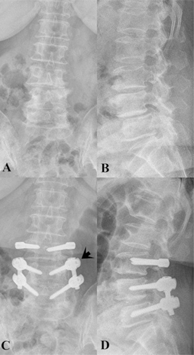
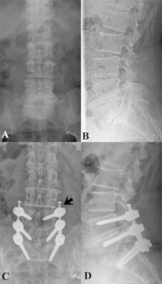
Two kinds of non-fusion dynamic stabilization systems, Dynesys-to-Optima (DTO) system and NFlex (Synthes Spine, Inc.) were used. The manufacturer of Dynesys had introduced DTO hybrid stabilization systems that permit fusion and non-fusion stabilization at adjacent segments(Figure 1).5 The NFlex system consists of polyaxial titanium alloy pedicle screws that are fixed to a semi-rigid polycarbonate urethane (PCU)-sleeved rod (Figure 2). The integrated PCU spacer is surrounded by a central titanium ring, to which a pedicle screw is locked. The rod can be attached to pedicle screws in the standard manner.6
Fig. 1.
DTO system. A 62-year-old female patient underwent hybrid surgery combining the Dynesys stabilization system at L3-4 and TLIF with PSF at L4-5, which had spinal instability, and transitioning to the DTO system system (arrow). Preoperative anteroposterior (A) and lateral (B) radiographs and postoperative 4-year anterolateral (C) and lateral (D) radiographs are shown.
Fig. 2.
NFlex system. A 67-year-old female had multilevel spinal stenosis with severe disc space narrowing at L5-S1. She underwent hybrid surgery, NFlex dynamic stabilization surgery at L4-5 and TLIF with PSF at L5-S1. Polycarbonate urethane (PCU) spacer was surrounded by a central titanium ring (arrow). Preoperative anteroposterior (A) and lateral (B) radiographs and postoperative 6-year anterolateral (C) and lateral (D) radiographs are shown.
Surgical procedure
All operations were performed in a neutral prone position by one senior surgeon. In the fusion surgery, a conventional or minimally invasive transforaminal lumbar interbody fusion (TLIF) procedure was used. Decompressive laminectomy with/without foraminal decompression, facetectomy, discectomy, and interbody fusion was performed followed by pedicle screw fixation (PSF) under fluoroscopic guidance. In the hybrid surgery, non-fusion dynamic stabilization system or rigid fixation was used according to the severity of degeneration at the corresponding segment. Intraoperatively, care was taken to preserve the facet joint integrity and to place the dynamic stabilization system screw lateral to the facet joints. The polyester cord at the non-fusion dynamic stabilization segment and titanium rod at the fusion segment was connected using the DTO system. In the NFlex system, the TLIF procedure at the fusion segment, decompression at the non-fusion dynamic stabilization segment and pedicle screw insertion at both segments was conducted, and then, the dynamic rod of NFlex was connected.
Radiologic evaluation
Preoperative disc degeneration was graded by the Pfirrmann disc degeneration grade system (I-V) on magnetic resonance images (MRIs).7 On the lateral radiographs, lumbar lordosis was measured using Cobb's method, and anterior disc height (ADH) and posterior disc height (PDH) were measured. The range of motion (ROM) was calculated from the difference between the flexion and extension dynamic radiographs. Every radiologic parameter was measured at the global angle (T12-S1) and each segmental angle, and it was classified at second above, above, operated, and below segment.
Radiologic ASP was defined as more than a 2 mm loss of posterior disc height at any segment, comparing preoperative and final follow-up radiographs.8 Fusion and pseudarthrosis was evaluated referencing literature.9 Radiologic abnormalities such as a radiolucent line around the pedicle screw and instrument failure like screw and rod fracture were evaluated at final follow-up on plain radiographs.
Clinical evaluation
Clinical parameters were retrospectively obtained from patients’ medical records at preoperation and final follow-up. Pain was measured by Visual Analog Scale (VAS, 0-10), and functional outcome was assessed by the Oswestry disability index (ODI, 0-100%). Additionally, medication, specifically pain analgesics, was evaluated at preoperation and last follow-up.
Statistical analysis
Mann-Whitney and Wilcoxon signed rank tests were used for continuous variables and Fisher's exact test was used for categorical variables. Statistical significance was defined as a p-value of less than 0.05. Analyses were performed with SPSS version 19.0 (IBM, Armonk, New York, USA).
Results
Patients’ characteristics
One hundred-eight patients underwent non-fusion dynamic stabilization surgery, and 87 patients underwent fusion surgery by one surgeon between 2003 and 2011. For two-segment surgery, 15 patients with hybrid surgery (Hybrid group) and 10 patients with fusion surgery (Fusion group) met the inclusion criteria (Table 1). Patients had a minimum 2-year follow-up period, and mean follow-up period was 48.8 ± 26.4 months in Hybrid group and 52.6 ± 25.6 months in Fusion group (p=0.33). Mean age at surgery was 60.7 ± 8.3 years and 63.9 ± 7.8 years in Hybrid and Fusion groups, respectively (p=0.47). For gender, the Hybrid and Fusion group had 11 and 5 female patients, respectively (p=0.24). Primary pathology was all lumbar stenosis associated with herniated nucleus pulposus (HNP) in 2 patients each, spondylolisthesis in 6 and 3 patients, and instability in 2 and 3 patients in the Hybrid and Fusion group, respectively (p=0.67). Operated segments were L3-4-5 and L4-5-S1 in 7 and 8 patients in Hybrid group, and L2-3-4, L3-4-5 and L4-5-S1 in 2, 6 and 2 patients in Fusion group. (p=0.10). The most common operated segment was L4-5 in Hybrid group, and L3-4 and L4-5 in Fusion group. Though non-fusion dynamic stabilization was most frequently applied at L4-5 (9 patients), 4 proximal segments of the operated segments were used in this system, that is, 11 distal segments were operated on using the non-fusion dynamic stabilization system. DTO was used in 5 patients (Figure 1) and NFlex system in 10 patients (Figure 2).
Table 1.
Patients’ characteristics
| Hybrid group (n = 15) | Fusion group (n = 10) | p-value | |
|---|---|---|---|
| Age (yrs) | 60.7 ± 8.3 | 63.9 ± 7.8 | 0.47 |
| Gender (F : M) | 11 : 4 | 5 : 5 | 0.24 |
| Primary pathology Lumbar stenosis With HNP With SPL With instability |
5 2 6 2 |
2 2 3 3 |
0.67 |
| Operated segment L2-3-4 L3-4-5 L4-5-S1 |
0 7 8 |
2 6 2 |
0.10 |
| Fusion segment L2-3 L3-4 L4-5 L5-S1 |
0 6 6 3 |
2 8 8 2 |
|
| Dynamic segment L3-4 L4-5 L5-S1 |
1 9 5 |
||
| F/U period (m) | 48.6 ± 26.4 | 52.6 ± 25.6 | 0.33 |
HNP: herniated nucleus pulposus, SPL: spondylolisthesis
MRI grade for disc degeneration
Pfirmann grade for disc degeneration was shown in Figure 3. The most common operated segment had the most degenerated disc status in both groups: mean score of 3.9 at L4-5 in Hybrid group, and 4.1 at L3-4 and 3.9 at L4-5 in Fusion group. The disc degeneration had a relatively high grade in the Fusion group, but each segment had no statistically significant difference from the Hybrid group. Sorting operated segments and adjacent segments, operated segments had a high grade of disc degeneration, but there was no statistical difference.
Fig. 3.

Preoperative disc degeneration on MRI. A: Each lumbar segment. The most common operated segment, L4-5 in the Hybrid group and L3-4 in the Fusion group, showed the most degenerated disc status. The disc degeneration had relatively high grade in the Fusion group, but each segment had no statistically significant difference from Hybrid group (all p>0.05). B: Adjacent segments. The operated segments have high grade of disc degeneration compared with adjacent segments. And there was no statistical difference between two groups (all p>0.05).
Radiologic changes on lateral radiographs
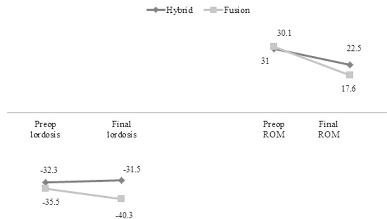
For the global angle at T12-S1, preoperative lumbar lordosis was -32.3° ± 18.2 in Hybrid group and -35.5° ± 11.5 in Fusion group (Figure 4). Additionally, final global lordosis was maintained at -31.5° ± 22.4 (p=0.86) but significantly increased (-40.3° ± 13.1, p=0.04) in the Hybrid and Fusion group, respectively. Moreover, global ROM was preserved in Hybrid group (p=0.42), but significantly restricted in Fusion group (p=0.01). The changes in lordosis and ROM in the Fusion group had statistical significance.
Fig. 4.
Global angles at T12-S1. In the Hybrid group, lumbar lordosis and ROM at T12-S1 was maintained between preoperation and the final follow-up without significant changes. In the Fusion group, global lordosis was significantly changed (p=0.04), but global ROM was restricted at the final evaluation (p=0.01).
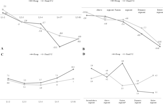
In the Hybrid group, segmental lordosis at each segment, operated segments and adjacent segments were preserved except for segmental lordosis at L4-5 (Figure 5A & B). Segmental ROM was generally decreased but preserved without statistical significance (Figure 5C). Segmental ROM at the fused segment decreased (p=0.02), but segmental ROM at the dynamic stabilized segment increased (p=0.05, Figure 5D). Segmental ROM at adjacent segments had no changes including below the segment.
Fig. 5.
Angle changes in the Hybrid group. A: Segmental lordosis at L1-2, L2-3, L3-4, L5-S1 was preserved (p=0.39, p=0.49, p=0.07, and p=0.58, respectively), but lordosis at L4-5 was significantly decreased (p=0.02). B: Segmental angle at dynamic stabilized segment was decreased but it had no statistical significance (p=0.09) and lordosis at fused segment and adjacent segments was maintained in Hybrid group. C: Segmental ROM was generally decreased at all segment without statistical significance (all p>0.05). D: Segmental ROM at fused segment was significantly deceased (p=0.02). Segmental ROM at dynamic stabilized segment was increased without significance (p=0.05).
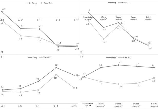
In the Fusion group, segmental lordosis at L2-3 was significantly increased (p=0.02, Figure 6A). Other segments including L3-4 and L4-5, the most common operated segment in the Fusion group had no lordotic change. The angle at the second above and the above adjacent segments were significantly changed to lordosis (p=0.04 and p=0.02, respectively, Figure 6B). Segmental ROM at each segment was generally decreased at the final follow-up, but the changes had no statistical significance (Figure 6C). Segmental ROM at the fused segments was significantly restricted (both p=0.04), and ROM at the above segment was also restricted (p=0.01, Figure 6D).
Fig. 6.
Angle changes in the Fusion group. A: Segmental lordosis at L2-3 was increased with statistical significance (p=0.02), but other segments had no changes in segmental lordosis. B: Segmental lordosis at the second above and above segment was significantly changed to lordosis (p=0.04 and p=0.02), but angle at the operated segments had no changes (p=0.38 and p=0.79). C: Segmental ROM at each segment was generally decreased at the final follow-up, but the changes had no statistical significance (L1-2, p=0.59, L2-3, p =0.73, L3-4, p=0.89, L4-5, p =0.50, and L5-S1, p=0.1). D: Segmental ROM at the fused segments was significantly restricted (both p=0.04) and ROM at above segment was also restricted (p=0.01).
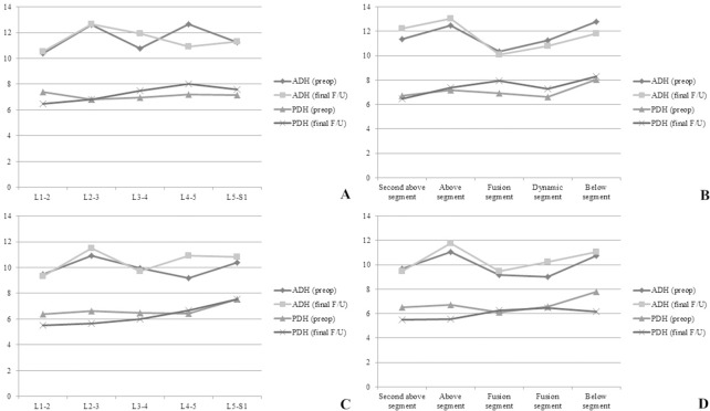
The changes in ADH and PDH are shown in Figure 7. In both groups, ADH has a relatively higher value than PDH, and the changes between preoperation and final follow-up were not significant. Preoperative disc height was not different between the two groups, but ADH at the second above segment and PDH at the above adjacent segment had a statistically higher value in the Hybrid group than that in the Fusion group at final follow-up (each p=0.04).
Fig. 7.
Disc height changes. A & B. Hybrid group. ADH and PDH at each segment and corresponding segment has no significant change (all p>0.05). C & D. Fusion group. ADH and PDH at each segment and corresponding segment both have no significant change in the Fusion group. However, ADH at the second above segment and PDH at the above adjacent segment have a statistically higher value in the Hybrid group than those in the Fusion group at final follow-up (each p=0.04).
As the definition of radiologic ASP, disc space narrowing was observed in 6 patients (40.0%) in the Hybrid group, and they were all located at proximal segments of the operated segments. In the Fusion group, disc space narrowing was observed in 7 patients (70.0%), and it was at 4 proximal and 3 distal segments of the fused segments. The development of ASP was not different between the Hybrid and Fusion group (p=0.22). Fusion was identified in 11 (73.3%) and 16 segments (80.0%) in the Hybrid and Fusion group, respectively, (p=0.66). Hence, pseudarthrosis was observed in 4 (26.6%) and 4 segments (20.0%) in the Hybrid and Fusion group, respectively. For radiologic abnormalities, screw and rod fractures were not observed, but a radiolucent line around a screw was identified in 2 patients with 3 screws instrumented with the NFlex system at the final radiograph.
Clinical outcomes
VAS for back and leg pain was significantly decreased at the final follow-up in both groups (Table 2), and each value at preoperation and final follow-up was not different between the two groups. ODI was also decreased in both groups, but ODI change in the Fusion group had no statistical significance (p=0.07). In addition, analgesic medication was also reduced after the operation in both groups, but patients in the Fusion group had taken more medicine during preoperation and postoperation. Clinical parameters did not have a difference according to disc space narrowing and pseudarthrodesis.
Table 2.
Clinical outcomes
| Hybrid group | Fusion group | p-value | ||
|---|---|---|---|---|
| VAS-back | Preop | 7.38 ± 1.44 | 7.11 ± 1.41 | 0.47 |
| Final F/U | 4.77 ± 1.73 | 3.78 ± 2.58 | 0.59 | |
| p-value | 0.002 | 0.02 | ||
| VAS-leg | Preop | 7.15 ± 1.40 | 7.44 ± 0.32 | 0.75 |
| F. F/U | 2.62 ± 2.50 | 3.89 ± 1.69 | 0.22 | |
| p-value | 0.001 | 0.02 | ||
| ODI (%) | Preop | 60.84 ± 9.65 | 65.00 ± 9.39 | 0.63 |
| Final F/U | 34.13 ± 6.55 | 34.44 ± 4.55 | 0.92 | |
| p-value | 0.008 | 0.07 | ||
| Analgesics medication | Preop | 2.87 ± 0.74 | 3.60 ± 1.07 | 0.07 |
| Final F/U | 1.60 ± 0.91 | 2.40 ± 1.07 | 0.04 | |
| p-value | 0.001 | 0.01 |
VAS; Visual Analogue Scale, ODI; Oswestry Disability Index.
Discussion
Hybrid surgery for two-segment degenerative spinal disease
This is the first clinical report with a minimum 2-year follow-up applying hybrid surgery combining a dynamic stabilization system and fusion for twosegment degenerative lumbosacral disease. Compared to fusion surgery, hybrid surgery could preserve global lumbar lordosis and ROM. Moreover, overall changes in segmental ROM were reduced angles that generally had lower values than those in the Fusion group. In the Hybrid group, clinical parameters in terms of VAS for back and legs had a corresponding result in the Fusion group. The functional outcome using ODI score showed a significant improvement in the Hybrid group and no significant change in the Fusion group. In this study, the incidence of disc space narrowing more than 2 mm at the posterior disc was not different between the two groups, but the last PDH of the above segment had a statistically higher value in the Hybrid group than in the Fusion group. During a mean 4-year follow-up, the application of hybrid surgery for two-segment could not only maintain original lumbar motion, but also had a tendency to delay disc degeneration at the above adjacent segment.
Biomechanical study of hybrid surgery
Mageswaran et al. conducted a biomechanical study comparing fusion and hybrid constructs and they found the dynamic stabilization system showed similar characteristics to the fusion construct because of greater stress in adjacent segments in hybrid construct. 4 Our biomechanical study using finite element analysis also revealed stiffness resulting from the Dynesys system which is the same as that from rigid fixation. 10 In contrast to the results of Mageswaran et al., Durani et al reported that a dynamic stabilization system could reduce the hypermobility caused by extended arthrodesis.11 In addition, other study assessed the intradiscal pressure (IDP) in monosegmental fusion at L5-S1 and hybrid surgery (Dynesys at L4-5 and rigid fixation at L5-S1).12 The IDP at the segment adjacent to the fusion was reduced when a dynamic stabilization system was added above the segment that underwent fusion. They concluded that hybrid surgery might have a possible preventative effect on degenerative disc changes at the adjacent segment.
Clinical application of hybrid surgery
For clinical application of soft stabilization using Graf bands, Imagama et al. compared degenerative changes between posterior lumbar interbody fusion (PLIF) only and PLIF with Graf band on MRI.13 The results were that the incidences of disc degeneration and spinal canal stenosis were significantly lower in PLIF with the Graf band group. In hybrid surgery using pedicle-based dynamic stabilization system, Maserati et al. reported preliminary results from the application of a DTO device.3 At a mean 8-month follow-up of 24 consecutive patients, improvement in VAS and no device-related complications were observed. Putizer et al. did a comparison study between mono-segmental fusion alone and Dynesys application adjacent to fusion in patients with asymptomatic but radiologically proven initial disc degeneration.14 A specially designed Dynesys strut (Allospine) was used in addition to a rigid rod, but there were a high number of implant failures and an increase in ASP. Chen et al. compared two-segment dynamic stabilization system (Dynesys) and hybrid stabilization system (FlexPLUS) and the hybrid stabilization system could better preserve lordosis at the operated segments and subsequently reduce the extent of compensatory hyperlordosis at the proximal adjacent segment.15
Study limitations
Because this study retrospectively analyzed twosegment lumbar surgery using hybrid surgery and pure fusion surgery and had a small number of patients in each group, no apparent result with low statistical power for generalization was obtained. The operated two segments showed severe disc degeneration in both groups compared to adjacent segments. L5-S1 segment, however, having similar disc degeneration with hybrid surgery underwent fusion in only 2 patients in the Fusion group. Initial disc degeneration was not different between the two groups on MRI, but a relatively poor disc grade would affect the patients’ result on plain radiographs in the Fusion group. If enough postoperative MRI evaluation was existed to compare the Pfirrmann's grade of disc degeneration in both groups, more details in degeneration severity should be suggested. Though two pedicle-based dynamic stabilization systems were used, the biomechanical effect might be different between each system. Because of the small number of patients, a comparison between the DTO and NFlex system was not performed. As an issue of the dynamic stabilization system, a radiolucent line around the screw was observed in 3 NFlex screws. Together with pseudarthrosis, hybrid surgery should be investigated on how it affects clinical outcomes. Disc degeneration was delayed at the above adjacent segment of the hybrid stabilization system, but the commercially used dynamic stabilization system still had a limitation in fully preventing ASP. More physiologic devices for motion preservation are needed as well as a prospective randomized controlled trial for twosegment lumbar surgery between pure dynamic stabilization surgery, hybrid surgery and pure fusion surgery designed to identify effective surgical treatment under similar surgical indications.
Conclusions
Hybrid surgery combining dynamic stabilization system and fusion can be an effective surgical method for multilevel degenerative spinal disease. Initial lumbar lordosis and ROM were preserved, and favorable clinical outcomes were obtained after hybrid surgery. The disc degeneration at the above adjacent segment of the hybrid stabilization system may be delayed; hence, further development of physiologic dynamic stabilization systems could be a promising surgical treatment.
Disclosures
The authors report no conflict of interest concerning the materials or methods used in this study or the findings described in this paper. No benefits in any form have been or will be received from any commercial party related directly or indirectly to the subject of this manuscript.
References
- 1.Lee MJ, Dettori JR, Standaert CJ, Brodt ED, Chapman JR. The natural history of degeneration of the lumbar and cervical spines: a systematic review. Spine (Phila Pa 1976). 2012;37(22 Suppl):S18–30. doi: 10.1097/BRS.0b013e31826cac62. [DOI] [PubMed] [Google Scholar]
- 2.Zhu Q, Larson CR, Sjovold SG, et al. Biomechanical evaluation of the Total Facet Arthroplasty System: 3-dimensional kinematics. Spine. 2007;32(1):55–62. doi: 10.1097/01.brs.0000250983.91339.9f. [DOI] [PubMed] [Google Scholar]
- 3.Maserati MB, Tormenti MJ, Panczykowski DM, Bonfield CM, Gerszten PC. The use of a hybrid dynamic stabilization and fusion system in the lumbar spine: preliminary experience. Neurosurgical focus. 2010;28(6):E2. doi: 10.3171/2010.3.FOCUS1055. [DOI] [PubMed] [Google Scholar]
- 4.Mageswaran P, Techy F, Colbrunn RW, Bonner TF, McLain RF. Hybrid dynamic stabilization: a biomechanical assessment of adjacent and supraadjacent levels of the lumbar spine. Journal of neurosurgery. Spine. 2012;17(3):232–42. doi: 10.3171/2012.6.SPINE111054. [DOI] [PubMed] [Google Scholar]
- 5.Stoll TM, Dubois G, Schwarzenbach O. The dynamic neutralization system for the spine: a multicenter study of a novel non-fusion system. Eur Spine J. 2002;11(Suppl 2):S170–8. doi: 10.1007/s00586-002-0438-2. [DOI] [PMC free article] [PubMed] [Google Scholar]
- 6.Coe JD, Kitchel SH, Meisel HJ, Wingo CH, Lee SE, Jahng TA. NFlex Dynamic Stabilization System : Two-Year Clinical Outcomes of Multi-Center Study. Journal of Korean Neurosurgical Society. 2012;51(6):343–9. doi: 10.3340/jkns.2012.51.6.343. [DOI] [PMC free article] [PubMed] [Google Scholar]
- 7.Pfirrmann CW, Metzdorf A, Zanetti M, Hodler J, Boos N. Magnetic resonance classification of lumbar intervertebral disc degeneration. Spine. 2001;26(17):1873–8. doi: 10.1097/00007632-200109010-00011. [DOI] [PubMed] [Google Scholar]
- 8.Kanayama M, Hashimoto T, Shigenobu K, et al. Adjacent-segment morbidity after Graf ligamentoplasty compared with posterolateral lumbar fusion. Journal of neurosurgery. 2001;95(1 Suppl):5–10. doi: 10.3171/spi.2001.95.1.0005. [DOI] [PubMed] [Google Scholar]
- 9.Lee CK, Vessa P, Lee JK. Chronic disabling low back pain syndrome caused by internal disc derangements. The results of disc excision and posterior lumbar interbody fusion. Spine. 1995;20(3):356–61. doi: 10.1097/00007632-199502000-00018. [DOI] [PubMed] [Google Scholar]
- 10.Jahng TA, Kim YE, Moon KY. Comparison of the biomechanical effect of pedicle-based dynamic stabilization: a study using finite element analysis. The spine journal. 2013;13(1):85–94. doi: 10.1016/j.spinee.2012.11.014. [DOI] [PubMed] [Google Scholar]
- 11.Durrani A, Jain V, Desai R, et al. Could junctional problems at the end of a long construct be addressed by providing a graduated reduction in stiffness? A biomechanical investigation. Spine. 2012;37(1):E16–22. doi: 10.1097/BRS.0b013e31821eb295. [DOI] [PubMed] [Google Scholar]
- 12.Cabello J, Cavanilles-Walker JM, Iborra M, Ubierna MT, Covaro A, Roca J. The protective role of dynamic stabilization on the adjacent disc to a rigid instrumented level. An in vitro biomechanical analysis. Archives of orthopaedic and trauma surgery. 2013;133(4):443–8. doi: 10.1007/s00402-013-1685-x. [DOI] [PubMed] [Google Scholar]
- 13.Imagama S, Kawakami N, Matsubara Y, Kanemura T, Tsuji T, Ohara T. Preventive effect of artificial ligamentous stabilization on the upper adjacent segment impairment following posterior lumbar interbody fusion. Spine. 2009;34(25):2775–81. doi: 10.1097/BRS.0b013e3181b4b1c2. [DOI] [PubMed] [Google Scholar]
- 14.Putzier M, Hoff E, Tohtz S, Gross C, Perka C, Strube P. Dynamic stabilization adjacent to singlelevel fusion: part II. No clinical benefit for asymptomatic, initially degenerated adjacent segments after 6 years follow-up. Eur Spine J. 2010;19(12):2181–9. doi: 10.1007/s00586-010-1517-4. [DOI] [PMC free article] [PubMed] [Google Scholar]
- 15.Chen H, Charles YP, Bogorin I, Steib JP. Influence of 2 different dynamic stabilization systems on sagittal spinopelvic alignment. Journal of spinal disorders & techniques. 2011;24(1):37–43. doi: 10.1097/BSD.0b013e3181d5349b. [DOI] [PubMed] [Google Scholar]


