Abstract
Genome engineering methods in E. coli allow for easy to perform manipulations of the chromosome in vivo with the assistance of the λ-Red recombinase system. These methods generally rely on the insertion of an antibiotic resistance cassette followed by removal of the same cassette, resulting in a two-step procedure for genomic manipulations. Here we describe a method and plasmid system that can edit the genome of E. coli without chromosomal markers. This system, known as Scarless Cas9 Assisted Recombineering (no-SCAR), uses λ-Red to facilitate genomic integration of donor DNA and double stranded DNA cleavage by Cas9 to counterselect against wild-type cells. We show that point mutations, gene deletions, and short sequence insertions were efficiently performed in several genomic loci in a single-step with regards to the chromosome and did not leave behind scar sites. The single-guide RNA encoding plasmid can be easily cured due to its temperature sensitive origin of replication, allowing for iterative chromosomal manipulations of the same strain, as is often required in metabolic engineering. In addition, we demonstrate the ability to efficiently cure the second plasmid in the system by targeting with Cas9, leaving the cells plasmid-free.
Genetic engineering through homologous recombination, known as recombineering, has provided new ways to manipulate DNA in vivo. In E. coli, λ-Red prophage assisted recombineering has facilitated new and easy methods for defined insertions, deletions, and point-mutations1,2,3. Perhaps the most popular method of recombineering described to date is that of Datsenko and Wanner for one-step gene inactivation1. In this method an antibiotic resistance cassette is inserted into the chromosome in place of the targeted gene. The target is determined by homology arms that are built into the PCR primers used for amplifying the antibiotic resistance marker and can be as short as 35 bp. This linear dsDNA is then introduced into cells that have the λ-Red genes bet, exo, and gam expressed to facilitate genome integration4. Recombinant cells are selected by antibiotic resistance and location of the marker confirmed by PCR. The antibiotic marker is flanked by flippase recombination targets (FRT) which allows removal of the marker, but leaves a single FRT scar site on the chromosome.
Methods for creating scarless chromosomal point mutations, gene insertions, or promoter replacements also proceed by initially inserting a dual-selectable marker2,5,6,7. After insertion of the dual-selectable marker, a second transformation is performed to remove the marker and insert the desired point mutation or gene. Since the targeted gene is initially removed from the chromosome, genes that are essential for cell survival must be expressed in-trans during the process, adding an additional layer of complexity. Alternatively, homing-endonuclease recognition sequences can be inserted into the chromosome as a counter-selectable marker8,9,10. A helper plasmid may then donate DNA through homologous recombination upon double strand break (DSB). After recombination the homing endonuclease recognition sequence is removed, while cells that do not undergo successful insertion are selected against by DSB. Again, essential genes must be expressed in-trans, though it was recently shown that clever designs allow this system to function on essential genes without in-trans expression10. While these methods have been used successfully, they suffer from drawbacks that include complicated cloning schemes to add DNA homology, scar sites that destabilize the chromosome, the requirement for multiple rounds of selection for a single manipulation, and low success rates.
Multiplex automated genome engineering (MAGE) was described several years ago as a tool for genome-wide manipulations using oligonucleotide mediated recombination11. This method utilizes short ssDNA fragments purchased in the form of oligonucleotides that were shown to recombine with high efficiency with assistance from the λ-red protein Bet12. Development of an automated system to continuously cycle a population of cells allowed for rapid changes on the genome scale. The power of this method was demonstrated by removing a single codon from the entire E. coli genome13. However, MAGE requires an automated system that is commercially unavailable. More recently co-selection MAGE was described as a means to achieve high-throughput genome engineering without the need for the automated system originally described14. For co-selection MAGE, several oligonucleotides are simultaneously transformed, including one which confers a selectable phenotype. Cells that possessed the selectable mutation were far more likely to have additional allelic replacements (AR) in targets within close proximity15. Utility of this system is limited by the availability of co-selectable markers at regions throughout the genome. A strain could be created which possesses such selectable markers located throughout the genome by recombineering with dsDNA. While possible, strain construction would be difficult and would prevent the use of project specific strains of E. coli.
The CRISPR/Cas9 system has been a transformational tool for genome engineering in a variety of organisms16,17. Cas9 is an endonuclease that targets a specific DNA sequence that can be easily programmed by a plasmid encoded target sequence of 20 bp18. The only requirement of the Streptococcus pyogenes Cas9 is that the protospacer be adjacent to the triplet NGG, known as the protospacer adjacent motif (PAM). There are 424,651 GG doublets on both strands of the E. coli chromosome19, making PAM site availability unlikely to limit Cas9 targeting. Cas9 cleavage of genomic DNA results in cell death because E. coli lacks the classical non-homologous end joining mechanism for DNA repair20. It was shown that the CRISPR Cas9 system can be used to successfully select for cells with point mutations that alter the guide RNA target sequence in both Streptococcus pneumoniae and E. coli21. In S. pneumoniae, a naturally recombinogenic strain, this system could be used to make deletions, insertions, and point mutations. In E. coli, expression of the λ-Red gene bet was required to introduce a mutation into rpsL. With Cas9 counterselection this mutation could be selected with an efficiency of 60% of surviving cells. Much more recently, this same two-plasmid system was combined with the λ-Red recombineering plasmid pKD46 and the ability to insert or delete genes from both ssDNA and dsDNA donors at a single chromosomal locus was demonstrated22. Others have shown that the E. coli genome can be edited using plasmid DNA as a donor with Cas9 counterselection23. We sought to extend utility of the Cas9 counterselection method and develop an easy to use plasmid system that could efficiently make point mutations, gene deletions, and short sequence insertions iteratively and with high efficiency.
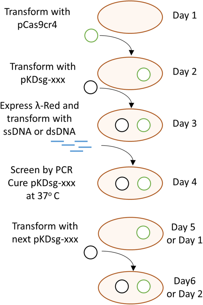
Here we describe the no-SCAR (Scarless Cas9 Assisted Recombineering) method for genome modifications in E. coli. This two-plasmid system contains all of the components required so that host specific modification are not needed. We demonstrate that this method enables the selection of gene deletions, point mutations, and short insertions in a single-step at many genomic locations. No-SCAR, outlined in Fig. 1, can increase the throughput and ease with which complex genome engineering projects are completed. In contrast to other recently described Cas9 counterselection methods we provide easy to perform instructions for sgRNA plasmid retargeting and demonstrate that both plasmids can be easily cured in a few days. Compared to other methods for scarless genome editing that use dual selection cassettes the no-SCAR system takes a similar amount of time and effort when only a single mutation is performed. However, for projects that require more than one mutation the system becomes much faster with subsequent mutations made in as little as three days.
Figure 1. General outline of the no-SCAR method.

On day 1 the pCas9cr4 plasmid is used to transform E. coli, followed by plating on LB + Cm, and growth at 37 °C. On day 2 the resulting strain can be transformed with pKDsg-xxx plasmid, where –xxx denotes the targeted gene, plated on LB + Spec and Cm, and incubated at 30 °C overnight. On day 3 the resulting strain is grown in SOB until OD ~0.5 and λ-red is induced with 50 mM L-arabinose. After 15–20 minutes the cells are made electrocompetent and transformed with ssDNA or dsDNA that confers a mutation to the protospacer or PAM sequence. After 1–2 hours of recovery the cells are plated on LB + Spec, Cm, and aTc, then incubated at 30 °C overnight. On day 4 colonies are screened by PCR and grown at 37 °C to cure the pKDsg-xxx plasmid. The next pKDsg-xxx plasmid is then used to transform the mutant strain on Day 5 and the process is repeated.
Methods
Plasmid construction and protospacer targeting
All plasmids listed in Table S1 were created using circular polymerase extension cloning (CPEC)24. The primers listed in Table S2 were used to create linear dsDNA products that were subsequently DpnI digested for at least 15 minutes and then gel-purified. The DNA was eluted in 6 ul of dH2O and mixed with corresponding linear product. These products were then used as template for a reaction with Q5 polymerase for 15 cycles. The product was then used to directly transform chemically competent E. coli DH5α or NEB Turbo cells. The following plasmids and maps have been deposited to Addgene (Cambridge, MA); pCas9cr4 (Plasmid #62655), pKDsg-ack (Plasmid #62654), and pKDsg-p15 (Plasmid #62656).
The 20 bp targeting sequences of the sgRNA were re-targeted by CPEC cloning of two linear PCR fragments. The re-targeting primers listed in Table S2 were approximately 40-mers that had overlapping protospacer sequences. The primer pair protospacerF and gamR were used to yield a 3 kb product. The protospacer R primer was paired with pKDseq1F to yield a product of about 4 kb. This design yielded PCR product with about 280 bp of overlapping homology between the gam and araC (Fig. 2), as well as 20 bp of overlap in the protospacer. PCR products were gel purified, mixed together in equal volumes and CPEC cloned with Q5 polymerase. The mixture was used to transform chemically competent DH5α or NEB Turbo cells (New England Biolabs), recovered for 1 hour in super optimal broth with catabolite repression (SOC), and then plated on LB with 50 mg L−1 spectinomycin (spec) and incubated at 30 °C. A step-by-step protocol for primer design and retargeting is available as part of the no-SCAR protocol at Addgene.
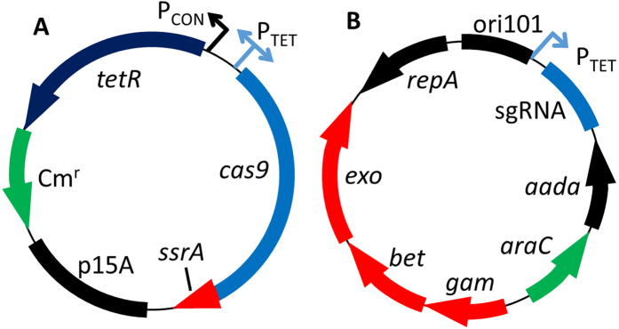
Figure 2. Schematic map of the no-SCAR plasmids.

(A) Schematic of the plasmid pCas9cr4 which has cas9 expressed under control of the PTET promoter and tetR constitutively expressed. (B) Schematic of the plasmid pKDsg-xxx which has the sgRNA expressed under control of the PTET promoter and the three genes that compose the λ-Red system under control of the arabinose inducible promoter ParaB.
Recombineering
Oligonucleotides used for recombineering should be designed using previously described parameters that maximize efficiency25. In our experience the most important factors for successful oligonucleotide recombineering are: proper length of oligo (60–90 bases), 2–4 phosphorothioate bonds to the 5′ end, targeting the lagging strand, and avoiding mismatch repair. The dsDNA recombineering presented in this work did not possess any special modifications. However, efficiency may be increased by adding a phosphate to the 5′ end of the strand that corresponds to the leading strand and/or adding 2–4 phosphorothioate bonds to the 5′ end of the strand corresponding to the lagging strand26,27.
E. coli MG1655 that possessed both the pCas9cr4 plasmid and pKDsgRNA plasmids were grown in 4 mL of super optimal broth (SOB) with 50 mg L−1 spec and 34 mg L−1 chloramphenicol (cm) at 30 °C. At an OD of approximately 0.5, λ-red was induced with the addition of 50 mM L-arabinose and incubated for 20 minutes. Cells were then made electrocompetent by the glycerol/mannitol density step gradient as described previously28. Cells were re-suspended in the glycerol/mannitol solution at 50 μl per one mL of culture. Oligonucleotides were added to 50 μl of cells at a final concentration of 10 μM and then transferred to a 0.1 mm electroporation cuvette. Electroporation was performed on a BioRad Gene Pulser and cells were immediately recovered in 1 mL final volume of SOC for 1–2 hours before plating on LB with 34 mg L−1 Cm, 50 mg L−1 Spec, and 100 ng mL−1 anhydrotetracycline (aTc). Plates were incubated at 30 °C overnight.
The ack mutation and deletion was screened by growth on M9 minimal medium plates with 2% glycerol, 10 mM chloroaceate, and 0.1% SOC. Colonies were patched from the overnight plates and onto the selective plates using a toothpick and incubated at 30 °C for two nights. The rpoB mutation was screened by patching colonies onto LB with 20 mg L−1 rifampicin. Plates were incubated at 37 °C overnight and scored for growth.
The dsDNA for insertion of the ssrA tag onto pfkA was PCR amplified with primers pfkA Down F and R using strain IB164329 as template.
Colony counts
Colony forming units were determined similarly to the miniaturized plating method described previously30. Briefly, 10 μL of recovered cultures, as well as dilutions of 10−1, 10−2, and 10−3, were spotted on plates in triplicate and incubated overnight at 30 °C.
Genotyping PCR
Transformants were genotyped by allele specific PCR31. Briefly, the 3′ end of the mutant genotyping primer listed in Table S1 annealed perfectly to the mutant genotype, while wild-type possessed mismatched DNA at the 3′ end. Hotstart Taq polymerase that did not possess 3′ to 5′ exonuclease activity was used for colony PCR of the putative mutants. Only those cells which had incorporated the mutation produced PCR product, since the mismatch between the 3′ end of the primer and wild-type genome did not allow for primer extension. The ack deletion was screened by using PCR primers up and downstream of the deletion. The pfkA ssrA tag was genotyped with the pfkA Down F and R.
Results
Inducible cell death by DNA double strand break
Coexpression of cas9 and single-guide RNA (sgRNA) that targets the E. coli chromosome is lethal due to the introduction of chromosomal DSB21,32,33. Even low levels of cas9 expression are likely to cause cell death and a plasmid-based system which is capable of maintaining both cas9 nuclease and genome targeting sgRNA has not been described. We hypothesized that low frequency recombineering events, such as sequence deletions and insertions, would benefit from the ability to co-maintain two plasmids. Methods described previously were successful with co-transformation of plasmid DNA and ssDNA, though this method is inherently less efficient because the cells would need to acquire both the ssDNA and plasmid21,34. Thus, we set out to design a system with tight control of cas9 and sgRNA expression to allow maintenance of both plasmids. Induction of cas9 and sgRNA expression will then cause cell death.
The previously described nuclease-null plasmid pdCas9-bacteria that possessed the S. pyogenes cas9 was first modified to re-activate nuclease activity creating plasmid pCas935. The pCas9 plasmid possessed the aTc inducible promoter PTET to drive expression of both tetR and cas9. In the absence of inducer, TetR represses transcription of both cas9 and tetR. However, even in the absence of aTc, co-transformation of pCas9 and sgRNA targeting the ack gene in E. coli had very low transformation efficiency, while transformation with an off-target sgRNA with pCas9 had an efficiency over two orders of magnitude higher. This suggested that in the absence of inducer leaky expression of cas9 and sgRNA caused cell death.
To maintain tighter control of cas9 expression an ssrA consensus tag was added to the C-terminus of cas9, creating plasmid pCas9cr1. The SsrA tag is recognized by ClpP protease and should speed degradation of Cas9 that results from leaky transcription. Next, plasmid pCas9cr2 was created by adding a constitutive promoter upstream of tetR to increase expression of the negative-regulator and maintain tighter transcriptional control. Lastly, the ribosome binding site (RBS) was modified to decrease the theoretical rate of translation initiation by tenfold36, creating plasmid pCas9cr4. The plasmid pCas9cr4 was still unable to co-transform with the sgRNA plasmid. However, when cells were first transformed with the pCas9cr4 plasmid and subsequently transformed with the sgRNA encoding plasmid, colonies were obtained in the absence of inducer. The sequential transformation allowed sufficient amounts of TetR to accumulate and effectively repress cas9 expression.
The sgRNA cassette targeting ack was moved from the previously described pgRNA-bacteria plasmid35 into the pKD46 plasmid that possessed the λ-Red system and the temperature sensitive origin of replication from pSC101 (Fig. 2)1. Initially the sgRNA cassette was placed downstream of the λ-Red genes and upstream of repA. However, initial experiments showed that this location was susceptible to recombination events that removed the sgRNA and caused counter-selection escape. Consequently, the sgRNA cassette was placed between the origin of replication and antibiotic marker, to limit the possibility of recombination since both of these components were essential to the plasmid. The ampicillin resistance gene bla was also replaced with the spec resistance gene aadA to avoid satellite colony formation associated with ampicillin resistance, creating plasmid pKDsg-ack. To further control the inducible cell death phenotype the PTET promoter was inserted in-place of the constitutive promoter that was driving expression of the sgRNA. In the absence of inducer these modifications enabled cell growth that was similar to the plasmid-free parent strain.
While most cells were killed by expression of cas9 and genome targeting sgRNA, some cells were able to escape the counterselection. To determine the rate of counterselection escape in cells that possessed both plasmids the number of CFU’s after plating was determined in the presence and absence of inducer. As summarized in Table 1, the rate of escape varied from 8.5 × 10⁻⁵ to 7.2 × 10⁻⁴ with four different guide RNA targets, with an average of 2.6 × 10⁻⁴. Even at the high end of the escape frequency, standard point mutations and deletions should be easily identified given the reported frequency of these allelic replacements11,12,37. This escape rate of 2.5 × 10⁻⁴ was similar to the rate previously determined in E. coli and Lactobacillus lactis21,34. In Jiang et al. the plasmids were constructed with the native S. pyogenes dual guide RNA, in which the plasmid encoded guide sequence was flanked by short palindromic repeats. In this case the escape mutants were found to have lost the guide sequence, likely the result of recombination events in the palindromic repeats. In the system presented here, single-guided RNA was used, which removed these palindromic repeats18. In all escape plasmids that were examined, the guide sequence remained intact. The chromosomal protospacer also remained unmodified, making the genetic basis for these escapes unclear. It is possible that point mutations in cas9 or elsewhere in the sgRNA plasmid alter the level of protein and transcript in the cell, resulting in the escape phenotype. In the cases that we’ve tested this escape phenotype is maintained when plasmids are purified from an escapee and used to transform wild-type MG1655. Nevertheless, this escape rate was low enough that our system could be successfully used to enrich for mutant populations as described below.
Table 1. Fold-change in CFU’s of sgRNA transformants when plated with and without aTc.
| Plasmid | Escape Rate1 |
|---|---|
| pKDsg-ack | 1.16 × 10⁻⁴ |
| pKDsg-apt | 8.49 × 10⁻⁵ |
| pKDsg-sspB | 7.16 × 10⁻⁴ |
| pKDsg-yqhD | 1.13 × 10⁻⁴ |
1Average of three independent experiments.
Point mutation selections
The system was first tested for the ability to counterselect against wild-type cells when inserting point mutations during oligonucleotide mediated recombineering. Oligonucleotides were designed to insert point mutations into the ack and rpoB genes of E. coli (Fig. 3a,b). The point mutations were designed to alter the PAM sequence or protospacer sequences within 12 bp of the PAM because this region was shown to be most important for Cas9 specificity38. The oligonucleotides were targeted to the lagging strand, since the efficiency of allelic replacement is over two orders of magnitude greater than the leading strand12. The ack mutation was designed to insert a nonsense mutation which gives resistance to the acetate analog chloroacetate. The rpoB mutation resulted in a missense point mutation, D516V, which confers resistance to rifampicin39.
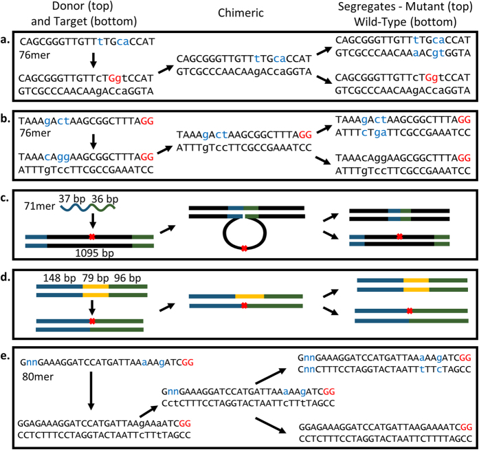
Figure 3. Schematic of the genomic DNA modifications demonstrated by the no-SCAR system.
The donor and target DNA are on the left, the chimeric genomic DNA produced after integration through a fully single-stranded DNA intermediate is in the middle, and the segregated chimeric DNA that results in populations of mutant and wild-type are on the right. (a,b) 21 bp of the targeted region for making point mutations into the rpoB and ack genes. The PAM site is shown in red, mutations are shown in lowercased blue text, and mutation target in lowercased black text. (c) Deletion of 1095 bp of ack using a 73mer oligo. (d) Insertion of 79 bp at the C-terminus of pfkA using dsDNA. Red × indicates the site of Cas9 targeting. (e) Insertion of degeneracies outside of the protospacer sequence.
For the ack mutation, the system was tested with two different transformation schemes. First, cells which possessed both the pKDsgRNA-ack and cas9 encoding plasmids were transformed with oligonucleotide. Alternatively, cells that possessed only the pKDsgRNA-ack encoding plasmid, which also carries the λ-Red genes, were co-transformed with pCas9cr4 and oligonucleotide. In both cases, cells were grown to mid-logarithmic phase, λ-Red was expressed for 20 minutes, and then electrocompetent cells were prepared. Cells were then transformed with oligonucleotides, recovered for 1–2 hours, and then spread onto plates with aTc to express cas9 and sgRNA. Controls were performed in which cells were transformed with oligonucleotides that did not match the sgRNA guide that it possessed. For the cells that possessed both plasmids before transformation with the oligonucleotides there were 60-fold (3.91 × 10⁵/6.67 × 103) more CFU’s mL−1 with ackmut2 than the control transformation (Table 2), suggesting efficient selection for only those cells that incorporated the desired mutation. Putative ack mutants and controls were patched onto M9 plates with 10 mM chloroacetate and glycerol as the sole carbon source. After a 48 hour incubation the results showed that 99 ± 1% of the putative point mutants grew on the selective plates while only 8.1 ± 0.9% of the control transformants grew. The colonies from the control plate that grew were escape mutants that became spontaneously resistant to chloroacetate. Thus, up to 8.1% of the point mutants could actually be escapes and not possess the desired mutation. However, allele specific PCR of 24 randomly selected chloroacetate resistant colonies showed that all 24 possessed the desired mutation (Figure S1), consistent with the colony counts presented in Table 2 which predicted that only one in 60 colonies were wild-type for ack.
Table 2. Efficiency of genome editing using the no-SCAR system.
| Plasmid | Mutation DNA | CFU’s1 | Positives2 | Control DNA | CFU’s1 | Positives2 | Fold-Change in CFU’s3 |
|---|---|---|---|---|---|---|---|
| pKDsg-ack | ack mut2 | 3.9 × 10⁵ | 99 ± 1% | rpoB mut | 6.6 × 10³ | 8 ± 1% | 61 ± 23 |
| pKDsg-ack | Ack CD | 5.5 × 10⁴ | 85 ± 1% | ack mut2 | 6.6 × 10³ | 8 ± 1% | 9.3 ± 1.9 |
| pKDsg-rpoB | rpoB | 1.7 × 10⁵ | 94 ± 4% | Ackmut2 | 1.1 × 10⁴ | 3 ± 1% | 16.6 ± 1.8 |
| pKDsg-pfkAE | dsDNA-ssrA | 8.0 × 10⁵ | 100% | dsDNA-off | 2.5 × 10⁵ | nd | 4.5 ± 1.8 |
1The CFU’s are the average of three independent experiments.
2The % of positive were obtained from chloroacetate and rifampicin selection plates.
3The Fold-Change in CFU’s is the average of the same three independent experiments ± SD.
The alternate protocol, in which both pCas9cr4 and the oligonucleotide were co-transformed, yielded a 4.6 fold change in CFU’s (2.3 × 10² / 0.5 × 10²), though significantly fewer colonies were obtained. Each colony was genotyped using allele specific PCR. The results showed that 21 of 24 colonies possessed the desired mutation. Thus, using this double transformation protocol it was possible to select for point mutations with high efficiency.
The rpoB mutation was tested by the single transformation protocol only. Cells that possessed both pCas9cr4 and pKDsgRNA-rpoB were transformed with the rpoBmut or ackmut2 and plated on LB with aTc. There were 15-fold more colonies with the on-target oligonucleotide than the off-target (1.7 × 10⁵/1.1 × 104). Colonies from both the experimental and control transformations were patched onto LB plates with rifampicin. After 1 day of incubation 94 ± 4% of the putative mutants grew while only 3 ± 1% of the control transformants grew. The number of positive colonies from the rifampicin plate was consistent with the colony counts which predicted that 1 of 16 (93.75%) colonies would be Cas9 escape mutants. To find the total mutation frequency, a similar experiment was performed in which the recovered cells were plated on medium with and without inducer. The total mutation frequency was found to be 2.3 ± 1.3% of cells that survived electroporation. This efficiency was slightly lower than the efficiencies reported elsewhere11,40, but still about two orders of magnitude above our escape rate.
In addition to the mutations described above, point mutations have been made in the genes apt, gyrA, ppc, pck, rpsL, pts, and yjgB. The success and efficiency of creating these point mutations suggested that the system is robust enough to make point mutations in any genomic location with an appropriate PAM site.
Oligonucleotide mediated deletions
Next, the system was tested for its ability to select for deletions of chromosomal DNA using oligonucleotides with upstream and downstream homology to the area of deletion. It was previously shown that up to 45 kbp could be deleted from the chromosome by a single 90mer oligonucleotide, though the frequency of recombination decreased as the size of deletion was increased11. We used the same ack targeting sgRNA as described in the previous section and a 71 bp oligonucleotide that was designed to delete 1095 bp of the ack gene (Fig. 3c), thereby removing the targeted protospacer. Again, both the single and double transformation protocols were tested. The single transformation protocol, in which the cells possessed both cas9 and sgRNA-ack encoding plasmids, yielded approximately 8-fold more CFU’s (5.5 × 10⁴/6.6 × 103) when transformed with on-target oligonucleotides than off-target. Colonies were patched from the original plate and onto the chloroacetate selection plate where 85.6 ± 6.4% of colonies grew. Colony PCR of 12 randomly selected colonies showed that all 12 possessed the deletion (Figure S2). For the double transformation protocol there were 2.4-fold more colonies with the on-target oligonucleotide than the control (2.2 × 10² / 0.5 × 10²). Colony PCR of the putative mutants showed that 60% had the deletion.
In addition to the deletion of ack, this method was used to successfully delete 500 bp of the sspB gene and 3 kbp spanning the dkgA-yqhD-yqhC locus. Colony PCR showed that 42% of the sspB and 16% of the larger dkgA-yqhD-yqhC (Figure S3) colonies possessed the correct deletions. The efficiency of oligonucleotide mediated deletions is known to be inversely related to the length of the deletion11. A deletion of 45 kbp was obtained at an efficiency of between 0.01–0.1 percent. Given the escape rate that was determined here, 2.6 × 10⁻⁴, even very large deletions should be possible with this method.
Short sequence insertions
Next, the ability to insert short sequence motifs was examined. It was previously demonstrated that oligonucleotides could be used to insert up to 30 bp into the chromosome11. The size of insertion using an oligonucleotide is limited due to size constraints of the oligonucleotide itself. As an alternative to ssDNA, dsDNA sequences could also be used to insert short fragments into the chromosome. The no-SCAR system maintains exo and gam from λ-Red which facilitate chromosomal integration through single stranded intermediates at the replication fork in a mechanism analogous to oligonucleotide recombineering26,27. Linear dsDNA has recently become available as gBlocks from Integrated DNA Technologies (Coralville, IA) and DNA strings from Life Technologies (Carlsbad, CA) for an economical price with delivery in just a few days. Fragments can be designed de novo and the homology required for targeting included in the sequence. This alleviates the need to incorporate homology on PCR primers, thereby allowing longer regions of homology to be used, which can increase the efficiency of insertion27. Since the mass of these fragments is typically only 200–500 ng, the fragment should be PCR amplified before transformation. To test the ability of our system to select for dsDNA mediated mutants, we PCR-amplified a 300 bp fragment that also contained a 79 bp ssrA tag insertion at the C-terminal of the pfkA gene (Fig. 3d). The fragment included 80 bp of upstream and 140 bp of downstream homology to the insertion site. After transformation there were approximately 4-fold more colonies after transformation with the on-target dsDNA (8.05 × 10⁻⁵/2.1 × 10−5). However, PCR genotyping of three independent experiments found that 36 out of 36 colonies possessed the 80 bp insertion (Figure S4). Thus, the efficiency of insertion was higher than the colony counts would predict. The insertions were further confirmed by sequencing of 5 clones, all of which had the predicted insertion (Figure S5). While we did not probe the upper limits of insertion size, it is known that recombination efficiency decreases with increasing insertion size27. However, it should be possible to identify insertions of over 1 kb using the no-SCAR system. Insertion of a 1.2 kb fragment with 45 bp homology arms had an insertion efficiency of 1.9 × 10⁻⁴ recombinants per viable cell26. Given our escape rate of 2.6 × 10⁻⁴, similar insertions should be easily identified after Cas9 counterselection.
Chromosomal degeneracies
The examples presented above demonstrated the ability to select for mutations that alter or remove sequences within 12 bp of the PAM site. We next sought to create an RBS library by including degeneracies in the oligonucleotide while targeting a PAM site that was outside of the 12 bp window of specificity (Fig. 3e). A PAM site located 13 bp downstream of the start codon was targeted, which allowed synonymous mutations to be inserted within 12 bp of the PAM site and disrupt Cas9 targeting. In addition, two N-degeneracies that were 26 and 27 bp from the PAM site were incorporated into the oligonucleotide used for mutagenesis. After transformation of the degenerate oligonucleotide, 23 putative mutants were sequenced (Figure S6). Of these 23 sequences 11 possessed the silent mutations and eight of these also possessed an altered RBS. There were three possible explanations for the three sequences that possessed the silent mutations but not the RBS alterations; the degeneracies could have simply matched the wild-type, oligo degradation by DNA Polymerase I or III could have removed the degeneracies41, or methyl-directed mismatch repair could have caused reversion to wild-type after oligo incorporation. Since mismatch repair is a possibility it may be prudent to design the mutations which fall outside of the protospacer target to also avoid mismatch repair. Though we have not tested the limits of how far away these additional mutations can be made, the possibility of DNA degradation and host mismatch repair will affect these efficiencies. Regardless, this experiment demonstrated that mutations can be made outside of the 15 bp window of the PAM site and adjacent sequence that is most important for Cas9 targeting. Moreover, the ability to insert degeneracies directly onto the chromosome and produce a genome encoded DNA library was demonstrated.
Plasmid curing
Upon completion of genome engineering projects removal of the no-SCAR plasmids allows for plasmid origins and markers to be used again for various purposes. The pKDsg-xxx plasmid can be cured by growth at 37 °C but pCas9cr4 did not possess an inherent property that allowed curing of the plasmid. We hypothesized that the pCas9cr4 plasmid could be cured by introducing DSB through Cas9 cleavage. To test this hypothesis the pKDsg-p15A plasmid was created which targeted the p15A origin of replication of pCas9cr4. Upon transformation of pKDsg-p15A into cells that contained pCas9cr4 the cells were recovered in SOC for 1 hour, then 100 ng mL−1 aTc was added and incubated for an additional 2 hours before plating on LB with Spec and aTc. The resultant colonies were patched onto LB plates with and without chloramphenicol (Cm) and grown at 37 °C. After overnight growth all patched colonies grew on the LB only plate and had no growth on the Cm plate, indicating loss of the pCas9cr4 plasmid. Furthermore, plasmid mini-preps did not yield either plasmid, confirming that both plasmids were cured. This technique could be used to cure other plasmids as well.
Comparison with other scar-free genome editing techniques
To demonstrate the speed and utility of this method a day-by-day comparison of the no-SCAR method with two other recently published methods for scarless genome modifications and the method of one-step gene inactivation from Datsenko and Wanner was performed (Table 3). The two recently described Cas9 counterselection techniques21,22 were not included because they do not demonstrate the ability to remove the sgRNA or cas9 encoding plasmids, making them unsuitable for iterative modifications. The scheme in Table 3 takes into account the time required for cloning and assumes that wild-type cells were used and λ-Red is not integrated into the chromosome. As demonstrated in Table 3, all four systems take a similar number of days for completion of a single modification when starting and ending with cells that are plasmid-free. When more than one modification is required the no-SCAR system was faster than the compared methods since the pCas9cr4 plasmid remained in the cells after the first iteration. For example, if 3 genome modifications were undertaken and all sgRNA cloned simultaneously, the no-SCAR method would take as little as 14 days, while the next fastest method would take as little as 18 days. Time savings continue to accumulate as additional mutations are made. Moreover, the no-SCAR method is the only one in which essential genes can be targeted without additional constraints since the wild-type gene is never removed from the chromosome.
Table 3. Comparison of genome editing techniques.
| No-SCAR | TetA-SacB Dual Selection5 | SceI counter-selection10 | Datsenko and Wanner1 | |
|---|---|---|---|---|
| Day 1 | 1) Transform pCas9cr4 2) Clone spacer (2 fragments)1,3 | Transform λ-Red plasmid | 1) Transform λ-Red plasmid 2) Clone Mutation Cassette (4 fragments)2,3 | 1) PCR donor DNA 2) Transform λ-Red plasmid pKD46 into target strain |
| Day 2 | Grow clones | Grow cells overnight | Screen and sequence clones | Transform Linear DNA |
| Day 3 | Isolate plasmid and transform into cells with pCas9cr4 | 1) Subculture and induce λ-Red 2) Transform TetA-SacB Cassette | PCR amplify Mutation Cassette and Transform | Screen AbR colonies Cure λ-Red |
| Day 4 | 1) Start culture and induce λ-Red 2) Transform linear DNA, induce Cas9 | Restreak colonies on counter-selection medium | Restreak Colonies | Transform pCP20 |
| Day 5 | 1) Screen colonies 2) Grow at 37 to cure pKDsgRNA4 | Identify sucroses clones and start overnight culture | Resuspend colonies and plate to induce DSB | Express Flp recombinase |
| Day 6 | Transform pKD-p15, induce Cas9 | 1) Start Culture and induce λ-Red 2) Transform with linear DNA and plate on counterselection medium | Patch colonies to screen for AbS | PCR screen or Patch for AbS Grow at 37 to cure plasmid |
| Day 7 | Patch colonies for cms, Grow at 37° | Incubate at 42° | Passage cells to cure plasmid4 | Plasmid free colonies |
| Day 8 | Plasmid free colonies | Screen by PCR or TetS | Plasmid free colonies | - |
| 1 mutation | 5 Days | 8 Days | 7 Days | 6 Days |
| 1 mutation w/ curing | 8 Days | 8 Days | 8 Days | 7 Days |
| 2 mutations w/ curing | 113 Days | 16 Days | 134 Days | 13 Days |
| 3 mutations w/ curing | 14 Days | 24 Days | 18 Days | 19 Days |
| Essential Genes | No additional requirements | Must express in-trans | Special considerations | N/A |
1Requires 2 fragment ligation independent cloning, with the primer designs given in the manuscript we have been 100% successful in producing these clones.
2Requires 4 fragment ligation independent cloning that should be sequence verified before transformation.
3When multiple mutations are desired cloning can be performed simultaneously on day 1, resulting in a faster turnaround time for subsequent mutations.
4For more than one mutation the strains obtained during day 5 can be cured of plasmid and immediately transformed with new pKDsgRNA, resulting in 3 day turnaround for each additional mutation.
5For more than one mutation the strain obtained on day 7 can be transformed with Linear DNA (Day 3) resulting in a 5 day turnaround for each additional mutation.
Discussion
Recombineering in E. coli with ssDNA was first described over ten years ago12. Based upon the belief that ssDNA tails were important intermediates in dsDNA recombineering, it was hypothesized that ssDNA alone may be capable of chromosomal recombination. Indeed, it was found that 70mer oligonucleotides were capable of introducing point mutations and deletions of 3.3 kb in the chromosome. Oligonucleotides that corresponded to Okazaki fragments, termed lagging strand, had a much higher efficiency for these allelic replacements. In subsequent years several additional modifications were identified that continued to increase the efficiency of ssDNA recombineering. Some of these modifications were manifested in the design of the oligonucleotides, such as increased length, use of phosphorothioate bonds, and limiting the secondary structure11. Other modifications were made to the host strain, including inactivation of the mismatch repair system40, removal of endogenous nuclease genes42, and alteration of replisome dynamics43. While these host strain modifications have increased the efficiency of ssDNA recombineering, they come at a cost of increased host-strain mutations and reduced cell fitness. A host-strain with these modifications is available through Addgene; however, using this pre-designed strain prevents the use of project-specific strains.
Disabling the host mismatch repair system was the first modification that was observed to have a significant impact on the efficiency of allelic replacement40. A mutS deletion strain increased the frequency of allelic replacement by over 100-fold40. Consequently, most studies have since relied on strains deficient in mismatch repair. Unfortunately, mutS deletion strains accumulate background mutation rates that are 100-fold higher than wild-type E. coli44. Though most of these background mutations are likely to be innocuous, some may have deleterious effects on cell fitness or desired phenotype. Recently a strain was developed which allowed for temperature sensitive control of host-mismatch repair gene expression45. While this strain or other mismatch repair deficient stains may be used with the no-SCAR system, they still suffer from the aforementioned drawbacks. We note that all of the work presented here was performed in strains with the mismatch repair system intact. The ack and rpoB point mutants were designed to include a C:C mismatch, which results in higher efficiencies due to mismatch repair evasion40. When possible, the oligonucleotide should be designed to include a C:C mismatch within six bp of the desired change37.
The no-SCAR method described here allows for single-step genome alterations without the use of a selectable marker. This method builds on previous work which demonstrated that Cas9 could counter-select against wild-type E. coli and enrich for a point-mutation with an efficiency of 60%21. This previous work relied on co-transformation of the guide RNA encoding plasmid along with mutation encoding oligonucleotides. The no-SCAR system was designed to stably maintain plasmids with cas9 and sgRNA that targets the host-cell chromosome. Several modifications were required to achieve a level of regulation that prevented constitutive cell death. This system used the PTET promoter to control transcription of both the sgRNA and cas9. The PTET promoter was repressed by the TetR regulator and it was essential that TetR be present in cells before both sgRNA and cas9 were introduced into the same cell.
A direct comparison of the plasmids described here with those described previously in Jiang et al. was not performed. One key difference between these systems was that the no-SCAR plasmids possess all system components, including λ-Red, and has no host specific requirements. The efficiency of the no-SCAR system was examined using two different transformation protocols. First, a protocol similar to that of Jiang et al., where the host cells possessed one plasmid and were then co-transformed with the second plasmid and mutation conferring oligonucleotide was tested. Second, cells that possessed both no-SCAR system plasmids were transformed with mutation-conferring oligonucleotides only. Both protocols were tested for making point mutations and gene deletions and in both cases a majority of transformants possessed the desired mutations. The efficiency of obtaining point mutations was similar for both protocols. For the less frequent recombination event of making gene deletions, the co-transformation protocol was slightly less efficient. Thus, to save time it is possible to only transform with the λ-Red encoding plasmid and then subsequently co-transform oligonucleotide and pCas9cr4. However, for less efficient modifications we suggest using cells that have both no-SCAR plasmids in order to maximize the likelihood of obtaining correct mutants.
The description of co-selection MAGE provided a framework for medium-throughput genetic manipulations in E. coli 15. In the absence of selectable markers available at well-spaced positions around the chromosome, the co-selection MAGE technique would be difficult to use for projects of large scale. The no-SCAR system can be used to make any gene or intergenic region with a PAM sequence a counter-selectable marker. Thus, the no-SCAR method integrates seamlessly with co-selection MAGE. However, we envision this system as a powerful tool to achieve medium to high-throughput genomic modifications in E. coli even without co-selection MAGE.
The no-SCAR method can be used for the sequential introduction of a potentially limitless number of modifications. In contrast to the traditional method of λ-Red recombination where the antibiotic marker is removed by Flp recombinase, this method does not leave recombinase recognition site scars, which can cause chromosomal instability and unwanted genomic rearrangements1. In contrast to recently described methods that combine Cas9 counter-selection with λ-Red recombineering, our system allows for the simple curing of the sgRNA encoding plasmid by means of its temperature sensitive origin of replication, allowing for transformation of a subsequent sgRNA encoding plasmid. Re-targeting the protospacer can be performed with very high efficiency using ligation independent cloning methods such as CPEC or Gibson assembly24,46. The first iteration of the no-SCAR method described here requires 5 days, including the cloning step required for sgRNA targeting. Subsequent iterations can be completed in as little as 3 days since the cells already contain the pCas9cr4 plasmid, which is faster than any other method published.
Continued development of this system could allow for simultaneous selection against more than one target. DNA synthesis and ligation independent cloning methods become difficult with highly repetitive sequences required for multiple sgRNA within a single plasmid. Moreover, the frequency of escape due to plasmid recombination and loss of protospacer sequences is also a challenge that must be addressed. In its current state, the no-SCAR system should facilitate genome engineering projects that require several to dozens of modifications.
Additional Information
How to cite this article: Reisch, C. R. and Prather, K. L. J. The no-SCAR (Scarless Cas9 Assisted Recombineering) system for genome editing in Escherichia coli. Sci. Rep. 5, 15096; doi: 10.1038/srep15096 (2015).
Supplementary Material
Acknowledgments
We thank Irene Brockman and Sue Zanne Tan for helpful suggestions and technical assistance and Emma Valentine for experimental assistance. We thank Barry Wanner for providing plasmid pKD46 and Stanley Qi for plasmids pdCas9 and pSgRNA. This work was supported by Shell Global Solutions (US) Inc. and a National Institute of Food and Agriculture Postdoctoral Fellowship to CRR (2013-67012-21022).
Footnotes
Author Contributions C.R.R. designed and performed the experiments. C.R.R. and K.L.J.P. interpreted the results and wrote the paper.
References
- Datsenko K. A. & Wanner B. L. One-step inactivation of chromosomal genes in Escherichia coli K-12 using PCR products. Proc. Natl. Acad. Sci. 97, 6640–6645 (2000). [DOI] [PMC free article] [PubMed] [Google Scholar]
- Heermann R., Zeppenfeld T. & Jung K. Simple generation of site-directed point mutations in the Escherichia coli chromosome using Red®/ET® Recombination. Microb. Cell Factories 7, 14 (2008). [DOI] [PMC free article] [PubMed] [Google Scholar]
- Sharan S. K., Thomason L. C., Kuznetsov S. G. & Court D. L. Recombineering: a homologous recombination-based method of genetic engineering. Nat. Protoc. 4, 206–223 (2009). [DOI] [PMC free article] [PubMed] [Google Scholar]
- Murphy K. C. Use of Bacteriophage λ Recombination Functions To Promote Gene Replacement in Escherichia coli. J. Bacteriol. 180, 2063–2071 (1998). [DOI] [PMC free article] [PubMed] [Google Scholar]
- Li X., Thomason L. C., Sawitzke J. A., Costantino N. & Court D. L. Positive and negative selection using the tetA-sacB cassette: recombineering and P1 transduction in Escherichia coli. Nucleic Acids Res. 41, e204 (2013). [DOI] [PMC free article] [PubMed] [Google Scholar]
- Stavropoulos T. A. & Strathdee C. A. Synergy between tetA and rpsL provides high-stringency positive and negative selection in bacterial artificial chromosome vectors. Genomics 72, 99–104 (2001). [DOI] [PubMed] [Google Scholar]
- Stringer A. M. et al. FRUIT, a Scar-Free System for Targeted Chromosomal Mutagenesis, Epitope Tagging, and Promoter Replacement in Escherichia coli and Salmonella enterica. PLoS ONE 7, e44841 (2012). [DOI] [PMC free article] [PubMed] [Google Scholar]
- Pósfai G., Kolisnychenko V., Bereczki Z. & Blattner F. R. Markerless gene replacement in Escherichia coli stimulated by a double-strand break in the chromosome. Nucleic Acids Res. 27, 4409–4415 (1999). [DOI] [PMC free article] [PubMed] [Google Scholar]
- Yu B. J. et al. Rapid and efficient construction of markerless deletions in the Escherichia coli genome. Nucleic Acids Res. 36, e84–e84 (2008). [DOI] [PMC free article] [PubMed] [Google Scholar]
- Kim J., Webb A. M., Kershner J. P., Blaskowski S. & Copley S. D. A versatile and highly efficient method for scarless genome editing in Escherichia coli and Salmonella enterica. BMC Biotechnol. 14, 84 (2014). [DOI] [PMC free article] [PubMed] [Google Scholar]
- Wang H. H. et al. Programming cells by multiplex genome engineering and accelerated evolution. Nature 460, 894–898 (2009). [DOI] [PMC free article] [PubMed] [Google Scholar]
- Ellis H. M., Yu D., DiTizio T. & Court D. L. High efficiency mutagenesis, repair, and engineering of chromosomal DNA using single-stranded oligonucleotides. Proc. Natl. Acad. Sci. 98, 6742–6746 (2001). [DOI] [PMC free article] [PubMed] [Google Scholar]
- Isaacs F. J. et al. Precise manipulation of chromosomes in vivo enables genome-wide codon replacement. Science 333, 348–353 (2011). [DOI] [PMC free article] [PubMed] [Google Scholar]
- Wang H. H. et al. Genome-scale promoter engineering by coselection MAGE. Nat. Methods 9, 591–593 (2012). [DOI] [PMC free article] [PubMed] [Google Scholar]
- Carr P. A. et al. Enhanced multiplex genome engineering through co-operative oligonucleotide co-selection. Nucleic Acids Res. (2012). 10.1093/nar/gks455. [DOI] [PMC free article] [PubMed] [Google Scholar]
- Charpentier E. & Marraffini L. A. Harnessing CRISPR-Cas9 immunity for genetic engineering. Curr. Opin. Microbiol. 19, 114–119 (2014). [DOI] [PMC free article] [PubMed] [Google Scholar]
- Doudna J. A. & Charpentier E. Genome editing. The new frontier of genome engineering with CRISPR-Cas9. Science 346, 1258096 (2014). [DOI] [PubMed] [Google Scholar]
- Jinek M. et al. A Programmable Dual-RNA–Guided DNA Endonuclease in Adaptive Bacterial Immunity. Science 337, 816–821 (2012). [DOI] [PMC free article] [PubMed] [Google Scholar]
- Keseler I. M. et al. EcoCyc: fusing model organism databases with systems biology. Nucleic Acids Res. 41, D605–D612 (2013). [DOI] [PMC free article] [PubMed] [Google Scholar]
- Bowater R. & Doherty A. J. Making Ends Meet: Repairing Breaks in Bacterial DNA by Non-Homologous End-Joining. PLoS Genet 2, e8 (2006). [DOI] [PMC free article] [PubMed] [Google Scholar]
- Jiang W., Bikard D., Cox D., Zhang F. & Marraffini L. A. CRISPR-assisted editing of bacterial genomes. Nat. Biotechnol. 31, 233–239 (2013). [DOI] [PMC free article] [PubMed] [Google Scholar]
- Pyne M. E., Moo-Young M., Chung D. A. & Chou C. P. Coupling the CRISPR/Cas9 system to lambda Red recombineering enables simplified chromosomal gene replacement in Escherichia coli. Appl. Environ. Microbiol. AEM.01248–15 (2015). 10.1128/AEM.01248-15. [DOI] [PMC free article] [PubMed] [Google Scholar]
- Jiang Y. et al. Multigene Editing in the Escherichia coli Genome via the CRISPR-Cas9 System. Appl. Environ. Microbiol. 81, 2506–2514 (2015). [DOI] [PMC free article] [PubMed] [Google Scholar]
- Quan J. & Tian J. Circular Polymerase Extension Cloning of Complex Gene Libraries and Pathways. PLoS ONE 4, e6441 (2009). [DOI] [PMC free article] [PubMed] [Google Scholar]
- Wang H. H. & Church G. M. Multiplexed genome engineering and genotyping methods applications for synthetic biology and metabolic engineering. Methods Enzymol. 498, 409–426 (2011). [DOI] [PubMed] [Google Scholar]
- Mosberg J. A., Lajoie M. J. & Church G. M. Lambda Red Recombineering in Escherichia coli Occurs Through a Fully Single-Stranded Intermediate. Genetics 186, 791–799 (2010). [DOI] [PMC free article] [PubMed] [Google Scholar]
- Maresca M. et al. Single-stranded heteroduplex intermediates in λ Red homologous recombination. BMC Mol. Biol. 11, 54 (2010). [DOI] [PMC free article] [PubMed] [Google Scholar]
- Warren D. J. Preparation of highly efficient electrocompetent Escherichia coli using glycerol/mannitol density step centrifugation. Anal. Biochem. 413, 206–207 (2011). [DOI] [PubMed] [Google Scholar]
- Brockman I. M. & Prather K. L. J. Dynamic knockdown of E. coli central metabolism for redirecting fluxes of primary metabolites. Metab. Eng. 28, 104–113 (2015). [DOI] [PMC free article] [PubMed] [Google Scholar]
- Sieuwerts S., De Bok F. a. m., Mols E., De Vos W. m. & Van Hylckama Vlieg J. e. t. A simple and fast method for determining colony forming units. Lett. Appl. Microbiol. 47, 275–278 (2008). [DOI] [PubMed] [Google Scholar]
- Newton C. R. et al. Analysis of any point mutation in DNA. The amplification refractory mutation system (ARMS). Nucleic Acids Res. 17, 2503–2516 (1989). [DOI] [PMC free article] [PubMed] [Google Scholar]
- Gomaa A. A. et al. Programmable Removal of Bacterial Strains by Use of Genome-Targeting CRISPR-Cas Systems. mBio 5, e00928–13 (2014). [DOI] [PMC free article] [PubMed] [Google Scholar]
- Vercoe R. B. et al. Cytotoxic Chromosomal Targeting by CRISPR/Cas Systems Can Reshape Bacterial Genomes and Expel or Remodel Pathogenicity Islands. PLoS Genet 9, e1003454 (2013). [DOI] [PMC free article] [PubMed] [Google Scholar]
- Oh J.-H. & van Pijkeren J.-P. CRISPR–Cas9-assisted recombineering in Lactobacillus reuteri. Nucleic Acids Res. 42, e131–e131 (2014). [DOI] [PMC free article] [PubMed] [Google Scholar]
- Qi L. S. et al. Repurposing CRISPR as an RNA-Guided Platform for Sequence-Specific Control of Gene Expression. Cell 152, 1173–1183 (2013). [DOI] [PMC free article] [PubMed] [Google Scholar]
- Salis H. M., Mirsky E. A. & Voigt C. A. Automated design of synthetic ribosome binding sites to control protein expression. Nat. Biotechnol. 27, 946–950 (2009). [DOI] [PMC free article] [PubMed] [Google Scholar]
- Sawitzke J. A. et al. Probing cellular processes with oligo-mediated recombination and using the knowledge gained to optimize recombineering. J. Mol. Biol. 407, 45–59 (2011). [DOI] [PMC free article] [PubMed] [Google Scholar]
- Hsu P. D. et al. DNA targeting specificity of RNA-guided Cas9 nucleases. Nat. Biotechnol. 31, 827–832 (2013). [DOI] [PMC free article] [PubMed] [Google Scholar]
- Jin D. J. & Gross C. A. Mapping and sequencing of mutations in the Escherichia coli rpoB gene that lead to rifampicin resistance. J. Mol. Biol. 202, 45–58 (1988). [DOI] [PubMed] [Google Scholar]
- Costantino N. & Court D. L. Enhanced levels of λ Red-mediated recombinants in mismatch repair mutants. Proc. Natl. Acad. Sci. 100, 15748–15753 (2003). [DOI] [PMC free article] [PubMed] [Google Scholar]
- Li X., Thomason L. C., Sawitzke J. A., Costantino N. & Court D. L. Bacterial DNA polymerases participate in oligonucleotide recombination. Mol. Microbiol. 88, 906–920 (2013). [DOI] [PMC free article] [PubMed] [Google Scholar]
- Mosberg J. A., Gregg C. J., Lajoie M. J., Wang H. H. & Church G. M. Improving Lambda Red Genome Engineering in Escherichia coli via Rational Removal of Endogenous Nucleases. PLoS ONE 7, e44638 (2012). [DOI] [PMC free article] [PubMed] [Google Scholar]
- Lajoie M. J., Gregg C. J., Mosberg J. A., Washington G. C. & Church G. M. Manipulating replisome dynamics to enhance lambda Red-mediated multiplex genome engineering. Nucleic Acids Res. 40, e170 (2012). [DOI] [PMC free article] [PubMed] [Google Scholar]
- Schaaper R. M. & Dunn R. L. Spectra of spontaneous mutations in Escherichia coli strains defective in mismatch correction: the nature of in vivo DNA replication errors. Proc. Natl. Acad. Sci. 84, 6220–6224 (1987). [DOI] [PMC free article] [PubMed] [Google Scholar]
- Nyerges Á. et al. Conditional DNA repair mutants enable highly precise genome engineering. Nucleic Acids Res. gku105 (2014). 10.1093/nar/gku105. [DOI] [PMC free article] [PubMed] [Google Scholar]
- Gibson D. G. et al. Enzymatic assembly of DNA molecules up to several hundred kilobases. Nat. Methods 6, 343–345 (2009). [DOI] [PubMed] [Google Scholar]
Associated Data
This section collects any data citations, data availability statements, or supplementary materials included in this article.


