Abstract
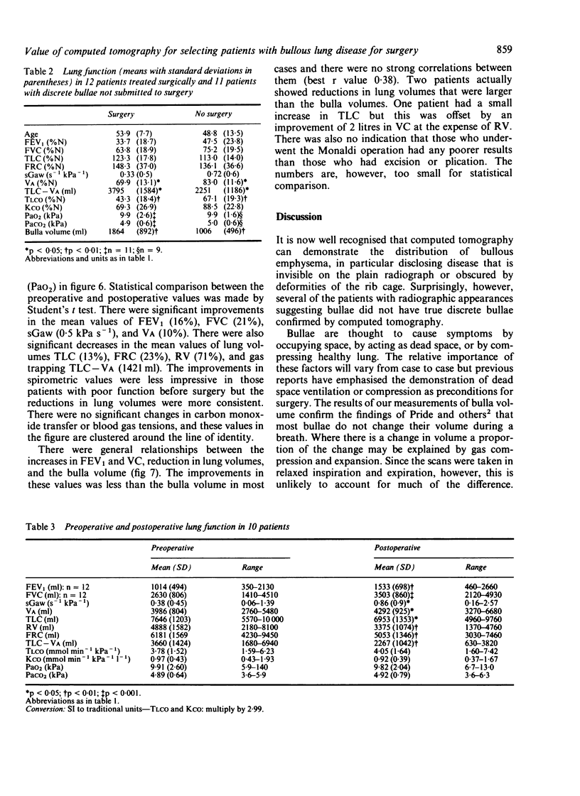
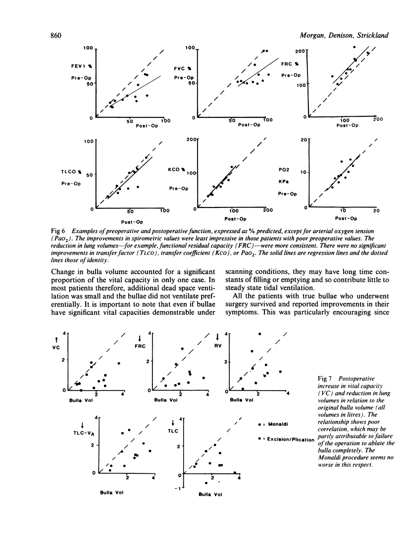
Computed tomography and lung function tests were performed on 43 patients who had evidence on the chest radiograph suggesting bullous emphysema. After computed tomography scan two groups of patients could be identified. Twenty patients had generalised emphysema, which was locally worse in the area of the suspected bulla; and 23 had well defined bullae, which were potentially operable. Results of lung function tests did not distinguish between the two groups. The volume and ventilation of the true bullae were measured by computed tomography and this confirmed that most of them did not contribute to ventilation (residual volume (RV)/total capacity (TLC) bulla = 89% (SD 10%). The patients with true bullae were considered suitable for surgery but only 12 had an operation. All the patients who underwent surgery survived and had a symptomatic improvement, which was accompanied by objective increases in spirometric volumes and by reductions in static lung volumes; there were no improvements in carbon monoxide transfer or blood gas tensions. It is concluded that computed tomography used alone can identify bullae that are amen-able to surgery and can measure their volume and ventilation. The surgical removal of such clearly identified bullae is safe and associated with symptomatic and functional improvement even when the preoperative FEV1 is less than 1 litre. This improvement is likely to be a consequence of reduction in lung volume and may not necessarily be associated with relief of compressed peribullous lung or the removal of dead space.
Full text
PDF

Images in this article
Selected References
These references are in PubMed. This may not be the complete list of references from this article.
- Carr D. H., Pride N. B. Computed tomography in pre-operative assessment of bullous emphysema. Clin Radiol. 1984 Jan;35(1):43–45. doi: 10.1016/s0009-9260(84)80232-9. [DOI] [PubMed] [Google Scholar]
- Denison D. M., Waller J. F. Interpreting the results of regional single-breath studies from the patient's point of view. Bull Eur Physiopathol Respir. 1982 Mar-Apr;18(2):339–351. [PubMed] [Google Scholar]
- Fiore D., Biondetti P. R., Sartori F., Calabró F. The role of computed tomography in the evaluation of bullous lung disease. J Comput Assist Tomogr. 1982 Feb;6(1):105–108. doi: 10.1097/00004728-198202000-00018. [DOI] [PubMed] [Google Scholar]
- Hayhurst M. D., MacNee W., Flenley D. C., Wright D., McLean A., Lamb D., Wightman A. J., Best J. Diagnosis of pulmonary emphysema by computerised tomography. Lancet. 1984 Aug 11;2(8398):320–322. doi: 10.1016/s0140-6736(84)92689-8. [DOI] [PubMed] [Google Scholar]
- Pearson M. G., Ogilvie C. Surgical treatment of emphysematous bullae: late outcome. Thorax. 1983 Feb;38(2):134–137. doi: 10.1136/thx.38.2.134. [DOI] [PMC free article] [PubMed] [Google Scholar]
- Potgieter P. D., Benatar S. R., Hewitson R. P., Ferguson A. D. Surgical treatment of bullous lung disease. Thorax. 1981 Dec;36(12):885–890. doi: 10.1136/thx.36.12.885. [DOI] [PMC free article] [PubMed] [Google Scholar]
- Pride N. B., Barter C. E., Hugh-Jones P. The ventilation of bullae and the effect of their removal on thoracic gas volumes and tests of over-all pulmonary function. Am Rev Respir Dis. 1973 Jan;107(1):83–98. doi: 10.1164/arrd.1973.107.1.83. [DOI] [PubMed] [Google Scholar]


