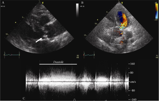
A 76-year-old Caucasian woman was admitted to the emergency room and referred for cardiac evaluation for dyspnea and abrupt onset of cough three weeks ago. She had a history of well-controlled arterial hypertension and was on adequate oral anticoagulant therapy for permanent atrial fibrillation. Previous thoracic injuries, connective tissue disorders or recent infections were excluded. No chest pain or syncope was reported. Blood pressure was 150/50 mmHg in both arms, heart rate was 90 beats/min. Physical examination revealed bilateral pleural effusion and mild peripheral edema with slight jugular venous distension. A loud “wood-sawing” or “sea-gull” diastolic murmur over the right upper sternal border and mesocardium was present, raising the suspicion of severe aortic regurgitation due to possible calcific degeneration and laceration of the aortic valve. The ECG showed atrial fibrillation with aspecific repolarization abnormalities. The laboratory tests showed mild elevation of C-reactive protein (22 mg/L, normal value < 5 mg/L), with normal white blood cell count (8620/µL) and normal procalcitonin (0.05 ng/mL). Brain-natriuretic-peptide (BNP) was increased (233 pg/mL). Chest X-ray revealed bilateral pleural effusion and calcification of the aortic arch. Transthoracic echocardiography (Figure 1) showed a thickened and calcific aortic valve with severe regurgitation due to prolapse of the non-coronary cusp. A concomitant moderate aortic stenosis (aortic valve area: 1.4 cm2) was present. Left ventricular size (end-diastolic diameter: 49 mm; end-diastolic volume: 78 mL) and ejection fraction (60%) were normal. A transesophageal echocardiography (TEE) was subsequently performed (Figure 2 and Movie 1 and 2), confirming the prolapse of the non-coronary cusp and showing its laceration with partial eversion of the medial portion in the outflow tract. Three-dimensional transverse section displayed the systolic motion of the flailing portion of the cusp. Moreover, TEE detected a large rupture of the posterior wall of the ascending aorta, immediately distal to the sino-tubular junction, communicating with a saccular dilation of 24 mm diameter, with fluctuating calcification inside. Color-doppler showed turbulent flow passing across the rupture within the pseudoaneurysm.
Figure 1. Transthoracic two-dimensional and color-Doppler echocardiography.
(A): Parasternal long-axis view shows the prolapsing non-coronary aortic cusp in left ventricle outflow tract (arrow); (B): color-Doppler from the apical five-chamber view shows severe aortic regurgitation; and (C): continuous Doppler across aortic valve displays a diastolic low-frequency jet due to the fluttering of the lacerated calcific cusp into the blood stream.
Figure 2. Transesophageal two- and three-dimensional echocardiography.
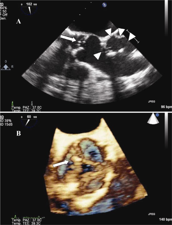
(A): Three-chamber two-dimensional views showing the prolapsed lacerated non-coronary cusp (arrow) and the saccular dilation communicating with the lumen through a large rupture of the aortic wall (arrowheads) (see also Movie 1); and (B): three-dimensional transverse section from aortic perspective shows systolic motion of the flailing cusp (arrow) (see also Movie 2).
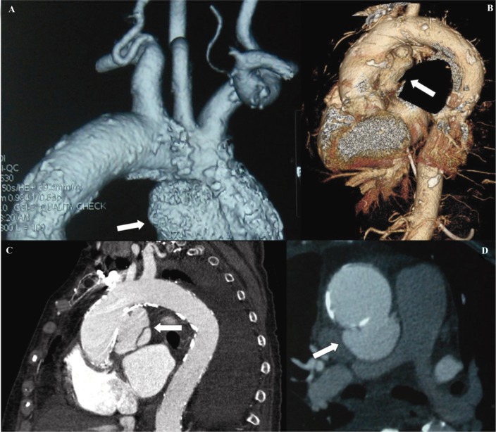
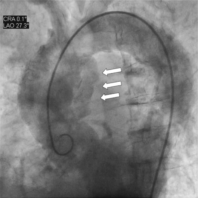
Multislice computed tomography (Figure 3) confirmed the presence of a large aortic pseudoaneurysm (3.1 × 2.9 cm, length 5.6 cm), probably originating from a penetrating atherosclerotic ulcer (PAU), which slightly compressed the right pulmonary artery. Diameters of proximal and distal ascending aorta, aortic arch and descending aorta were 3.7 cm, 3.1 cm, 2.6 cm and 2.6 cm, respectively. Coronary angiography excluded significant lesions; aortic angiography (Figure 4 and Movie 3) showed the saccular dilation of the posterior ascending aorta and confirmed the entity of aortic regurgitation.
Figure 3. Three-dimensional reconstruction of multi-slice computed tomography shows the large pseudoaneurysm on the posterior wall of ascending aorta (arrow) (A&B); multislice-computed tomography section displays the saccular pseudoaneurysm and its mild compression of the right pulmonary artery (arrow) (C&D).
Figure 4. Aortic angiography demonstrates the pseudoaneurysm (arrows) and the severe aortic regurgitation (see also Movie 3).
The patient was therefore referred for cardiac surgery. Intraoperative direct examination revealed a large aortic pseudoaneurysm originating from an ulcerated atherosclerotic plaque. The aortic wall was resected and replaced with a 30 mm intervascular graft. Aortic valve had three cusps with significant calcifications and no sign of acute or healed infective endocarditis. Non-coronary cusp showed two fenestrations and was lacerated at the site of a nodular calcification (Figure 5). The valve was replaced with a Hancock II 25 mm bioprosthesis. Histological examination confirmed the macroscopic findings. The patient's post-operative course was uneventful. A normal postoperative appearance of the ascending aorta graft was proven by multislice computed tomography while pre-discharge transthoracic echocardiography demonstrated a normally functioning bioprosthetic aortic valve and a normal left ventricular ejection fraction (68%). At three-year follow-up, the patient is alive in NYHA class I.
Figure 5. Native aortic valve cusps after surgery excision.
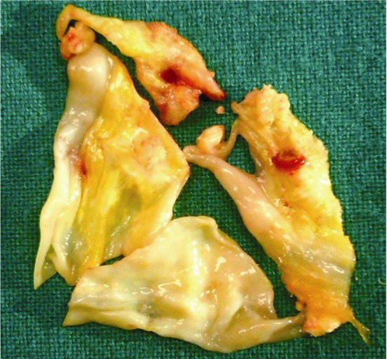
The laceration of the non-coronary cusp occurred at the site of the calcified portion. Close to the breaking point there is a typical degenerative fenestration.
Aortic cusps rupture and aortic pseudoaneurysm are both uncommon findings, usually as a consequence of aortic aneurysm, dissection, iatrogenic injury, blunt chest trauma, connective tissue disorders or infection. Asymptomatic spontaneous rupture of the ascending aorta resulting in aortic pseudoaneurysm is a very rare condition. Calcification of the aorta and atherosclerotic change, especially in longstanding hypertension, play an important role;[1] PAU is an alternative cause.[2] More than aortic rupture, spontaneous rupture of tricuspid aortic valve is extremely rare, with less than 10 cases described.[3] Age-related fenestrations of the cusps seems to be the primary cause since, with their progressive enlargement, the residual lunular tissue is reduced to a cord-like structure that may break, particularly if calcific degeneration made it more fragile.[3]
Of note, in the present case, the clinical suspicion of calcific degeneration and laceration of the aortic valve was raised by the presence of a peculiar diastolic murmur. The presence of a retroverted portion of valve tissue vibrating in the regurgitant stream determines indeed a loud “wood-sawing” or “sea-gull” murmur.[4] The subsequent diagnostic work-up, which included 2D and 3D transesophageal echocardiography, multislice computed tomography and aortic angiography, confirmed the diagnosis and revealed the presence of spontaneous and asymptomatic rupture of the ascending aorta resulting in aortic pseudoaneurysm.
To the best of our knowledge, the present case is the first to describe an association between aortic pseudoaneurysm and aortic cusp rupture, both of spontaneous etiology. They could be the expression of a widespread degenerative age-related process with a common pathogenesis secondary to the atherosclerotic disease.
References
- 1.Hirai S, Hamanaka Y, Mitsui N, et al. Spontaneous rupture of the ascending thoracic aorta resulting in a mimicking pseudoaneurysm. Ann Thorac Cardiovasc Surg. 2006;12:223–227. [PubMed] [Google Scholar]
- 2.Braverman AC. Penetrating atherosclerotic ulcers of the aorta. Curr Opin Cardiol. 1994;9:591–597. doi: 10.1097/00001573-199409000-00014. [DOI] [PubMed] [Google Scholar]
- 3.Blaszyk H, Witkiewicz AJ, Edwards WD. Acute aortic regurgitation due to spontaneous rupture of a fenestrated cusp: report in a 65-year-old man and review of seven additional cases. Cardiovasc Pathol. 1999;8:213–216. doi: 10.1016/s1054-8807(99)00009-5. [DOI] [PubMed] [Google Scholar]
- 4.McKusick VA. A grade 6 systolic murmur. N Engl J Med. 1999;341:1472–1473. doi: 10.1056/NEJM199911043411913. [DOI] [PubMed] [Google Scholar]


