Abstract
Objectives:
The aim of this study was to compare the retention of conventional mandibular complete dentures with that of mandibular complete dentures having lingual flanges constructed with flexible acrylic resin “Versacryl.”
Materials and Methods:
The study sample comprised 10 completely edentulous patients. Each patient received one maxillary complete denture and two mandibular complete dentures. One mandibular denture was made of conventional heat-cured acrylic resin and the other had its lingual flanges made of flexible acrylic resin Versacryl. Digital force-meter was used to measure retention of mandibular dentures at delivery and at 2 weeks and 45 days following denture insertion.
Results:
The statistical analysis showed that at baseline and follow-up appointments, retention of mandibular complete dentures with flexible lingual flanges was significantly greater than retention of conventional mandibular dentures (P < 0.05). In both types of mandibular dentures, retention of dentures increased significantly over the follow-up period (P < 0.05).
Conclusions:
The use of flexible acrylic resin lingual flanges in the construction of mandibular complete dentures improved denture retention.
Keywords: Complete denture, flexible acrylic, lingual flange, retention
INTRODUCTION
Despite the current decline in the rate of complete edentulism,[1,2] the need for complete denture treatment will probably continue.[3] Thus, research and efforts to improve the outcome of treatment with complete dentures should also continue. Prosthodontists and general dental practitioners recognize that wearing complete dentures is troublesome for some patients and associated with a wide range of problems. However, complaints related to complete denture retention and stability are the most frequent.[4] Such problems become even worse with mandibular dentures due to a number of anatomical and physiological factors.[5] Unsatisfactory denture retention has its implication on the prognosis of treatment with complete dentures. Oral discomfort, defective speech, difficulty during chewing, and irritation of the supporting tissues are examples of the problems that are associated with inadequate denture retention. It was also found that patients’ use and satisfaction with complete dentures is highly dependent on the quality of denture retention.[6,7] Many authors provided tips and recommendations for dentists to improve the quality of complete denture retention.[8,9,10] Also, denture adhesives and dental implants were used to enhance denture retention. One possible and simple way of enhancing denture retention is by extension of denture flanges to engage an existing soft-tissue undercut.[11,12] However, extension of conventional denture bases into soft-tissue undercuts should be kept minimal due to rigidity of acrylic resin. The introduction of resilient denture liners[13] and flexible acrylic resin[14] increased the chance for denture bases to be extended into deeper soft-tissue undercuts to gain further retention without risking the health of the supporting tissues or creating pain and difficulty during denture removal or insertion.
Some authors reported the use of permanent soft liners in the retromylohyoid eminence to aid denture retention.[15,16] Lowe[17] used flexible acrylic resin to create flexible denture flanges for patients exhibiting undercut tuberosities. This technique can also be used to aid retention of mandibular complete dentures by creating flexible lingual flanges that engage the lingual undercuts of the mandible. However, it is not yet clear from the current literature to what extent would denture retention be improved by the use of flexible acrylic resin in fabrication of the lingual flanges of mandibular complete dentures. The aim of this study was to examine the hypothesis that there is no significant difference in the retention force between mandibular dentures of conventional construction and mandibular dentures constructed with flexible acrylic resin lingual flanges.
MATERIALS AND METHODS
This study was approved by the Research Ethics Committee of Faculty of Oral and Dental Medicine, Cairo University, Egypt.
Patient selection
Ten completely edentulous patients were selected from the out-patient clinic of the Removable Prosthodontics Department, Faculty of Oral and Dental Medicine, Cairo University. All patients were presented with explanation about the objectives, implications, and possible complications of this study and invited to sign an informed consent.
The inclusion criteria were as follows:
Completely edentulous male patients
Age range between 45 and 65 years
Patients having had no previous complete dentures
Patients who had their last remaining teeth extracted at least 3 months before recruitment in the study
Patients with well-developed edentulous ridge that was covered with healthy firm mucosa
Patients with normal Angle Class 1 maxillomandibular relationship
Patients who were free from systemic diseases that affect the neuromuscular control, such as Parkinson's disease.
The exclusion criteria were as follows:
Patients with resorbed ridges
Patients with xerostomia and patients undertaking medications that affect salivary flow (e.g., diuretics). Similarly, patients with systemic diseases that may affect the amount or consistency of saliva (e.g., uncontrolled diabetes mellitus) were excluded.
Construction of the dentures
Complete dentures were provided by the first author in the Department of Removable Prosthodontics, Faculty of Oral and Dental Medicine, Cairo University. One expert dental technician constructed all the dentures.
Before construction of the dentures, full medical and dental history was taken from each patient, following which an extraoral and intraoral examination and an orthopantomogram (OPG) were performed.
Complete dentures were constructed for each patient following the guidelines of the British Society for the Study of Prosthetic Dentistry.[18]
Each patient received one maxillary denture and two mandibular dentures. One mandibular denture was made entirely of conventional heat-cured acrylic resin and the other mandibular denture was made of conventional heat-cured acrylic resin with thermoplastic flexible acrylic resin “Versacryl” (Keystone Industries GmbH, Sigen, Germany) at the lingual flange area.
Primary impressions were made by irreversible hydrocolloid alginate impression material (Cavex alginate, dust free, high consistency; Cavex Holland BV, Haarlem, Netherlands) using perforated stock trays. Self-cured acrylic resin (cold cure denture base material; Acrostone, Cairo, Egypt) special trays were constructed for making the secondary impressions. The special trays were trimmed 2 mm short of the tissue reflection area and checked for border extension and adaptation inside the patient's mouth. The secondary impressions were taken using putty and medium addition type rubber base impression material (Speedex, Coltène/Whaledent Company, Altstätten, Switzerland). In the first step of making the secondary impression, the putty body rubber base impression material was used for border molding following the conventional methods and the manufacturer's instructions. The medium body rubber base impression material was used to make a wash impression in the second step of making the secondary impression. The lower secondary impressions were boxed and poured twice to obtain two stone master casts in order to construct two sets of dentures.
At the wax-up stage of the mandibular dentures, the lingual flanges for both types of dentures were designed to engage 2 mm of the undercut area of the mylohyoid ridge. At the insertion appointment, patients were provided with verbal and written instructions about how to deal with their new dentures.
Figure 1 shows a mandibular complete denture with flexible acrylic resin lingual flanges after processing and finishing.
Figure 1.
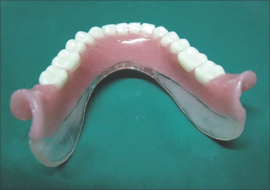
The mandibular complete denture with “Versacryl” lingual flanges
Allocation of patients
This study implemented a cross-over design. The selected patients were randomly allocated to two equal treatment groups, with five patients in each group as follows:
Group 1: Patients in this group received conventional maxillary and mandibular complete dentures. The patients used these dentures for 45 days. Then, the conventional lower denture was replaced by one with flexible lingual flanges. The same patients were followed up again for 45 days
Group 2: Patients in this group received a conventional maxillary complete denture and a mandibular complete denture with flexible lingual flanges. The patients used these dentures for 45 days. Afterward, the lower denture with flexible lingual flanges was replaced by one with a conventional construction. The same patients were followed up again for 45 days.
Follow-up of patients
With each type of mandibular denture, the patient was followed up for 45 days. At each review appointment, patient's complaints were noted. The supporting tissues, the denture surfaces and borders, the occlusion and articulation of the dentures, were all examined. Then the dentures were adjusted in the light of clinical examination and patient's complaints.
Retention of dentures was assessed and recorded during the follow-up period.
Assessment of denture retention
In both groups, retention of mandibular dentures was tested at the time of delivery and at 2 weeks and 45 days following denture insertion.
A digital force-meter (Extech instruments 475040, Nashua, New Hampshire, USA) was used to measure denture resistance to vertical displacement (i.e., retention) by applying a pulling force on a metal hook located in the geometric center of each mandibular denture.
Based on geometrical principles,[19] identification of the geometric center for each mandibular denture was carried out as follows:
All undercuts in the fitting surface of the denture were blocked by base plate wax
A mix of plaster was then poured into the fitting surface of the denture and another mix was used to construct a base
The centers of the retromolar pads and the midline were marked on the polished surface of the denture [Figure 2]
In the next step, a cardboard was cut out so as to form a triangle which was placed on the plaster base to occupy the space in between the three aforementioned markings
Three lines bisecting the three angles of the triangle were then drawn on the cardboard. The intersection of these three lines was considered the geometric center of the denture [Figure 3]
Following the former step, a pin was passed through the cardboard at the identified geometric center to mark a point on the plaster base. A plastic rod was then fixed to the base and suspended upward from the marked point to maintain the location of the geometric center.
Figure 2.
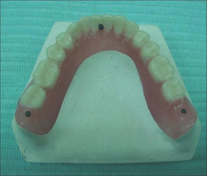
The markings on the mandibular denture
Figure 3.
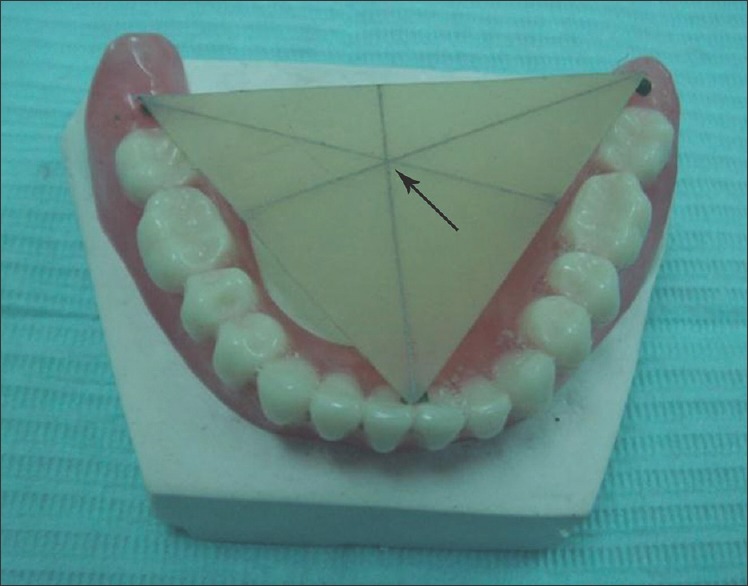
Identification of the geometric center of the mandibular denture on the cardboard (arrow)
Three “V” shaped grooves were created on the polished surface of the lower denture. One was made on the lingual flange at the midline region just below the central incisors. The other two grooves were created at the retromolar pad area just distal to the second molar of both sides.
A wrought wire of 1 mm in diameter was then bent at its center and adjusted so as not to encroach on the tongue space and to run 2 cm above the occlusal plane from the retromolar pad groove of one side to the retromolar pad groove of the other side.
Afterward, a second wrought wire of the same diameter was adjusted to extend from the groove at the lingual flange upward, so that it is 2 cm above the occlusal plane.
The two wrought wires were then bent toward each other until they met at the identified geometric center.
One end of the second wire was adapted in the created groove just below the central incisors and the other end was shaped to form a c-shaped loop around the first wire.
The free ends of the two wires were then fixed to the polished surface of the lower denture by self-cured acrylic resin [Figure 4a and b]. Excess acrylic resin was then removed and the denture surface was refinished and polished.
Figure 4.
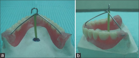
(a and b) The second wire adjusted to meet the first wire at the geometric center with its free end bent into a c-shaped loop
Retention of mandibular dentures was assessed as follows:
Each patient was asked to sit comfortably in a dental chair with his head on the headrest and the occlusal plane is parallel to the floor of the room.
The lower denture was then inserted inside the patient's mouth. Before insertion of dentures with flexible lingual flanges, the dentures were immersed in warm water bath (50°C) for 5 min to soften the flexible flange, so that they can be easily adjusted to adapt to the undercut area of the mylohyoid ridge.
After denture insertion, tongue freedom and loop position were checked and 3 min seating time was allowed before taking the measurements.
The metallic probe of the digital force-meter was then attached to the c-shaped metal hook created at the geometric center of the mandibular dentures and a vertical pulling force was applied to measure denture retention. Retention strength was measured in grams. Three readings were taken and the average value was recorded.
Figure 5 shows one of the participating patients during evaluation of denture retention by a digital force-meter.
Figure 5.
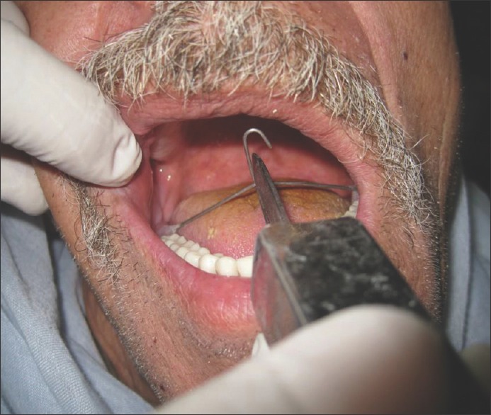
Clinical evaluation of denture retention by a digital force-meter
Data were collected, tabulated, and statistically analyzed.
RESULTS
Over the study period, no patient was lost to follow-up and no major adjustments were required for the constructed dentures.
Mean and standard deviation of the retention force for the two study groups at the follow-up appointments are summarized in Table 1.
Table 1.
Mean and standard deviation of retention force measured in grams for study groups with both denture materials at different follow-up periods
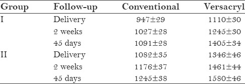
The t-test was used to test the hypothesis that there is no significant difference in retention force between mandibular dentures of conventional construction and mandibular dentures with flexible lingual flanges. The statistical test indicated that retention of dentures with flexible lingual flanges was significantly higher than retention of conventional dentures at the different follow-up appointments (P < 0.05).
In order to examine the effect of follow-up time on the retention force of conventional mandibular dentures and dentures with flexible lingual flanges, one-way analysis of variance (ANOVA) followed by pair-wise Newman–Keuls post-hoc tests were used. The statistical analysis showed that in both types of dentures, retention force after 45 days recorded significantly the highest mean value (P <0.05), whereas retention force at the time of denture delivery was significantly the lowest (P < 0.05).
DISCUSSION
The provision of a retentive and stable denture can be considered the principal goal of general dental practitioners when dealing with complete dentures. Research findings indicate that retention and stability of mandibular complete dentures are strong determinants of patients’ satisfaction with newly delivered dentures.[7] Extension of the lingual flanges of the mandibular denture to engage the sublingual undercut of the mandible was used and recommended as a mean to maximize retention of mandibular complete dentures.[11,12,20,21] While acrylic resin is the most commonly used denture base material, this study reported the use of flexible acrylic resin “Versacryl” to construct the lingual flanges of mandibular complete dentures. The flexible acrylic resin was introduced first in 1950 as an alternative to conventional acrylic resin denture base material.[14,22] It is a flexible biocompatible thermoplastic denture base material with unique physical and esthetic properties. The flexible acrylic can create any part of a denture to be made adjustable, simply by using warm water to soften the material so as to conform to the contours of the soft and hard tissues. Also, the flexible acrylic can be extended into undercut areas to mechanically retain the denture. Interference with soft tissue undercuts would be facilitated by the use of flexible acrylic flanges so that the denture base can be inserted and removed smoothly. Furthermore, the softness of flexible acrylic imparts a feeling of comfort to the patient.[14] However, the flexible acrylic has a number of disadvantages[23,24] and may lose the desired flexibility in the long term.
Many brands of thermoplastic or flexible denture materials are available in the market, such as Valplast (Valplast Int. Corp., Westbury, New York, USA), Flexiplast (Bredent, Senden, Germay), Flexite (The Flexible Company, Mineola, New York, USA), and Lucitone® FRS™ (DENTSPLY International, Woodbridge, Ontario, Canada). In this investigation, Versacryl (Keystone Industries GmbH, Sigen, Germany) was used as a thermoplastic material to construct the lingual flanges of the mandibular complete dentures. Versacryl has the physical properties of the thermoplastic materials, as indicated by the manufacturer. When inserted and adapted to the mouth after immersion in warm water (50°C) for 5 min, Versacryl will cool to body temperature and take on the desired rigidity to fulfill its function.[25]
Despite the relatively old age of the thermoplastic denture base materials, reports to evaluate its impact on denture retention are scarce. Antonelli and Hottel[26] reported the use of flexible acrylic flanges to construct stable, retentive, well-adapted, and comfortable complete dentures record bases. Singh et al.[27] found that flexible denture bases produced better patient satisfaction and comfort, compared to conventional acrylic resin denture bases.
In this clinical study, the use of flexible lingual flanges in the construction of mandibular complete dentures has resulted in improved denture retention, compared to conventionally made dentures with acrylic resin flanges. This may be attributed to the physical properties of the flexible acrylic which allowed effective engagement with the lingual pouch undercut and close adaptation to the supporting tissues.[14,27] It can be argued that an intimate adaptation of the flexible denture flanges to the underlying tissues with the existence of a thin film of saliva in between the two objects would increase the effectiveness of adhesion forces and enhance the peripheral seal around the denture borders.[8,28] Thus, effective mechanical and physical factors interplayed to produce better retention for mandibular complete dentures with flexible acrylic resin flanges. However, this is a short-term study and further studies are recommended to evaluate the long-term quality of retention of mandibular complete dentures with flexible lingual flanges and its impact on patients’ satisfaction.
Finally, it can be noted that over the follow-up period, there was an improvement in denture retention for both types of dentures. This finding can be explained by the improved fit of the denture bases due to the medium-term remodeling of the soft tissues underlying the dentures in order to maintain the mucosal contact with denture bases.[28]
CONCLUSION
The use of flexible acrylic resin lingual flanges in the construction of mandibular complete dentures resulted in improved denture retention. The hypothesis that there was no significant difference in retention force between mandibular dentures of conventional construction and mandibular dentures constructed with flexible acrylic resin lingual flanges was, therefore, rejected.
Financial support and sponsorship
Nil.
Conflicts of interest
The authors declare no financial relationship with any commercial firm that may pose a conflict of interest for this study.
REFERENCES
- 1.Wu B, Liang J, Landerman L, Plassman B. Trends of edentulism among middle-aged and older Asian Americans. Am J Public Health. 2013;103:e76–82. doi: 10.2105/AJPH.2012.301190. [DOI] [PMC free article] [PubMed] [Google Scholar]
- 2.Slade GD, Akinkugbe AA, Sanders AE. Projections of U.S. Edentulism prevalence following decades of decline. J Dent Res. 2014;93:959–65. doi: 10.1177/0022034514546165. [DOI] [PMC free article] [PubMed] [Google Scholar]
- 3.Douglass CW, Shih A, Ostry L. Will there be a need for complete dentures in the United States in 2020? J Prosthet Dent. 2002;87:5–8. doi: 10.1067/mpr.2002.121203. [DOI] [PubMed] [Google Scholar]
- 4.Bilhan H, Geckili O, Ergin S, Erdogan O, Ates G. Evaluation of satisfaction and complications in patients with existing complete dentures. J Oral Sci. 2013;55:29–37. doi: 10.2334/josnusd.55.29. [DOI] [PubMed] [Google Scholar]
- 5.Ribeiro JA, de Resende CM, Lopes AL, Farias-Neto A, Carreiro Ada F. The influence of mandibular ridge anatomy on treatment outcome with conventional complete dentures. Acta Odontol Latinoam. 2014;27:53–7. doi: 10.1590/S1852-48342014000200001. [DOI] [PubMed] [Google Scholar]
- 6.Fenlon MR, Sherriff M, Walter JD. An investigation of factors influencing patients’ use of new complete dentures using structural equation modelling techniques. Community Dent Oral Epidemiol. 2000;28:133–40. doi: 10.1034/j.1600-0528.2000.028002133.x. [DOI] [PubMed] [Google Scholar]
- 7.Fenlon MR, Sherriff M. An investigation of factors influencing patients’ satisfaction with new complete dentures using structural equation modelling. J Dent. 2008;36:427–34. doi: 10.1016/j.jdent.2008.02.016. [DOI] [PubMed] [Google Scholar]
- 8.Jacobson TE, Krol AJ. A contemporary review of the factors involved in complete denture retention, stability, and support. Part I: Retention. J Prosthet Dent. 1983;49:5–15. doi: 10.1016/0022-3913(83)90228-7. [DOI] [PubMed] [Google Scholar]
- 9.McCord JF, Grant AA. Identification of complete denture problems: A summary. Br Dent J. 2000;189:128–34. doi: 10.1038/sj.bdj.4800703. [DOI] [PubMed] [Google Scholar]
- 10.Scott BJ, Hunter RV. Creating complete dentures that are stable in function. (265-7).Dent Update. 2008;35:259–62. doi: 10.12968/denu.2008.35.4.259. [DOI] [PubMed] [Google Scholar]
- 11.Bocage M, Lehrhaupt J. Lingual flange design in complete dentures. J Prosthet Dent. 1977;37:499–506. doi: 10.1016/0022-3913(77)90162-7. [DOI] [PubMed] [Google Scholar]
- 12.von Krammer R. Principles and technique in sublingual flange extension and complete mandibular dentures. J Prosthet Dent. 1982;47:479–82. doi: 10.1016/0022-3913(82)90293-1. [DOI] [PubMed] [Google Scholar]
- 13.Soni A. Management of severe undercuts in fabrication of complete dentures. N Y State Dent J. 1994;60:36–9. [PubMed] [Google Scholar]
- 14.Rickman LJ, Padipatvuthikul P, Satterthwaite JD. Contemporary denture base resins: Part 2. (180-2).Dent Update. 2012;39:176–8. doi: 10.12968/denu.2012.39.3.176. 184 passim. [DOI] [PubMed] [Google Scholar]
- 15.Whitsitt JA, Battle LW, Jarosz CJ. Enhanced retention for the distal extension-base removable partial denture using a heat-cured resilient soft liner. J Prosthet Dent. 1984;52:447–8. doi: 10.1016/0022-3913(84)90467-0. [DOI] [PubMed] [Google Scholar]
- 16.Mendez M, Lee C. Use of a permanent soft denture liner in the retromylohyoid eminence and knife-edge ridge areas of the mandible to aid in retention and stability. Gen Dent. 2013;61:e14–5. [PubMed] [Google Scholar]
- 17.Lowe LG. Flexible denture flanges for patients exhibiting undercut tuberosities and reduced width of the buccal vestibule: A clinical report. J Prosthet Dent. 2004;92:128–31. doi: 10.1016/j.prosdent.2004.04.026. [DOI] [PubMed] [Google Scholar]
- 18.Ogden A. London: Quintessence Publishing Co. Ltd. for the British Society for the Study of Prosthetic Dentistry; 1996. BSSPD Guidelines in Prosthetic and Implant Dentistry; pp. 7–11. [Google Scholar]
- 19.Weisstein EW. “Geometric Centroid.” MathWorld--A Wolfram Web Resource. [Last accessed on 2015 July 31]. Available from: http://mathworld.wolfram.com/GeometricCentroid.html .
- 20.Azzam MK, Yurkstas AA, Kronman J. The sublingual crescent extension and its relation to the stability and retention of mandibular complete dentures. J Prosthet Dent. 1992;67:205–10. doi: 10.1016/0022-3913(92)90454-i. [DOI] [PubMed] [Google Scholar]
- 21.Chang JJ, Chen JH, Lee HE, Chang HP, Chen HS, Yang YH, et al. Maximizing mandibular denture retention in the sublingual space. Int J Prosthodont. 2011;24:460–4. [PubMed] [Google Scholar]
- 22.Price CA. A history of dental polymers. Aust Prosthodont J. 1994;8:47–54. [PubMed] [Google Scholar]
- 23.Sunitha NS, Jagadeesh KN, Kalavathi SD, Kashinath KR. ”Flexible dentures” - An alternate for rigid dentures? J Dent Sci Res. 2010;1:74–9. [Google Scholar]
- 24.Sharma A, Shashidhara HS. A review: Flexible removable partial dentures. J Dent Med Sci. 2014;13:58–62. [Google Scholar]
- 25.Keystone Industries. [Last accessed on 2015 July 31]. Available from: https://keystoneind.wordpress.com/tag/versacryl/
- 26.Antonelli JR, Hottel TL. The “flexible augmented flange technique” for fabricating complete denture record bases. Quintessence Int. 2001;32:361–4. [PubMed] [Google Scholar]
- 27.Singh JP, Dhiman RK, Bedi RP, Girish SH. Flexible denture base material: A viable alternative to conventional acrylic denture base material. Contemp Clin Dent. 2011;2:313–7. doi: 10.4103/0976-237X.91795. [DOI] [PMC free article] [PubMed] [Google Scholar]
- 28.Darvell BW, Clark RK. The physical mechanisms of complete denture retention. Br Dent J. 2000;189:248–52. doi: 10.1038/sj.bdj.4800734. [DOI] [PubMed] [Google Scholar]


