Abstract
Forensic odontology is the application of dental principles to legal issues. Sex determination is a subdivision of forensic odontology and it is very important especially when information relating to the deceased is unavailable. Sex determination becomes the first priority in the process of identification of a person by a forensic investigator in the case of mishaps, chemical and nuclear bomb explosions, natural disasters crime investigations, and ethnic studies. This article reviews upon the various methods used in sex determination.
KEY WORDS: Forensic odontology, sex determination, investigations
Forensic odontology is the application of dental principles to legal issues.
Dental identification involves either comparative method or postmortem dental profiling. The main advantage of dental evidences is that it can be preserved indefinitely after the death. The unique pattern of tooth enables the analysis of antemortem and postmortem dental variables. The main problem in dental identification is mostly related to acquiring an interpreting antemortem records. Sometimes because of the time duration there will be variation while comparing these records.
Sex determination is a subdivision of forensic odontology and it is very important especially when information relating to the deceased is unavailable. Sex determination becomes the first priority in the process of identification of a person by a forensic investigator in the case of mishaps, chemical and nuclear bomb explosions, natural disasters crime investigations, and ethnic studies.
Various methods have been used for the identification of sex. Sex determination can be done either by Morphological analysis (of the tooth, skull and other soft tissues of oral and paraoral region) or molecular analysis.
Sex Determination Analysis
Sex determination analysis can be done either by morphological analysis or by molecular analysis.
Morphological analysis can be done on hard tissues (odontometric, orthometric, and miscellaneous) of oral and paraoral regions or soft tissue (lip prints-Cheiloscopy, palatal rugae pattern-Rugoscopy).
Morphological Analysis
Hard tissue analysis
Odontometric methods
In hard tissue analysis odontometric method involves (a) mesiodistal (MD) dimensions and buccolingual (BL) dimension of teeth (b) mean canine index (MCI) (dental index), and (c) distinct tooth morphology.
Sexual dimorphism exists in the shape and size of the tooth. Tooth size can be measured best during early permanent dentition because it's the stage when the tooth is subject to less external and internal stimuli.[1]
Mesiodistal dimensions and buccolingual dimension of teeth Tooth dimension is the most simple and reliable method to analyze sexual dimorphism.
Many studies report that the MD dimensions of teeth in male are more than that of female.[2] Various theories have been given to explain canine sexual dimorphism. According to Moss, it is because of the (1) Greater thickness of enamel in males due to the long period of amelogenesis compared to females or (2) because of Y chromosomes producing slower male maturation.[3]
Many authors (Garn et al. Iscan and Kedici) recommend the BL dimension as a reliable measurement than other variables in the determination of sex.[4]
Garn et al. in their study reported that BL dimension in male dentition were larger than female which was statistically significant.[5]
An earlier study has recommended that MD dimension was better suited for identification of sex than BL dimension. Since arch size influences the tooth size, the larger jaw size in males can be related to the larger MD dimension of tooth compared to female.[6]
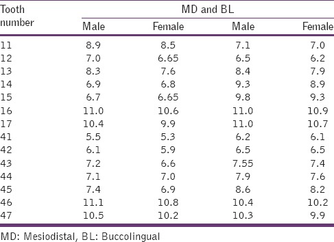
Though studies have concluded that MD dimension to be a better predictor of sex than BL dimension, certain discrepancies occur while measuring the MD dimension due to close proximal contacts. Therefore, both MD and BL dimensions aid in as a more reliable tool in determining sex[7] [Table 1].
Table 1.
Difference in MD and BL dimension of tooth among male and female

Iscan and Kedici emphasized that more accurate results can be attained when all available teeth are included in sex determination.
Mean canine index (dental index)
Mean canine index is also known as dental index. Since canine exhibit the greater sexual dimorphism and are also highly resistant to disease and postmortem insults, Rao et al. developed the MCI which was derived as follows:
Mean canine index = Mesiodistal crown width of mandibular canine.
Mandibular inter-canine arch width.
The cut-off point, or standard MCI value, obtained by Rao et al. was 0.274. If the MCI value of a skull specimen is less than or equal to the standard MCI, the individual is categorized as female; a value more than the standard MCI would group the person as male.[8]
Distinct tooth morphology
Nonmetric features like a distal accessory ridge, number of cusp in mandibular first molar can be used in sex determination. Distal accessory ridge in canine is more pronounced in male compared to female.[9] Female exhibit lesser number of cusp in the mandibular first molar compared to male (distobuccal or distal cusp).[10] This feature can be attributed to the evolutionary reduction in the size of the lower jaw in females.[11]
Orthometric method
Orthometric method involves morphology of skull and mandible with a constellation of six traits and frontal sinus dimensions. Williams and Rogers found sex could be predicted correctly in 96% of cases using different features of skull and mandible.[12]
Morphology of skull and mandible[8]
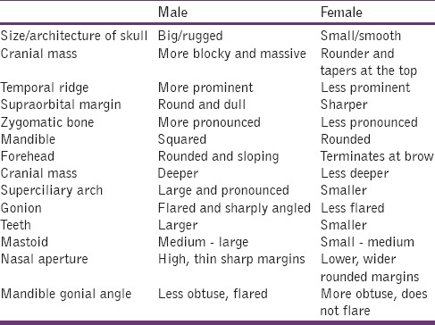
Constellation of six traits are mastoid, supraorbital ridge, size and architecture of skull, zygomatic extensions, nasal aperture, and mandible gonial angle and it was said that the determination of sex using only these six traits shows accuracy of 94%[8] [Table 2].
Table 2.
Depicts the difference in skull morphology among male and female

Frontal sinus dimensions
Sinuses are mucosa-lined air spaces within the bones of the face and skull. Frontal sinuses are situated between the internal and external laminae of the frontal bone.[13] Frontal sinuses are absent at birth and fully developed around 8 years and reaches full size after puberty.[14,15] Frontal sinuses are important parameters in the determination of sex as it presents a distinctive differences in shape, measurements, and symmetry.[16]
Uthman et al. in their study of evolution of frontal sinuses and frontal measurements using spiral computed tomography scanning of 90 patients concluded that frontal sinus measurements are valuable aid in differentiating sex and stated that, including skull measurements along with frontal sinus measurements improved the accuracy.[17]
Belaldavar et al. showed a greater mean values of frontal sinus height, width, and area in male compared to female.[18]
Soft tissue analysis
The analysis of soft tissue includes the study of lip prints (Cheiloscopy) and study of palatal rugae patterns (Rugoscopy).
Cheiloscopy
The word chelios comes from the Greek word meaning lip. The study of lip prints is called Cheiloscopy.[19] Lip prints can be identified even at 6th week of intrauterine life.[20] These prints do not change after that. Therefore, lip prints are unique patterns on lip which helps in identification of a person.[21]
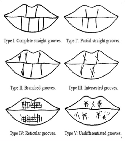
The 10 mm wide area in the middle part of lower lip is used as the best-suited area of study.[22] These lip prints are classified by Suzuki and Tsuchihashi as follows:[23]
Type I - Clear-cut grooves running vertically across the lip
Type I′ - The grooves are straight but disappear half-way instead of covering the entire breadth of the lip
Type II - The grooves are branched
Type III - The grooves intersect
Type IV - The grooves are reticulate
Type V - Undetermined.
Vahanwala et al. in their study concluded that sex of the individual can be identified by lip prints as follows:[24]
Type I, I’ pattern dominant: Female
Type I and II patterns are dominant: Female
Type III pattern dominant: Male
Type IV patterns: Male
Type V varied patterns: Male.
Similar findings were reported by many other authors[23,25,26] [Figure 1].
Figure 1.

The pattern of lip prints by Suzuki and Tsuchihashi (1970)[23]
Rugoscopy
Palatal Rugoscopy or Rugoscopy is the study of the pattern on the palatal rugae to identify a person.[27] Trobo Hermosa, a Spanish investigator in 1932 first proposed on palatal Rugoscopy.[28] Due to its internal position, stability, perennity that is, it persists throughout life, it is selected in forensic for human identification.[29]
Thomas et al. classified the palatal rugae pattern based on their number, length, and shape. Based on length it is classified as follows:[28]
Primary rugae (5–10 mm)
Secondary rugae (3–5 mm)
Fragmentary rugae (<3 mm).
Based on the shape it is classified as:[28]
Straight - Runs directly from the origin to termination
Curvy - A simple crescent shape which was curved gently
Circular - A definite continuous ring formation
Wavy - Serpentine form.
Various studies have compared the rugae pattern in male and female. A study on Japanese population concluded that female had fewer rugae than male. Shetty et al. compared the rugae pattern between Indian and Tibetan population. The result of their study showed that Indian male possessed more primary palatal rugae on left side when compared to female. And also more curved rugae was observed in Indian male compared to Tibetan male and more wavy rugae was observed in Tibetan female when compared to Indian females. Study results of Bharat et al. among males and females showed a specific pattern of palatal rugae pattern among both sex of coastal Andhra population.[28]
Miscellaneous methods
Stenberg and Borrman in 1998 stated that dental prostheses labeled with at least the patient's name and further unique identifiers such as sex, phone number, address, job and national identity number may play an important role in forensic casework's.[29] These labeled prosthesis can be used as antemortem record for forensic identification. Denture labeling can be classified as inclusion system and marking system. Inclusion system uses metal, nonmetal, micro label, and chips. Marking system uses spirit based pen or pencil. Borrman et al. in 1999 suggested that due to the biologically inert nature, durability and ability to withstand even elevated temperature, lead paper as the best-suited denture markers that helps in forensic identification.[29,30]
Molecular Analysis
Since morphological patterns vary with time and external factors, the best-suited method in the identification of gender is by molecular analysis of DNA. The extracted DNA from the teeth of an unidentified person can be compared with the antemortem DNA samples. DNA stored in blood, hairbrush, clothes, cervical smear or biopsy sample can provide a good source of antemortem DNA. The different types of DNA are nuclear DNA and Mitochondrial DNA. Extraction of DNA can be done either cryogenic grinding which involves cooling the whole tooth to extreme low temperature using liquid nitrogen, and grind the tooth to extract the DNA. The lesser destructive method for DNA isolation involves opening of root canals and scrapping the pulp area with a notched medical needles. The extracted DNA can be analyzed by various methods like restriction fragment length polymorphism, polymerase chain reaction (PCR), microarrays, etc.
Barr bodies
Murray Barr termed the deeply stained chromatin material in nuclei of cells in female as Barr bodies. These structures play an important role in the determination of sex of an individual. The chromatin materials represent inactivation of one of the X chromosome in each somatic cell in females occurring during early embryonic development. This process is called as lyonization named after Lyon.[31]
Barr bodies are basophilic structures measuring 0.8 × 1.1 microns. They exhibit various shapes such as spherical, rectangular, plano-convex, biconvex, and triangular. In electron microscopy, they resemble as various alphabetical letter such as V, W, S, or X.
Das et al. in their study stated that up to 4 week after death sex can be determined from the study of X and Y chromosomes.
As Barr bodies are seen with the nucleus, they can be visualized by various special staining procedures like papanicolaou stain. Negative results can be attained under certain pathological conditions as they can be associated with variations in size and shape of Barr bodies.[32]
F-bodies
Y chromosome contains F-bodies. These F-bodies can be used to identify sex. Various studies have been undertaken to identify F-bodies from pulpal tissue.[33] Casperson et al. stained pulpal tissue with quinacrine mustard, specific for Y chromosome. He also demonstrates that Y chromosome fluoresced more brightly than other chromosomes when they were stained with quinacrine and viewed under ultraviolet light. He suggested that alkylating agents like quinacrine acts on the DNA portion rich in guanine and accumulate there.[33] This method can be applicable in forensic for sex determination. Dried blood stains, saliva, hair, and extracted dental pulp can serve as sample for the test. Seno and Ishizu carried out the detection of Y chromosome in the nuclei of dental pulp.[34] Their study result was that over 30% of the male pulpal tissue showed positivity for F-bodies. F-bodies could be examined even in teeth as old as 5 months after extraction.[35]
Nayar et al. in their study of pulp tissue in sex determination using fluorescent microscopy concluded that sex determination by fluorescent staining of the Y chromosome is a reliable technique in teeth with healthy pulps or caries within enamel or up to half the way of dentin. Teeth with caries involving pulp cannot used for sex determination.[34]
Sex determining region “Y” gene
The abbreviation of SRY is the sex determining region “Y” gene. These gene codes for the sex-determining region Y protein, which is responsible for further development as male. Females have 2X chromosomes (46XX) and males have 1X and 1Y chromosome (46XY). SRY is located on the short (p) arm of the Y chromosomes at the position 11.3. More accurately, from base pair 2,786,854 to base pair 2,787,740. Therefore, SRY gene can be used as a sex-typing marker in forensic samples.[36,37] Many studies have shown the amplified SRY gene in various samples to determine sex.
False positive results can be attained in certain syndromes, maternal – fetal microchimerism and dissimilar sex between donor and recipient during transplantation (chimerism).[38,39]
George et al. identified gender by amplification of SRY gene using real-time PCR from isolated epithelial cells of removable partial denture. They concluded that saliva-stained acrylic dentures can act as a source of forensic DNA and co-amplification of SRY gene with other routine sex typing markers will give unambiguous gender identification.[40]
Reddy et al. studied the epithelial cells adherent to toothbrush as a source of DNA for sex determination using real-time PCR. All male sample in their study showed positive results and out of 15 female samples four were wrongly identified as males.[41]
Amel gene
Amelogenin is the protein involved in amelogenesis. Developing human enamel has about 30% protein, 90% of which are amelogenins. AMEL gene is involved in the formation of amelogenin. AMEL X gene is present in 106 bps and AMEL Y is present in 112bps of the DNA. Therefore, the female has two identical AMEL genes or alleles, whereas the male has two different AMEL genes. This can be used to determine the sex of the remains with very small samples of DNA.[10]
Conclusion
Sex determination in forensic odontology can be done by either on morphological Analysis or molecular analysis. Morphological variations linking to sex can be done on either hard or soft tissue. Analysis of hard tissue includes variation in morphology of tooth dimension (odontometric method) and variations in morphological traits of the skull (orthometric method) and few miscellaneous methods like denture labels. Cheiloscopy and Rugoscopy come under soft tissue analysis. Molecular analysis involves the study of DNA from extracted pulp, cartilage, hair, skin. Buccal mucosa, epithelium attached to denture and toothbrush. Barr bodies, F-bodies, SRY gene, AMEL gene can be studied to determine sex from these samples. A thorough knowledge and usage of the appropriate evidence from forensic scene enables proper identification of the individual.
Footnotes
Source of Support: Nil
Conflict of Interest: None declared.
References
- 1.Doris JM, Bernard BW, Kuftinec MM, Stom D. A biometric study of tooth size and dental crowding. Am J Orthod. 1981;79:326–36. doi: 10.1016/0002-9416(81)90080-4. [DOI] [PubMed] [Google Scholar]
- 2.Khangura RK, Sircar K, Singh S, Rastogi V. Sex determination using mesiodistal dimension of permanent maxillary incisors and canines. J Forensic Dent Sci. 2011;3:81–5. doi: 10.4103/0975-1475.92152. [DOI] [PMC free article] [PubMed] [Google Scholar]
- 3.Acharya AB, Mainali S. Univariate sex dimorphism in the Nepalese dentition and the use of discriminant functions in gender assessment. Forensic Sci Int. 2007;173:47–56. doi: 10.1016/j.forsciint.2007.01.024. [DOI] [PubMed] [Google Scholar]
- 4.Harris EF, Nweeia MT. Tooth size of Ticuna Indians, Colombia, with phenetic comparisons to other Amerindians. Am J Phys Anthropol. 1980;53:81–91. doi: 10.1002/ajpa.1330530112. [DOI] [PubMed] [Google Scholar]
- 5.Iscan MY, Kedici PS. Sexual variation in bucco-lingual dimensions in Turkish dentition. Forensic Sci Int. 2003;137:160–4. doi: 10.1016/s0379-0738(03)00349-9. [DOI] [PubMed] [Google Scholar]
- 6.Townsend GC, Brown T. Tooth size characteristics of Australian aborigines. Occas Pap Hum Biol. 1979;1:17–38. [Google Scholar]
- 7.Lakhanpal M, Gupta N, Rao NC, Vashisth S. Tooth dimension variations as a gender determinant in permanent maxillary teeth. JSM Dent. 2013;1:1014. [Google Scholar]
- 8.Rajendran R, Sivaparthasundharam B. Shafer's Textbook of Oral Pathology. 6th ed. New Delhi: Elsevier; 2009. [Google Scholar]
- 9.Scott GR, Turner CG., 2nd . Cambridge: Cambridge University Press; 1997. The anthropology of modem human teeth: Dental morphology and its variation in recent human populations. [Google Scholar]
- 10.Hemanth M, Vidya M, Nandaprasad, Karkera BV. Sex determination using dental tissue. Medico-Legal Update 2008-07 – 2008-12. 8:2. [Google Scholar]
- 11.Anderson DL, Thompson GW. Interrelationships and sex differences of dental and skeletal measurements. J Dent Res. 1973;52:431–8. doi: 10.1177/00220345730520030701. [DOI] [PubMed] [Google Scholar]
- 12.Neville B, Damm DD, Allen CM, Bouquot J. Oral and Maxillofacial Pathology. 3rd ed. St. Louis: Saunders Elsevier Publications; 2009. [Google Scholar]
- 13.Schwartz JH, editor. Skeleton Keys: An Introduction to Human Skeletal Morphology, Development and Analysis. New York: Oxford University Press; 1995. The skull; pp. 23–78. [Google Scholar]
- 14.Montovani JC, Nogueira EA, Ferreira FD, Lima Neto AC, Nakajima V. Surgery of frontal sinus fractures: Epidemiologic study and evaluation of techniques. Braz J Otorhinolaryngol. 2006;72:204–9. doi: 10.1016/S1808-8694(15)30056-2. [DOI] [PMC free article] [PubMed] [Google Scholar]
- 15.Silva RF, Pinto RN, Ferreira GM, Daruge Júnior E. Importance of frontal sinus radiographs for human identification. Braz J Otorhinolaryngol. 2008;74:798. doi: 10.1016/S1808-8694(15)31396-3. [DOI] [PMC free article] [PubMed] [Google Scholar]
- 16.Riepert T, Ulmcke D, Schweden F, Nafe B. Identification of unknown dead bodies by X-ray image comparison of the skull using the X-ray simulation program FoXSIS. Forensic Sci Int. 2001;117:89–98. doi: 10.1016/s0379-0738(00)00452-7. [DOI] [PubMed] [Google Scholar]
- 17.Uthman AT, Al-Rawi NH, Al-Naaimi AS, Tawfeeq AS, Suhail EH. Evaluation of frontal sinus and skull measurements using spiral CT scanning: An aid in unknown person identification. Forensic Sci Int. 2010;197:124.e1–7. doi: 10.1016/j.forsciint.2009.12.064. [DOI] [PubMed] [Google Scholar]
- 18.Belaldavar C, Kotrashetti VS, Hallikerimath SR, Kale AD. Assessment of frontal sinus dimensions to determine sexual dimorphism among Indian adults. J Forensic Dent Sci. 2014;6:25–30. doi: 10.4103/0975-1475.127766. [DOI] [PMC free article] [PubMed] [Google Scholar]
- 19.Kasprzak J. Cheiloscopy. In: Siegel JA, Saukko PJ, Knupfer GC, editors. Encyclopedia of Forensic Sciences. 2nd ed. I. London: Academic Press; 2000. pp. 358–61. [Google Scholar]
- 20.Venkatesh R, David MP. Cheiloscopy: An aid for personal identification. J Forensic Dent Sci. 2011;3:67–70. doi: 10.4103/0975-1475.92147. [DOI] [PMC free article] [PubMed] [Google Scholar]
- 21.Tsuchihashi Y. Studies on personal identification by means of lip prints. Forensic Sci. 1974;3:233–48. doi: 10.1016/0300-9432(74)90034-x. [DOI] [PubMed] [Google Scholar]
- 22.Dongarwar GR, Bhowate RR, Degwekar SS. Cheiloscopy-method of person identification and sex determination. Scientific Reports. 2013;2:612. [Google Scholar]
- 23.Malik R, Goel S. Chelioscopy: A deterministic aid for forensic sex determination. J Indian Acad Oral Med Radiol. 2011;23:17–9. [Google Scholar]
- 24.Vahanwala S, Nayak CD, Pagare SS. Study of lip prints as aid to sex determination. Medicoleg Update. 2005;5:93–8. [Google Scholar]
- 25.Sivapathasundharam B, Prakash PA, Sivakumar G. Lip prints (cheiloscopy) Indian J Dent Res. 2001;12:234–7. [PubMed] [Google Scholar]
- 26.Sharma P, Saxena S, Rathod V. Cheiloscopy: The study of lip prints in sex identification. J Forensic Dent Sci. 2009;1:24–7. [Google Scholar]
- 27.Pueyo VM, Garrido BR, Sánchez JS. Vol. 23. Masson: Barcelona; 1994. Odontología Legal Y Forense; pp. 277–92. [Google Scholar]
- 28.Bharath ST, Kumar GR, Dhanapal R, Saraswathi T. Sex determination by discriminant function analysis of palatal rugae from a population of coastal Andhra. J Forensic Dent Sci. 2011;3:58–62. doi: 10.4103/0975-1475.92144. [DOI] [PMC free article] [PubMed] [Google Scholar]
- 29.El-Gohary MS, Saad KM, El-Sheikh MM, Nasr TM. A new denture labeling system as an ante-mortem record for forensic identification. Mansoura J Forensic Med Clin Toxicol. 2009;XVII:79–86. [Google Scholar]
- 30.Gijallapudi M, Anam C, Mamidi P, Saxena A, Kumar G, Rathinam J. The new ID proof: A case report of denture labeling. J Orofac Res. 2013;3:63–5. [Google Scholar]
- 31.Anoop UR, Ramesh V, Balamurali PD, Nirima O, Premalatha B, Karthikshree VP. Role of barr bodies obtained from oral smears in the determination of sex. Indian J Dent Res. 2004;15:5–7. [PubMed] [Google Scholar]
- 32.Das N, Gorea RK, Gargi J, Singh JR. Sex determination from pulpal tissue. JIAFM. 2004;26:50–4. [Google Scholar]
- 33.Caspersson T, Zech L, Johansson C. Analysis of human metaphase chromosome set by aid of DNA-binding fluorescent agents. Exp Cell Res. 1970;62:490–2. doi: 10.1016/0014-4827(70)90586-0. [DOI] [PubMed] [Google Scholar]
- 34.Nayar A, Singh HP, Leekha S. Pulp tissue in sex determination: A fluorescent microscopic study. J Forensic Dent Sci. 2014;6:77–80. doi: 10.4103/0975-1475.132527. [DOI] [PMC free article] [PubMed] [Google Scholar]
- 35.Seno M, Ishizu H. Sex identification of a human tooth. Int J Forensic Dent. 1973;1:8–11. [PubMed] [Google Scholar]
- 36.Temel SG, Gulten T, Yakut T, Saglam H, Kilic N, Bausch E, et al. Extended pedigree with multiple cases of XX sex reversal in the absence of SRY and of a mutation at the SOX9 locus. Sex Dev. 2007;1:24–34. doi: 10.1159/000096236. [DOI] [PubMed] [Google Scholar]
- 37. [Last accessed on 2011 Apr 04]. Available from: http://www.ghr.nlm.nih.gov/gene/sry .
- 38.von Wurmb-Schwark N, Bosinski H, Ritz-Timme S. What do the X and Y chromosomes tell us about sex and gender in forensic case analysis? J Forensic Leg Med. 2007;14:27–30. doi: 10.1016/j.jcfm.2005.09.003. [DOI] [PubMed] [Google Scholar]
- 39.Costa JM, Benachi A, Gautier E, Jouannic JM, Ernault P, Dumez Y. First-trimester fetal sex determination in maternal serum using real-time PCR. Prenat Diagn. 2000;1:1070–4. doi: 10.1002/pd.219. [DOI] [PubMed] [Google Scholar]
- 40.George R, Sriram G, Saraswathi T, Sivapathasundharam B. Isolation of epithelial cells from acrylic removable dentures and gender identification by amplification of SRY gene using real time PCR. J Forensic Dent Sci. 2010;2:32–6. doi: 10.4103/0974-2948.71055. [DOI] [PMC free article] [PubMed] [Google Scholar]
- 41.Reddy VS, Sriram G, Saraswathi T, Sivapathasundharam B. Isolation of epithelial cells from tooth brush and gender identification by amplification of SRY gene. J Forensic Dent Sci. 2011;3:27–32. doi: 10.4103/0975-1475.85293. [DOI] [PMC free article] [PubMed] [Google Scholar]


