

Dear Editor,
Two recent papers (Aspelund et al. 2015; Louveau et al. 2015) reported the presence in mice of lymphatics in the cerebral dura mater, the most external of the meningeal layers covering the brain. This datum was reported as a novel discovery in the fields of neuroanatomy and neuroimmunology. However, for the sake of clarity, we would like to highlight the following issues:
The presence of lymphatics in human dura mater has already been described by other authors: the first, to our knowledge, was Mascagni (1787) in his ‘Vasorum lymphaticorum corporis humani historia et ichonographia’ (Fig.1). More recent reports (Lecco, 1953; Li et al. 1996) have also confirmed this historic observation.
The most recent studies (Aspelund et al. 2015; Louveau et al. 2015) did not confute the dogma that lymphatics in the central nervous system (CNS) do not exist. In fact, dura mater is not a component of the neuraxis but covers, along with the other meningeal layers, both the brain and the spinal cord.
The so-called blood–brain barrier (BBB) (an anatomical structure that, despite its name, is present in all parts of the neuraxis, with some small exceptions) prevents the formation of transudate (interstitial fluid) in the nervous tissue, thus preventing any change in the volume of these structures that would interfere with the functioning of the nervous cells; e.g. disruption of the BBB is the main cause of cerebral oedema, a very severe pathological condition. As transudate does not form in the CNS, it is not necessary to have any lymphatic vessel in this part of the human body.
In the two most recent papers (Aspelund et al. 2015; Louveau et al. 2015), the experiments were performed on mice. Lecco (1953) examined the dura mater of 30 human subjects and found lymphatics structures in four of them. Hence he concluded that these lymphatics probably developed in the dura mater of these subjects for functional reasons, independently of the age of the subjects.
Dural lymphatics, when present, can take part in the absorption of cerebrospinal fluid (CSF), produced by the choroid plexus, a dedicated region of the CNS cavities (‘ventricles’) covered by ependymal cells, which lack the BBB, and the formation of a transudate is a prerequisite for formation of this fluid. Lymphatic cells may migrate through the ependymal cell layer to the CSF, in turn reaching the dural lymphatics.
Fig 1.
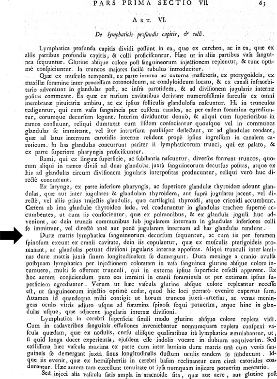
This picture shows the reproduction of the first page of the chapter of the book ‘Vasorum lymphaticorum corporis humani historia et ichnographia’ in which Paolo Mascagni first described the presence of lymphatics in the cerebral dura mater. The arrow shows the incipit of the sentence in which Mascagni describes the course of these lymphatic vessels.
We agree with the authors of the two most recent studies (Aspelund et al. 2015; Louveau et al. 2015) who believe that a better knowledge of dural lymphatics can shed light on the circulation of lymphatic cells from the arachnoid liquid to the venous bloodstream; and, as these cells may be a hallmark of disease, such as neuroinflammatory or neurodegenerative pathologies, the study of these cells may open up new scenarios in the context of CNS disease pathogenesis, early diagnosis, follow up and therapy. We would also like to thank the authors of both papers (Aspelund et al. 2015; Louveau et al. 2015) for having directed their attention to a sometimes forgotten chapter of human anatomy. However, in our opinion, their work has not resulted in a new discovery, nor can they claim that these lymphatics are present in the entire central nervous system, but only in its covering layers. We would like to make these observations known for the benefit of our students and colleagues.
References
- Aspelund A, Antila S, Proulx ST, et al. A dural lymphatic vascular system that drains brain interstitial fluid and macromolecules. J Exp Med. 2015;212:991–999. doi: 10.1084/jem.20142290. [DOI] [PMC free article] [PubMed] [Google Scholar]
- Lecco V. Di una probabile modificazione delle fissure linfatiche della della parte dei seni venosi della dura madre. Arch Ital Otol Rinol Laringol. 1953;64:287–296. [PubMed] [Google Scholar]
- Li J, Zhou J, Shi Y. Scanning electron microscopy of human cerebral meningeal stomata. Ann Anat. 1996;178:259–261. doi: 10.1016/S0940-9602(96)80059-8. [DOI] [PubMed] [Google Scholar]
- Louveau A, Smirnov I, Keyes TJ, et al. Structural and functional features of central nervous system lymphatic vessels. Nature. 2015;523:337–341. doi: 10.1038/nature14432. [DOI] [PMC free article] [PubMed] [Google Scholar]
- Mascagni P, editor. Vasorum lymphaticorum corporis humani historia et ichnographia. Siena: Pazzini Carli; 1787. De lymphaticis profundis capitis et colli. (ed.). Pars Prima Section VII, Art. VI. [Google Scholar]