Summary
Background
Arterial dissection is defined as the cleavage of the arterial wall by an intramural hematoma. Reports of dissection of the celiac and/or superior mesenteric artery are rare; as far as we know, only 24 cases of spontaneous isolated celiac trunk dissection, and 71 cases of spontaneous isolated superior mesenteric artery dissection have been reported.
Case Report
The case presents a 48-year-old male with a sudden-onset epigastric pain. A Computed Tomography Angiography of the thoracoabdominal aorta was applied and dissections of both the celiac artery and SMA were determined. A conservative therapeutic approach was preferred and the patient was discharged with anticoagulant and antihypertensive therapy.
Conclusions
Although rare, spontaneous isolated celiac artery and superior mesenteric artery dissections must be kept in mind in the differential diagnosis of the epigastric pain in the emergency room. Contrast-enhanced Computed Tomography Angiography examination is the method of choice in the diagnosis.
MeSH Keywords: Celiac Artery; Dissection; Mesenteric Artery, Superior
Background
Arterial dissection is defined as the cleavage of the arterial wall by an intramural hematoma. Isolated extra-aortic arterial dissection mostly occurs in renal and carotid arteries but a spontaneous isolated visceral artery dissection happens very rarely. Most of the reported cases involve the superior mesenteric artery (SMA). Reports of dissection of the celiac artery or hepatic artery are even rarer; as far as we know only 24 cases of celiac trunk and 71 cases of SMA dissection can be found in literature [1,2].
Acute or chronic pain in the epigastric region is the general presenting symptom for celiac artery dissection cases. Computed tomography (CT) angiography is a useful technique for the diagnosis of celiac artery dissection. In addition, intramural thrombus and splenic infarctions are some of the radiological findings to accompany celiac artery dissection. There is no consensus on the most appropriate management for celiac artery dissection. Surgical repair, interventional radiology, and conservative therapy are all possible choices [3,4].
Case Report
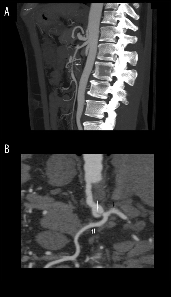
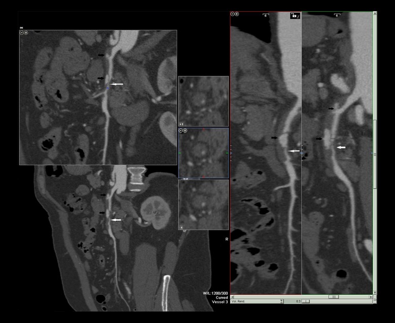
A 48-year-old male presented with acute epigastric pain. The pain radiated to both sides of his back. He vomited once. There were no other symptoms. At presentation, complete blood count (CBC) and biochemical tests were all normal. On physical examination there was only mild epigastric tenderness. He was a smoker and had a history of hypertension. There was no evident pathology in the abdominal sonographic examination. Contrast-enhanced CT was used to exclude aortic dissection. The thoracoabdominal aorta was seen to be normal. The diameter of the abdominal aorta was 17–18 mm. However, dissection of both the celiac trunk and SMA was determined. The dissection of the celiac artery was approximately 14 mm long, and located proximal to the common hepatic artery and splenic artery bifurcation. It extended to the orifice of the left gastric artery. There was also focal widening in the dissected part of the artery and the diameter was 13 mm (Figure 1). In addition, another dissection was found in SMA, beginning just after the orifices of the first branches of SMA and extending for approximately 1.5 cm, accompanied by millimetric contrast accumulation in the wall due to ulceration in the proximal part of the artery. SMA was visualized as 50–60% narrowed because of the dissection, and it was classified as a Sakamoto type IIb dissection of SMA (Figure 2). The ascending and descending aorta, inferior mesenteric artery and renal arteries were normal. Conservative treatment was the method of choice in that case. The pain subsided eventually and on CT 7 days later, there was no intermittent change. The patient was discharged home on hospital day 7 with medical therapy. Further follow-up CT examinations were planned.
Figure 1.

Axial (A) and coronal reformat maximum intensity projection (B) images showing dissection of the celiac artery (white arrow) located proximally to common hepatic (double arrow) and splenic arteries (black arrow) bifurcation.
Figure 2.
Sagittal reformat maximum intensity projection and curved planar reformat images showing Sakamoto type IIb dissection of the superior mesenteric artery (white arrow) and millimetric contrast accumulation in the wall due to ulceration (black arrows).
Discussion
Isolated visceral artery dissections are rarely seen and were first described by Bauersfeld in 1947. SMA is the most common location. Spontaneous dissection of the celiac artery is rarer than SMA dissection and the first reported case was described in 1959 [5].
The initial symptom in most patients is acute or chronic epigastric pain. Patients with ruptured aneurysms may present acutely with bleeding. Chronic dissection has symptoms such as postprandial abdominal pain and weight loss. The average age of patients is approximately 55 years. Both celiac trunk and SMA dissections are more common in males than in females. Risk factors are described as hypertension, cystic medial necrosis, abdominal aortic aneurysm, fibro-muscular dysplasia, and trauma, pregnancy, and connective tissue disorders. The definitive cause has not yet been fully clarified [6]. Computed tomography angiography is the method of choice for the evaluation of vascular structures and related pathologies. It is as accurate as conventional angiography with less morbidity, and it is less expensive. In addition, CT scan can provide high quality images of the dissection site and provide information about the extent of the lesion, presence of aneurysm formation or intramural hematomas [7,8].
SMA dissections were classified by Sakamoto et al. and Yun et al. into three main groups; type I: true and false lumina are both patent, entry and re-entry sites can be seen; type II: true lumen is patent, however, there is no re-entry flow from the false lumen; type IIa: false lumen is visible but re-entry site cannot be seen; type IIb: false luminal flow is not seen, (false lumen is thrombosed), true luminal narrowing is present; and type III: SMA dissection and SMA is also occluded [9]. The current case was classified as type IIb.
Open surgery, endovascular surgery, and interventional radiology are invasive options for the management of visceral artery dissections. Indications for surgical intervention in SMA dissection are increased size of the aneurysm, intraluminal thrombosis, abnormal blood flow through the vessel, and persistent symptoms despite anticoagulation. In celiac artery dissection surgical repair is recommended in the presence of complications such as occlusive lesions, aneurysm formation, and arterial rupture, or extension of the dissection into the hepatic arteries. Conservative treatments include anticoagulants, anti-platelet, and antihypertensive therapy. If a conservative approach is chosen, CTA monitoring on regular basis is generally required [5,8].
In the current case, the patient was followed up with antihypertensive and anticoagulant therapy and CT angiographic follow-up examinations were planned.
Conclusions
In conclusion, spontaneous celiac and SMA artery dissection must be kept in mind in the differential diagnosis of epigastric pain in the emergency room. It is a rare situation but may have been underestimated in the past because of unavailability or the rarity of angiographic CT examinations. With improvements in the use of contrast-enhanced CT in emergency rooms, the diagnosis will eventually become more common.
Footnotes
Conflict of interest
The authors declare that they have no conflict of interest.
References
- 1.Nordanstig J, Gerdes H, Kocys E. Spontaneous isolated dissection of the celiac trunk with rupture of the proximal splenic artery: a case report. Eur J Vasc Endovasc Surg. 2009;37(2):194–97. doi: 10.1016/j.ejvs.2008.10.009. [DOI] [PubMed] [Google Scholar]
- 2.Mousa AY, Coyle BW, Affuso J, et al. Nonoperative management of isolated celiac and superior mesenteric artery dissection: case report and review of the literature. Vascular. 2009;17(6):359–64. doi: 10.2310/6670.2009.00053. [DOI] [PubMed] [Google Scholar]
- 3.Woolard JD, Ammar AD. Spontaneous dissection of the celiac artery: a case report. J Vasc Surg. 2007;45(6):1256–58. doi: 10.1016/j.jvs.2007.01.048. [DOI] [PubMed] [Google Scholar]
- 4.Takayama T, Miyata T, Shirakawa M, Nagawa H. Isolated spontaneous dissection of the splanchnic arteries. J Vasc Surg. 2008;48(2):329–33. doi: 10.1016/j.jvs.2008.03.002. [DOI] [PubMed] [Google Scholar]
- 5.Obon-Dent M, Shabaneh B, Dougherty KG, Strickman NE. Spontaneous celiac artery dissection case report and literature review. Tex Heart Inst J. 2012;39(5):703–6. [PMC free article] [PubMed] [Google Scholar]
- 6.D’Ambrosio N, Friedman B, Siegel D, et al. Spontaneous isolated dissection of the celiac artery: CT findings in adults. Am J Roentgenol. 2007;188(6):W506–11. doi: 10.2214/AJR.06.0315. [DOI] [PubMed] [Google Scholar]
- 7.McGuinness B, Kennedy C, Holden A. Spontaneous coeliac artery dissection. Australas Radiol. 2006;50(4):400–1. doi: 10.1111/j.1440-1673.2006.01610.x. [DOI] [PubMed] [Google Scholar]
- 8.Ozturk TC, Yaylaci S, Yesil O, et al. Spontaneous isolated celiac artery dissection. J Res Med Sci. 2011;16(5):699–702. [PMC free article] [PubMed] [Google Scholar]
- 9.Katsura M, Mototake H, Takara H, Matsushima K. Management of spontaneous isolated dissection of the superior mesenteric artery: Case report and literature review. World J Emerg Surg. 2011;6:16. doi: 10.1186/1749-7922-6-16. [DOI] [PMC free article] [PubMed] [Google Scholar]


