Abstract
The congenital H-type fistula between the anorectum and genital tract besides a normal anus is a rare entity in the spectrum of anorectal anomalies. We described a girl with an anovestibuler H-type fistula and left vulvar abscess. A 40-day-old girl presented symptoms after her parents noted the presence of stool at the vestibulum. On the physical examination, anus was in normal location and size, and had normal sphincter tone. A vestibuler opening was seen in the midline just below of the hymen. A fistulous communication was found between the vestibuler opening and the anus, just above the dentate line. There was a vulvar abscess which had a left lateral vulvar drainage opening 15 mm left lateral to the perineum. After the management of local inflammation and abscess, the patient was operated for primary repair of the fistula. A protective colostomy wasn’t performed prior the operation. A profuse diarrhea started after 5 hours of postoperation. After the diarrhea, a recurrent fistula was occurred on the second postoperative day. A divided sigmoid colostomy was performed. 2 months later, and anterior sagital anorectoplasty was reconstructed and colostomy was closed 1 month later. Various surgical techniques with or without protective colostomy have been described for double termination repair. But there is no consensus regarding surgical management of double termination.
INTRODUCTION
The congenital H-type fistula between the anorectum and genital tract without anal atresia is a rare entity in the spectrum of anorectal anomalies. This type of anomaly has been termed as “double termination” of the alimentary tract in girls[1-3]. Most of the fistulas localized between the anorectum and vestibule of the vagina, and this type anomaly was described as Perineal Canal[1,2,4-6]. We described a 40-day-old girl with a congenital anovestibuler H-type fistula complicated with a vulvar abscess.
CASE REPORT
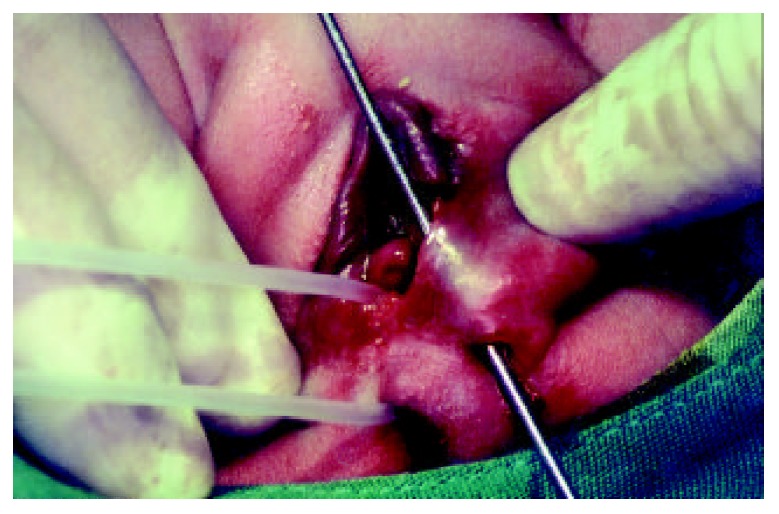
A 40-day-old girl referred to our university medical center with a passage of stool through vestibulum as well as through the normal anal passage from birth. She had been swelling and tenderness on her left vulvar region for one week. On the physical examination, anus was at the normal location and size, and it had normal sphincter tone. A vestibuler opening was found in the midline just below of the hymen. With insertion of an 8 Fr catheter through the vestibuler opening, a fistulous communication was detected between the vestibule of the vagina and the anus at the dentate line. Nearly 3 mm to the left side of this opening, there was a second opening about 3 mm in diameter. A metal sound was inserted through this opening and it was found that it had a subcutaneous continuity with a vulvar abscess. This abscess was detected to drain through a cutaneous opening 15 mm lateral to the perineum (Figure 1). No associated anomaly was detected.
Figure 1.

8 Fr catheter was placed in the perineal canal and metal sound was passed through vestibuler and cutaneous openings of the abscess.
After the management of local inflamation and abscess, the patient was operated for primary repair of the fistula. The bowel preparation was performed with saline enemas. A protective colostomy was not performed either prior or during the definitive operation. We used the technique of anterior sagittal anorectoplasty described by Kulshrestha et al[1] in 1998. At the operation, a metal sound was passed through the fistula from the vestibule of the vagina into the anal canal in the lithotomy position. Then the perineal body that remained over the sound vertically was incised. This incision included skin, subcutaneous tissue, a few fibers of external sphincter muscle, wall of the fistulous tract and anal canal mucosa. The mucosa of the opened fistulous tract was dissected off the underlying muscle. Then, perineal reconstruction was performed by stitching the vestibule, perineal body, sphincter muscle and the anorectum with interrupted 4/0 PDS sutures. Perineal skin was closed with 4/0 Prolene. Five hours following the transfer of the patient to the ward, a profuse diarrhea started resulting in 16 defecations on the first postoperative day. Unfortunately, this caused a partial breakdown of the wound on the second postoperative day, which caused the recurrence of the fistula. And the divided sigmoid colostomy was performed.
Two months later, we operated with the same technique and this attempt was successful. One month following the repair of the fistula, we closed her colostomy and started a rectal dilatation program. Her first year postoperative follow-up was uneventful.
DISCUSSION
The incidence of double termination among female anorectal malformations ranged between 4%-14% in different Asian series[1,5-8]. On the basis of Wingspread classification, these H-type fistulas were divided into three groups according to their level. Low type double termination included cases in which the fistula was lying between the anal canal and the vestibule and this was named as “perineal canal”. In the intermediate type, communication was found between the rectum and the vestibule. High type of double termination consisted of a fistula between the rectum and the vagina[1]. Our case had a low type double termination and an abscess which was detected in her vulvar region complicated her anomaly. When we reviewed the cases reported in the literature, we found out that Rintala et al[2] reported vulvar abscess formation in three girls with double termination. In another report by Brem et al[9] abscess formation was claimed to be secondary to the infection in either a congenital blind-ending sinus or an existing fistula leading to additional openings.
On the other hand, high type of vestibuler fistula demands a more specific operation and it may be named as vestibulorectal pull-through procedure. Without protective colostomy, this type of repair is reported to have a fairly high recurrence rate[7]. Various surgical techniques have been described for low-type double termination repair. These techniques were utilized either with or without protective colostomy in different series. Chatterjee described a simple perineal procedure without colostomy and with good results[3]. Tsuchida et al[5] did not use diversion during definitive procedure they named as “pull-through of the anterior wall of the rectum” in 3 of his 7 cases with perineal canal. Their results were satisfactory for both diverted and non-diverted cases. However, they were not as successful as above with their cases managed with another different simple repair technique and without diversion. Kulshrestha et al[1] described anterior sagittal anorectoplasty in 1998 as a surgical technique for all types of double termination without colostomy and without no complications. This series which were the most recent and had a successful outcome for the repair of perineal canal directed us to prefer this technique in our series. Furthermore, avoiding a colostomy in using this technique was another major factor for our selection.
Unfortunately, our case’s fistula recurred. This may have resulted not from insufficiency of the technique, but from some factors that did not take place in Kulshrestha’s series. A major reason for breakdown of the repair may be due to the unexpected and unwanted profuse diarrhea of the patient that started after 5 hours of the post-operation. The sufficiency of the technique may be thought of when one thinks that in the redo operation, it was proved to be a good result.
In conclusion, it is believed that definitive repair can be performed without protective colostomy when no abscess formation is present in the vulvar region. If an abscess is detected, it should be well managed before the operation.
Footnotes
Edited by Xu XQ
References
- 1.Kulshrestha S, Kulshrestha M, Prakash G, Gangopadhyay AN, Sarkar B. Management of congenital and acquired H-type anorectal fistulae in girls by anterior sagittal anorectovaginoplasty. J Pediatr Surg. 1998;33:1224–1228. doi: 10.1016/s0022-3468(98)90155-5. [DOI] [PubMed] [Google Scholar]
- 2.Rintala RJ, Mildh L, Lindahl H. H-type anorectal malformations: incidence and clinical characteristics. J Pediatr Surg. 1996;31:559–562. doi: 10.1016/s0022-3468(96)90496-0. [DOI] [PubMed] [Google Scholar]
- 3.Chatterjee SK, Talukder BC. Double termination of the alimentary tract in female infants. J Pediatr Surg. 1969;4:237–243. doi: 10.1016/0022-3468(69)90398-4. [DOI] [PubMed] [Google Scholar]
- 4.Rao KL, Choudhury SR, Samujh R, Narasimhan KL. Perineal canal-repair by a new surgical technique. Pediatr Surg Int. 1993;8:449–450. [Google Scholar]
- 5.Tsuchida Y, Saito S, Honna T, Makino S, Kaneko M, Hazama H. Double termination of the alimentary tract in females: a report of 12 cases and a literature review. J Pediatr Surg. 1984;19:292–296. doi: 10.1016/s0022-3468(84)80190-6. [DOI] [PubMed] [Google Scholar]
- 6.Wakhlu A, Pandey A, Prasad A, Kureel SN, Tandon RK, Wakhlu AK. Anterior sagittal anorectoplasty for anorectal malformations and perineal trauma in the female child. J Pediatr Surg. 1996;31:1236–1240. doi: 10.1016/s0022-3468(96)90241-9. [DOI] [PubMed] [Google Scholar]
- 7.Chatterjee SK. Double termination of the alimentary tract--a second look. J Pediatr Surg. 1980;15:623–627. doi: 10.1016/s0022-3468(80)80513-6. [DOI] [PubMed] [Google Scholar]
- 8.Bagga D, Chadha R, Malhotra CJ, Dhar A. Congenital H-type vestibuloanorectal fistula. Pediatr Surg Int. 1995;10:481–484. [Google Scholar]
- 9.Brem H, Guttman FM, Laberge JM, Doody D. Congenital anal fistula with normal anus. J Pediatr Surg. 1989;24:183–185. doi: 10.1016/s0022-3468(89)80245-3. [DOI] [PubMed] [Google Scholar]


