Significance
The orange carotenoid protein (OCP) is critical for the antenna-associated energy-dissipation mechanism of cyanobacteria under high light conditions. We show that light activation causes a global conformation change, the complete separation of the two domains of the OCP. Such a conformational change has been postulated to be a prerequisite for interaction with the antenna. We also identify local structural changes in residue solvent accessibility and roles for structural water molecules in activation of the OCP. By combining small-angle scattering, hydrogen-deuterium exchange, and X-ray hydroxyl radical footprinting studies, we were able to construct a model of the structural changes during the activation of the OCP with an unprecedented level of detail.
Keywords: orange carotenoid protein, photoprotection, X-ray footprinting, hydrogen deuterium exchange, SAXS
Abstract
Photoprotective mechanisms are of fundamental importance for the survival of photosynthetic organisms. In cyanobacteria, the orange carotenoid protein (OCP), when activated by intense blue light, binds to the light-harvesting antenna and triggers the dissipation of excess captured light energy. Using a combination of small angle X-ray scattering (SAXS), X-ray hydroxyl radical footprinting, circular dichroism, and H/D exchange mass spectrometry, we identified both the local and global structural changes in the OCP upon photoactivation. SAXS and H/D exchange data showed that global tertiary structural changes, including complete domain dissociation, occur upon photoactivation, but with alteration of secondary structure confined to only the N terminus of the OCP. Microsecond radiolytic labeling identified rearrangement of the H-bonding network associated with conserved residues and structural water molecules. Collectively, these data provide experimental evidence for an ensemble of local and global structural changes, upon activation of the OCP, that are essential for photoprotection.
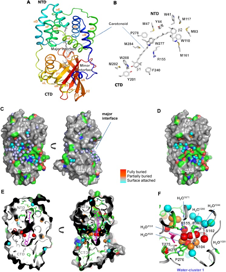
Photosynthetic organisms have evolved a protective mechanism known as nonphotochemical quenching (NPQ) to dissipate excess energy, thereby preventing oxidative damage under high light conditions (1). In plants and algae, NPQ involves pH-induced conformation changes in membrane-embedded protein complexes and enzymatic interconversion of carotenoids (2, 3). Cyanobacteria, in contrast, use a relatively simple NPQ mechanism governed by the water soluble orange carotenoid protein (OCP). The OCP is composed of an all α-helical N-terminal domain (NTD) consisting of two discontinuous four-helix bundles and a mixed α/β C-terminal domain (CTD), which is a member of the widely distributed nuclear transport factor 2-like superfamily (Fig. S1A) (4, 5). There are two regions of interaction between the NTD and CTD (4, 5): the major interface, which buries 1,722 Å of surface area, and the interaction between the N-terminal alpha-helix (αA) and the CTD (minor interface) (Fig. S1A). A single noncovalently bound keto-carotenoid [e.g., echinenone (ECN)] spans both domains in the structure of the resting (inactive) form of the protein (OCPO).
Fig. S1.
Structure of the OCP. (A) Crystal structure of Synechocystis OCP (PDB ID code 3MG1) consisting of two domains, NTD and CTD as described in the main text introduction, which form major and minor interfaces. (B) Amino acid residues within 3.9 Å of the carotenoid are shown by sticks. (C) Surface-bound water molecules at the major interface are shown in slate-colored spheres in Synechocystis OCP (PDB ID code 3MG1). This layer of water molecules fully or partially eclipses other water molecules, which are either conserved or found to be at the same location (within 0.5 Å) in the crystal structures of A. maxima and Synechocystis OCP (Table S6). The coloring indicates their depth from the surface of the OCP as shown in Fig. 4 A and B. Removal of the slate-colored spheres, exposing partially buried water, is shown in orange in D. The fully buried waters (red spheres) are invisible in the surface diagram of OCP. (E) Cross-sectional view to show the position of fully and partially buried structural waters in OCPO. (F) Details of water–protein H-bonding network in water cluster 1 at the major interface. The absolutely conserved R155 is closely surrounded (<3.2 Å, capable of forming H-bond) by a number of buried (HOH1151,1200, and 1671) water molecules, which are involved in dense residue-water interactions as discussed in the main text. Similar H-bonding networks are also observed in the water clusters 2 and 3 (Table S6).
The NTD and CTD of the OCP have discrete functions. The isolated NTD acts as an effector domain that binds to the antenna whereas the CTD has been proposed to play a sensory/regulatory role in controlling the OCP’s photoprotective function (6). Exposure to blue light converts OCPO to the active (red) form, OCPR (7). OCPR is involved in protein–protein interactions with the phycobilisome (PB) (5) and the fluorescence recovery protein (FRP), which converts OCPR back to OCPO (8). The OCPR form is therefore central to the photoprotective mechanism, and determining the exact structural changes that accompany its formation are critical for a complete mechanistic understanding of the reversible quenching process in cyanobacteria. Although crystal structures exist of both the (inactive) OCPO (4, 5) and the active NTD (effector domain) form of the protein (9), crystallization of the activated, full-length OCPR has not been achieved. To identify the protein structural changes that occur after absorption of light by the OCP’s ECN chromophore, we undertook a hybrid approach to structurally characterize OCPR in solution.
In Synechocystis OCPO, the 4-keto group on the “β1” ring of ECN is H-bonded to two conserved residues, Y201 and W288, in a hydrophobic pocket in the core of the CTD (Fig. S1B) (5). The other end of the carotenoid is positioned between the two four-helix bundles of the NTD. Several conserved residues within 3.9 Å of the carotenoid are known to interact with its extensive conjugation and result in fine tuning of the spectral characteristics of the OCP (Fig. S1B) (4, 5); these residues have been implicated in photochemical function via mutagenesis studies (5). A recent study of the OCP bound to the carotenoid canthaxanthin (OCP-CAN) showed that photoactivation of the OCP results in a substantial translocation (12 Å) of the carotenoid deeper into the NTD (9). Mutational analyses of the full-length OCP and biochemical studies on the constitutively active NTD [commonly known as the red carotenoid protein (RCP)] suggested that the NTD and CTD at least partially separate, resulting in the breakage of an interdomain salt-bridge (R155–E244) upon photoactivation (9–12). Together, the previous studies suggest that large-scale protein structural changes in the OCP accompany carotenoid translocation upon light activation; however, such changes in the context of the full-length protein have yet to be experimentally demonstrated. Here, we report use of X-ray radiolytic labeling with mass spectrometry (XF-MS) and hydrogen/deuterium exchange with mass spectrometry (HDX-MS), which detect residue-specific changes (13–15), to investigate the structural changes that occur during OCP photoactivation. In conjunction with small angle X-ray scattering (SAXS), which enables characterization of global conformational changes in the solution state (16), we show that dissociation of the NTD and CTD is complete in photoactivated OCP. This separation is accompanied by an unfolding of the N-terminal α-helix that is associated with the CTD in the resting state. We also pinpoint changes in specific amino acids and structurally conserved water molecules, providing insight into the signal propagation pathway from carotenoid to protein surface upon photoactivation. Collectively, these data provide a comprehensive view of both global and local intraprotein structural changes in the OCP upon photoactivation that are essential to a mechanistic understanding of cyanobacterial NPQ.
Results and Discussion
Global Structural Changes upon Photoactivation of the OCP as Observed by SEC, CD, and SAXS.
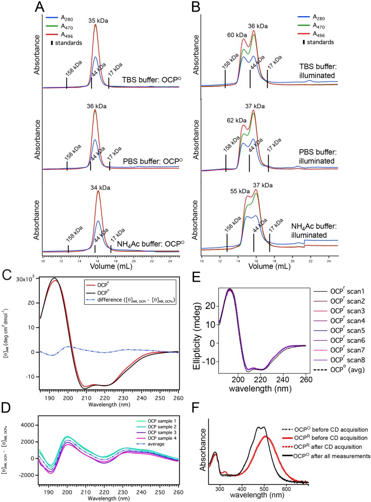
Size-exclusion chromatography (SEC) of purified OCPO [Synechocystis OCP containing ECN (OCP-ECN)] in darkness yielded a single (nearly Gaussian) peak in its chromatogram (Fig. S2A). An ∼1:1 ratio of the 470:496-nm absorbance in the peak is consistent with the UV-visible spectrum of OCPO (7). Relative to globular standards, the estimated molecular mass (MW) of OCPO is 36 kDa (Fig. S2A), consistent with the expected monomeric MW (35.4 kDa). In contrast, SEC of OCP preilluminated with blue light (OCPR) yielded a heterogeneous elution profile with an additional peak eluting before the OCPO monomer peak (Fig. S2B). The A470:496 ratio (<<1) of the early eluting peak is consistent with the visible absorbance spectrum of OCPR (7), and its relative elution volume (corresponding to a predicted globular mass of 63 kDa) suggests a larger size (Stokes radius) for OCPR. Similar results were obtained in the buffer used for XF- and HDX-MS studies (20 mM phosphate, pH 7.4, 100 mM NaCl).
Fig. S2.
(A and B) Size-exclusion chromatograms for dark (OCPO) and illuminated OCP samples applied to a Superdex 200 10/300 GL column in different buffers. Experiments were performed in the following buffers: TBS (50 mM Tris⋅HCl, 200 mM NaCl); PBS (20 mM potassium phosphate, pH 7.4, 100 mM NaCl); 200 mM ammonium acetate buffer. Elution volumes for three globular standards are indicated (vertical lines) on the baseline of each chromatogram. Estimated MWs for each elution peak, as determined from a calibration to the globular standards in each buffer system, are noted on the chromatograms. (C) Circular dichroism spectra of OCPO and OCPR in the far-UV range. The spectra of OCP in darkness (OCPO, black solid line) and after illumination (OCPR, dashed red line) are shown as the average result of four independent experiments (4 × 8 scans per spectrum). Changes in the average mean-residue-ellipticity values between OCPO and OCPR are shown additionally as a difference spectrum (blue dashed line). (D) Far-UV CD difference spectra (OCPR – OCPO) calculated from four independent experiments. Illumination has a small but reproducible effect on the far-UV CD spectra. (E) Time-dependent OCPR CD data collection of OCP samples before and after CD measurements. A representative dataset showing successive scans of the OCPR sample (∼2 min per scan) and the lack of significant thermal reversion to the OCPO form during the full time course of the measurement. An OCPO spectrum (average of eight scans) is also shown for reference. (F) Time-dependent UV-visible (UV-Vis) absorbance spectra of OCP samples before and after CD measurements. UV-Vis spectra collected during the CD experiment: OCPO (black dotted line) was immediately illuminated to OCPR (red solid line) after CD measurements on OCPO. The OCPR spectrum after CD acquisition (dotted red line) was acquired after ∼20 min of spectral averaging (eight scans) at 4 °C in the CD spectrometer. Thermal reversion of OCP after the OCPR measurements resulted in recovery of the OCPO form (black solid line).
Light scattering was used to examine whether the larger apparent size of OCPR was resulting from a change in the oligomeric state (i.e., dimerization). Dynamic light scattering, a metric dependent on the protein’s shape, showed a 10% increase in the radius of hydration of the OCP upon activation (Table S1). At the same time, the static light scattering intensity, a shape-independent metric, did not change, indicating no change in the MW of the protein. Therefore, structural changes within the monomer, and not changes in the oligomeric state, were responsible for the altered elution time observed by analytical SEC.
Table S1.
Light scattering derived structural parameters
| Rh, nm | %Pd | Scattering detector, V | Molecular mass, kDa* | |
| OCPO | 3.252 ± 0.031 | 6.9 ± 2.4 | 0.285 ± 0.007 | 63.5 ± 2* |
| OCPR | 3.572 ± 0.024 | 7.2 ± 2.3 | 0.287 ± 0.002 | 65.7 ± 1* |
Because both OCPO and OCPR absorb at a wavelength that overlaps with the laser in the light scattering detector (658 nm), the molecular mass from static scattering will be inaccurate (Wyatt Technologies).
Circular dichroism (CD) spectroscopy was used to determine whether secondary structure changes contributed to the larger size of OCPR. Only small differences in the far-UV (185–260 nm) CD spectra are apparent after illumination of OCP (Fig. S2C). The small difference between the OCPO and OCPR spectra was found to be consistently reproducible across multiple independent measurements (Fig. S2D), and careful monitoring of the time-dependent CD signal and sample absorbance ensured that data were collected on the OCPR form without significant contribution from OCPO (Fig. S2 E and F). Three different algorithms (CDSSTR, CONTINLL, and SELCON3) were used to calculate the predicted secondary structure of OCPO and OCPR from the experimental CD curves (Table S2) (17). The helical (47.0%), beta strand (13.2%), turn (17.1%), and unordered (22.8%) content of OCPO predicted by the average of the results from all methods is in good agreement with the crystal structure (48.3% helix, 15.9% strand, 14.6% turn, 21.2% unordered). For OCPR, all three algorithms predict a small decrease (∼2%) in α-helical content and a small increase (∼1%) in disordered structure (Table S3). A small increase in beta-strand content (∼1–3%) is predicted using the CDSSTR and CONTINLL methods, but not SELCON3. Overall, these results are consistent with the loss of only a small portion of the protein’s α-helical structure (2% α-helical content = ∼6 residues) after illumination, with the majority of the secondary structure remaining otherwise unchanged.
Table S2.
Fractions of secondary structure content calculated by multiple methods and basis sets for OCPO and OCPR
| Method | Basis | OCPo secondary structure content | OCPr secondary structure content | ||||||
| Helix | Sheet | Turn | Unrd | Helix | Strand | Turn | Unrd | ||
| CDSSTR | SP29 | 47.7 ± 1.8 | 13.4 ± 1.4 | 17.9 ± 0.8 | 20.8 ± 0.4 | 45.5 ± 1.8 | 15.3 ± 1.2 | 17.3 ± 0.7 | 21.8 ± 0.4 |
| CDSSTR | SP37 | 47.6 ± 1.5 | 12.9 ± 1.0 | 17.3 ± 0.5 | 22.2 ± 1.1 | 44.6 ± 1.9 | 15.9 ± 1.2 | 16.7 ± 0.5 | 22.9 ± 0.8 |
| CDSSTR | SP43 | 50.7 ± 1.8 | 11.6 ± 0.9 | 13.7 ± 0.5 | 23.9 ± 0.9 | 47.6 ± 1.8 | 13.7 ± 0.9 | 14.8 ± 1.0 | 24.0 ± 0.6 |
| CDSSTR | SDP42 | 47.9 ± 1.7 | 13.5 ± 1.1 | 19.2 ± 0.9 | 19.2 ± 0.9 | 45.3 ± 1.9 | 16.8 ± 1.2 | 19.0 ± 1.0 | 18.9 ± 0.8 |
| CDSSTR | SDP48 | 50.9 ± 1.8 | 12.0 ± 1.3 | 15.8 ± 1.5 | 20.9 ± 0.8 | 48.0 ± 1.9 | 15.2 ± 0.9 | 16.7 ± 0.8 | 20.0 ± 1.3 |
| CONTINLL | SP29 | 46.3 ± 2.0 | 13.9 ± 1.3 | 18.7 ± 0.6 | 21.3 ± 0.6 | 44.5 ± 1.9 | 14.9 ± 1.0 | 17.7 ± 0.9 | 23.0 ± 0.9 |
| CONTINLL | SP37 | 43.9 ± 1.4 | 14.7 ± 0.9 | 18.7 ± 0.6 | 22.8 ± 0.7 | 40.6 ± 1.1 | 17.4 ± 0.7 | 17.2 ± 0.9 | 24.8 ± 1.1 |
| CONTINLL | SP43 | 47.2 ± 1.3 | 12.2 ± 1.1 | 16.2 ± 0.2 | 24.4 ± 0.4 | 45.6 ± 1.6 | 13.6 ± 1.1 | 14.6 ± 0.6 | 26.2 ± 0.7 |
| CONTINLL | SDP42 | 44.1 ± 1.2 | 15.4 ± 0.9 | 19.1 ± 0.5 | 21.5 ± 0.6 | 41.6 ± 1.2 | 16.8 ± 0.5 | 17.9 ± 0.7 | 23.8 ± 1.2 |
| CONTINLL | SDP48 | 47.1 ± 1.4 | 12.6 ± 1.1 | 15.5 ± 0.5 | 24.8 ± 0.3 | 45.2 ± 1.6 | 14.6 ± 0.9 | 14.3 ± 0.6 | 25.9 ± 0.9 |
| SELCON3 | SP29 | 45.3 ± 1.8 | 13.2 ± 1.0 | 18.7 ± 0.9 | 22.8 ± 1.2 | 43.7 ± 1.7 | 13.1 ± 0.8 | 18.2 ± 0.8 | 24.2 ± 0.7 |
| SELCON3 | SP37 | 45.5 ± 1.7 | 13.3 ± 1.2 | 17.9 ± 0.8 | 23.7 ± 0.2 | 43.2 ± 1.6 | 13.8 ± 0.9 | 17.8 ± 1.0 | 24.8 ± 0.8 |
| SELCON3 | SP43 | 47.5 ± 1.6 | 12.6 ± 1.0 | 14.6 ± 0.2 | 25.6 ± 1.6 | 45.9 ± 1.7 | 13.1 ± 0.9 | 13.9 ± 0.5 | 27.1 ± 1.2 |
| SELCON3 | SDP42 | 45.4 ± 1.7 | 13.8 ± 0.9 | 18.2 ± 0.7 | 23.2 ± 0.6 | 43.4 ± 1.7 | 13.9 ± 0.8 | 17.7 ± 1.0 | 24.5 ± 1.0 |
| SELCON3 | SDP48 | 47.5 ± 1.6 | 12.7 ± 0.9 | 14.8 ± 0.4 | 25.4 ± 1.6 | 45.9 ± 1.7 | 13.3 ± 0.8 | 14.1 ± 0.3 | 26.8 ± 1.1 |
| Average | 47.0 ± 1.6 | 13.2 ± 1.1 | 17.1 ± 0.6 | 22.8 ± 0.8 | 44.7 ± 1.7 | 14.7 ± 0.9 | 16.5 ± 0.8 | 23.9 ± 0.9 | |
Fractions of content are listed as an average result from the analysis of CD spectra from four independent OCPO/OCPR datasets. The total helix and strand content shown here represent the sum of regular and disordered helix/sheet content output by the secondary structure analysis packages for each of these elements.
Table S3.
Differences in secondary structure content in the OCPO and OCPR forms as calculated from the results in Tables S1–S4
| Method | Basis | Δ(OCPr-OCPo) secondary structure content | |||
| ΔHelix | ΔStrand | ΔTurn | ΔUnrd | ||
| CDSSTR | SP29 | −2.3 | 1.9 | −0.6 | 1.0 |
| CDSSTR | SP37 | −3.0 | 3.0 | −0.6 | 0.7 |
| CDSSTR | SP43 | −3.1 | 2.0 | 1.1 | 0.1 |
| CDSSTR | SDP42 | −2.6 | 3.3 | −0.2 | −0.3 |
| CDSSTR | SDP48 | −2.8 | 3.2 | 0.9 | −0.9 |
| CONTINLL | SP29 | −1.8 | 1.0 | −1.0 | 1.7 |
| CONTINLL | SP37 | −3.3 | 2.7 | −1.5 | 2.0 |
| CONTINLL | SP43 | −1.6 | 1.4 | −1.6 | 1.8 |
| CONTINLL | SDP42 | −2.5 | 1.5 | −1.2 | 2.3 |
| CONTINLL | SDP48 | −2.0 | 2.0 | −1.2 | 1.2 |
| SELCON3 | SP29 | −1.7 | −0.1 | −0.5 | 1.5 |
| SELCON3 | SP37 | −2.3 | 0.5 | −0.1 | 1.2 |
| SELCON3 | SP43 | −1.6 | 0.5 | −0.7 | 1.5 |
| SELCON3 | SDP42 | −2.0 | 0.1 | −0.5 | 1.3 |
| SELCON3 | SDP48 | −1.5 | 0.6 | −0.7 | 1.4 |
| Average | -2.2 | 1.6 | -0.6 | 1.1 | |
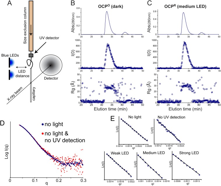
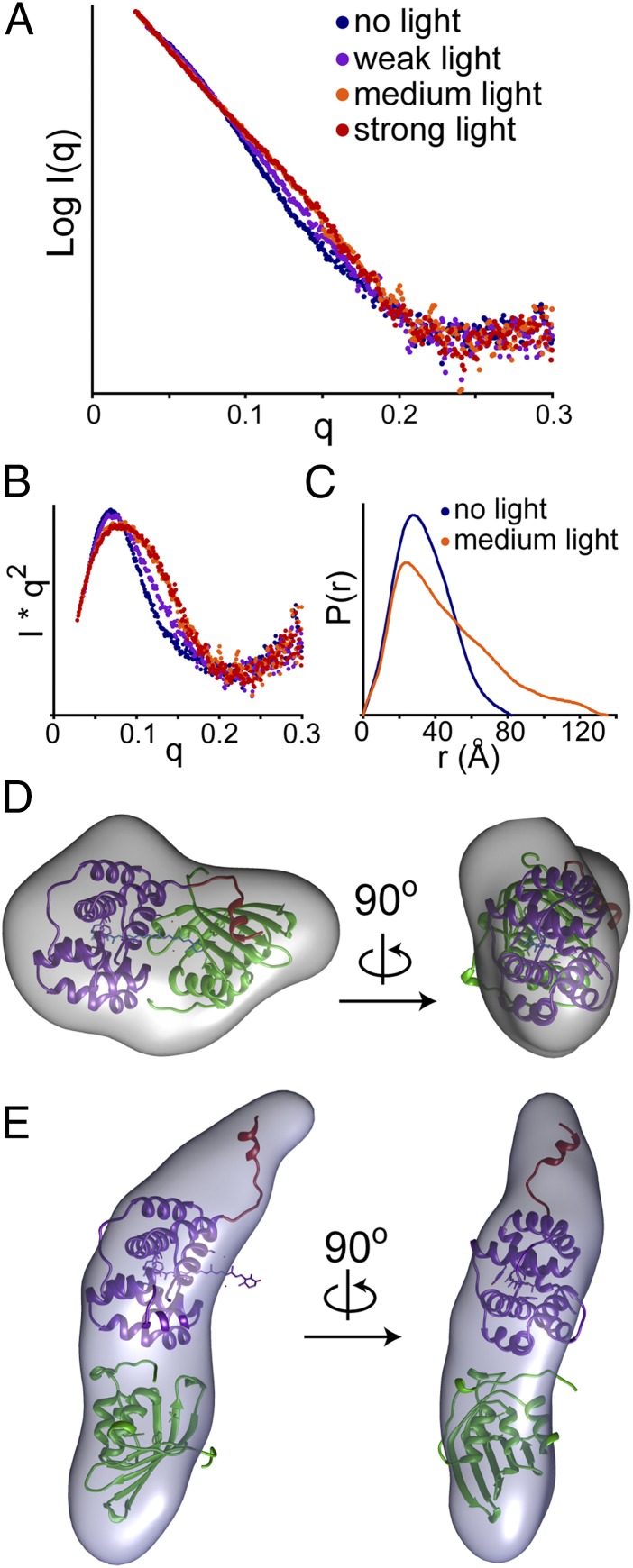
To obtain more detailed information on the changes in tertiary structure that occur during the transition to the light activated state OCPR, we used tandem SEC-SAXS with online illumination of the eluting OCP (Fig. S3A). This experimental configuration enabled us to circumvent sample aggregation and vary light intensity to obtain datasets with different levels of illumination (SI Methods). The SAXS patterns of the OCP with and without illumination are shown in Fig. 1A. With illumination, there is a distinct change in the scattering pattern, most apparent between q = 0.1–0.2. The radius of gyration data from Guinier and real space (GNOM) analysis indicated a large difference in the global structure, with OCPR showing a ∼30% and ∼50% increase in the radius of gyration (Rg) and maximal dimension (Dmax), respectively (Table S4). Kratky plots for both the OCPO and OCPR converged near baseline, indicating that, although there are large global changes, the protein remains folded (Fig. 1B) (16). The real space pairwise distance distribution [P(r)] plot showed that, whereas OCPO has a profile consistent with a globular protein, OCPR is much more elongated (Fig. 1C).
Fig. S3.
(A) SEC-SAXS data collection setup for collecting data on OCPR. The eluting OCP from the size exclusion passes the UV detector and is illuminated in the tubing connecting the UV detector to the capillary, as well as at the first portion of the capillary, above the X-ray beam path. The capillary block was enclosed in a circulating water bath, held at 8 °C. The LED distance was varied to acquire data for low (20 cm), medium (7 cm), and high illumination (2 cm). OCPO was collected by covering the lines with aluminum foil and blocking all background light near the capillary. An additional control OCP was run bypassing the UV detector, to test for any background activation of OCP from the UV detector. (B and C) SEC-SAXS traces of OCPO and OCPR. The UV absorbance at 280 nm, scattering intensity at zero angle, and radius of gyration are shown for each point throughout the SEC-SAXS run for OCPO (B, kept in the dark) and OCPR (C, medium LED intensity). (D) SAXS profiles of OCP in the absence of illumination either with (blue) and without (red) online UV detection during SEC-SAXS. (E) The Guinier fits of the merged SEC-SAXS datasets for OCP show good linearity. I(0) and Rg are shown in Table S4.
Fig. 1.
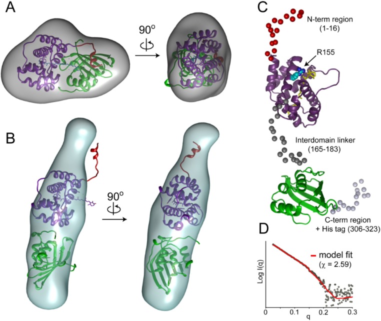
Global structural changes in the OCP upon activation monitored by SAXS. (A) SAXS data for OCP in the absence of light (blue), weak illumination (purple), medium illumination (orange), and strong illumination (red) (see SI Methods for specific illumination levels). Guinier plots are shown in Fig. S3E. Kratky plots (B) and corresponding distance distribution plots (C) of the scattering data are shown with the same coloring as in A. Ab initio bead reconstructions (gray volumes) are shown for OCP in the absence of light (D) and under medium illumination (E) using GASBOR (20). Similar results were obtained using DAMMIN, another bead modeling algorithm (Fig. S4 A and B) (21). (D) A monomer of OCPO from the crystal structure is docked into the volume envelope with the far N-terminal helix (red), N-terminal domain (NTD; purple), and C-terminal domain (CTD; green) (PDB ID code 3MG1) (5). (E) The two domains of OCP along with the N-terminal helix were modeled to approximate the volume envelope of OCPR. The (nontranslocated) carotenoid is shown in sticks.
Table S4.
SAXS derived structural parameters
| Rg (Guinier), Å | I(0) | Peak Abs280 | Rg (GNOM), Å | Dmax, Å | |
| Dark (no UV) | 25.9 ± 0.2 | 363 ± 2 | * | * | * |
| Dark | 25.8 ± 0.2 | 775 ± 6 | 0.527 | 25.7 ± 0.1 | 82 |
| Weak LED | 27.6 ± 0.2 | 813 ± 6 | 0.576 | 29.6 ± 0.2 | 115 |
| Medium LED | 33.3 ± 0.4 | 785 ± 8 | 0.52 | 37.0 ± 0.3 | 135 |
| High LED | 34.5 ± 0.4 | 937 ± 9 | 0.574 | 38.0 ± 0.2 | 135 |
UV was not collected and the SAXS data was collected at a different q range, therefore real space analysis was not comparable.
Native mass spectrometry has shown that the OCPO forms dimers, which are also consistently observed in crystal structures (4, 5), and a change in the monomer/dimer distribution upon activation (18). By SAXS, the scattering intensity at zero angle, I(0), is the same for OCPO and OCPR (775 ± 6 and 785 ± 8, respectively), indicating no difference in the MW of the protein (Table S4). We used SAXSMOW (19), a method for determination of the MW of globular proteins from the raw SAXS data, which resulted in an estimate of ∼38.2 kDa for the OCP. The contrast to previous studies may be due to weak dimerization that is observed only at higher protein concentrations (>1 mg/mL), as previously suggested (5).
Because the changes in the SAXS patterns of the OCPR could be attributed to structural changes, and not oligomeric state changes, we next applied ab initio bead modeling to obtain more information about the global structure of OCPR (20, 21). The filtered shape envelope calculated from the SAXS profile of OCPO was consistent with the shape and size of the OCP monomer in the crystal structure (Fig. 1D) (5). The envelope for OCPR showed a narrower and longer shape, in which the isolated NTD and CTD of the OCP could be readily fit (Fig. 1E). To obtain more structural information on OCPR, we applied the program Coral (22), which uses iterative rigid body modeling, with the aim of generating a model consistent with experimental SAXS data. The positions of the NTD and CTD of the OCP were treated as isolated rigid bodies, and the N/C-terminal extensions and interdomain linker sequences were added as flexible residues. Although the relative orientation of the two domains varied, the resulting models of the OCPR were in good agreement with the experimental SAXS profile, with the lowest χ value of 2.59 (Fig. S4 C and D). In contrast, Coral models sampling different linker and N/C-terminal extensions, without allowing NTD and CTD separation, resulted in very poor fits to the SAXS data (χ > 11). These data constitute, to our knowledge, the first coarse-grained/low resolution view of the overall organization of OCPR, demonstrating that the two domains fully dissociate upon photoactivation.
Fig. S4.
Changes in the shape of OCP upon light activation by SAXS. (A) SAXS based ab initio shape reconstruction of OCPO using DAMMIN. The NTD and CTD from the crystal structure are shown in purple and green, respectively (PDB ID code 3MG1). The N-terminal helix is shown in red. (B) Shape reconstructions of OCPR using DAMMIN. (C) The program Coral was used for rigid body modeling of the two domains to match the SAXS pattern for OCPR. The models show that, in OCPR, the NTD and CTD are separated by ∼16 Å. The carotenoid ligand is shown in yellow, and the position of R155 is shown in cyan. (D) Models from the 20 Coral runs were consistent with the scattering curve (χ = 3.1 ± 0.2), with a χ of 2.59 for the best fit model.
Local Structural Changes upon Photoactivation of the OCP as Observed by XF-MS and HDX-MS.
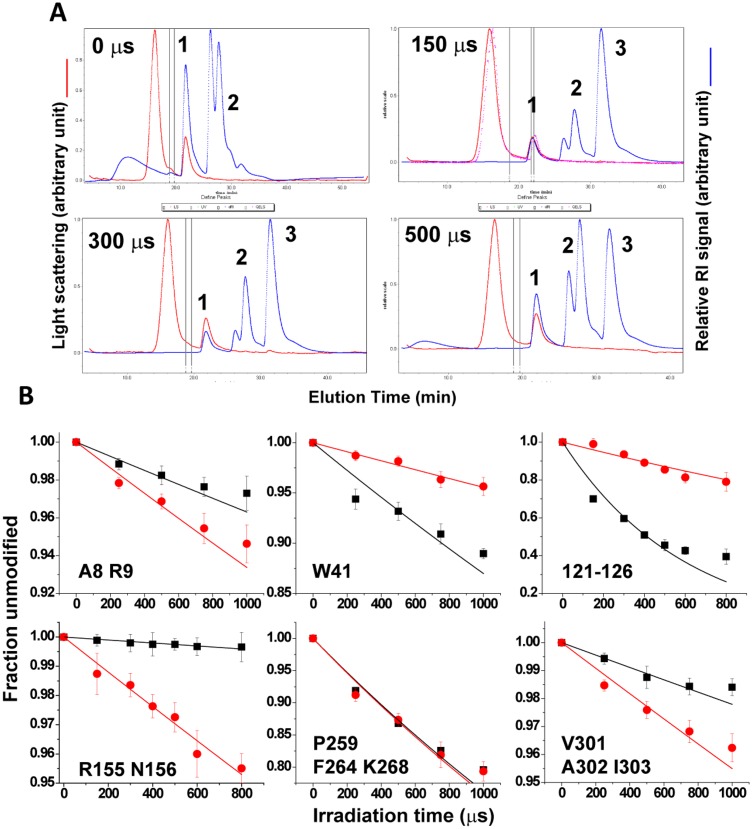
With XF-MS, microsecond X-ray pulses ionize both bulk and bound water rapidly and isotropically to generate hydroxyl radicals in situ, which then react with proximal residues to yield stable modification products (13, 23). X-ray radiolysis resulted in reproducible side-chain modifications in OCPO and OCPR samples without any significant structural perturbation, as evaluated by UV absorption and SEC-multiangle light scattering (MALS) (Fig. S5A). Postirradiation, bottom-up liquid chromatography (LC)-MS analysis using trypsin and Glu-C digestion generated greater than 94% sequence coverage. A progressive increase in the hydroxyl radical dose resulted in residue-specific dose–response plots and provided side chain-specific hydroxyl radical reactivity rates (Fig. S5B and Table S5). The rate of side-chain labeling is governed by both the intrinsic reactivity of the amino acid and the solvent accessibility to the side-chain. However, the ratio of the measured reactivity rates for the same residues in the OCPO vs. OCPR gave a ratiometric account of the change in accessibility, which was independent of the intrinsic side-chain reactivity (Fig. 2A).
Fig. S5.
(A) SEC-MALS: Chromatogram of MALS (red) and differential refractive index (dRI) (blue) from SEC of “zero” and X-ray irradiated OCPO samples using Ethan LC (GE Healthcare Life Science) coupled to Dawn Heleos MALS detector with an embedded dynamics light scattering detector and Optilab rEX dRI detector (Wyatt Technology Corp.). The data shown are directly from the Astra software. The scattering signal indicates no change in peak characteristics of the major component, OCP-monomer (peak “1”) upon X-ray irradiation. The dRI signal indicates presence of buffer components (peak “2” in all of the samples) and methionine amide (peak “3” in the irradiated samples). (B) Representative XF-MS dose–response plots: peptide modification as a function of X-ray irradiation dose for residue and residues clusters of OCPO, (black squares) and illuminated OCPR, (red circles). Error bars represent the SE from three independent measurements. The solid lines represent single-exponential fits to the dose-dependent data. The ratio of the modification rates (R) indicates the change in relative accessibilities, which are shown in Table S6.
Table S5.
Peptides and modification sites detected by XF-MS, and the ratio(s) of hydroxyl radical reactivity for OCPR versus OCPO
| Seq. no.* | Peptide sequence† | Site of modification‡ | Ratio “R“ of hydroxyl radical reactivity kOCPR/kOCPO§ |
| 2–9 | PFTIDSAR | P2, F3 (+16 Da)¶ | 0.74 ± 0.15 |
| A8, R9 (+16 Da) | 1.85 ± 0.39 | ||
| 10–27 | GIFPNTLAADVVPATIAR | Residues 13–22 (+16 Da) | 1.93 ± 0.25 |
| 28–49 | FSQLNAEDQLALIWFAYLEMGK | W41 (+48 Da) | 0.35 ± 0.05 |
| W41, F42, Y44, M47 (+16 Da) | 0.47 ± 0.09 | ||
| M47 (+16 Da) | 0.76 ± 0.16 | ||
| 50–69 | TLTIAAPGAASMQLAENALK | A54, A55, P56 (+16 Da) | 0.76 ± 0.09 |
| M61 (+16 Da) | 1.06 ± 0.22 | ||
| 70–89 | EIQAMGPLQQTQAMCDLANR | M74 (+16 Da) | 0.69 ± 0.13 |
| M74P76M83 (+16 Da) | 0.67 ± 0.02 | ||
| 97–106 | TYASWSPNIK | Y98W101P103 (+16 and +32 Da) | 1.95 ± 0.30 |
| 107–112 | LGFWY | F109W110Y111 (+16 and +32 Da) | 0.90 ± 0.13 |
| 113–155 | LGELMEQGFVAPIPAGYQLSANANAVLATIQGLESGQQITVLR | M117 (+16 Da) | 0.69 ± 0.13 |
| 119–146 | QGFVAPIPAGYQLSANANAVLATIQGLE | F121,P124,P126 (+16 Da)# | 0.17 ± 0.05 |
| 147–160 | SGQQITVLRNAVVD | R155, N156 (+16 Da) | 10.11 ± 3.96 |
| 156–167 | NAVVDMGFTAGK | M161 (+16 Da) | 0.68 ± 0.12 |
| F163 (+16 Da) | 0.78 ± 0.46 | ||
| K167 (+16 Da) | 0.70 ± 0.21 | ||
| 172–185 | IAEPVVPPQDTASR | P175P178P179R185 (+16 Da) | 0.58 ± 0.06 |
| 192–215 | GVTNATVLNYMDNLNANDFDTLIE | M202 (+16 Da) | 1.1 ± 0.18 |
| Residues 119–215 (+16 Da)# | 0.89 ± 0.08 | ||
| 221–235 | GALQPPFQRPIVGKE | Residues 226–231 (+16 Da)# | 0.58 ± 0.07 |
| 243–249 | EECQNLK | No modification | || |
| 255–268 | GVTEPAEDGFTQIK | P259, F264, K268 (+16 Da) | 1.02 ± 0.11 |
| 273–289 | VQTPWFGGNVGMNIAWR | P276, W277, F278 (+32 Da) | 2.02 ± 0.23 |
| M284 (+16 Da) | 1.54 ± 0.17 | ||
| 290–297 | FLLNPEGK | F190 (+16 Da) | 2.16 ± 0.33 |
| L291, L292 (+16 Da) | 0.59 ± 0.05 | ||
| P294 (+16 Da) | 0.58 ± 0.11 | ||
| 298–310 | IFFVAIDLLASPK | I298, F299 (+16 Da) | 1.21 ± 0.27 |
| V301, A302, I303 (+16 Da) | 2.08 ± 0.30 | ||
| P309, K310 (+16 Da) | 0.46 ± 0.07 |
The 94% sequence coverage was obtained from the bottom-up LC-electrospray ionization (ESI)-MS analysis of OCPO and OCPR using trypsin and Glu-C digestion.
Sequences of digested fragments identified by mass spectrometry analysis described in SI Methods.
Positions of modified residues identified by mass spectrometry analysis described in SI Methods.
The ratio (R) of hydroxyl radical reactivity rate between OCPO and OCPR from three independent measurements. The rate constants (k s−1) were estimated by using a nonlinear fit of hydroxyl radical modification data to a first order decay as described in SI Methods. R is a quantitative measure of the change in the solvent accessibility.
Mass shift due to side chain modification is show within the parentheses.
Side chain modification at multiple residues within the specified sequence number.
No ratio of hydroxyl radical reactivity.
Fig. 2.
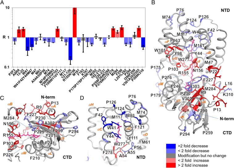
Local structural changes in the OCP upon activation monitored by XF-MS. (A) Solvent accessibility changes in response to photoactivation measured by the ratio (R) of hydroxyl radical reactivity rates for residues obtained from the dose–response plot (Fig. S5B). The rate for each site indicated is summarized in Table S5. The coloring indicates changes in rates of modifications upon transition from OCPO to OCPR. Error bars represent the SE from three independent measurements. (B) Modified residues are represented by sticks on the X-ray crystal structure of OCPO (PDB ID code 3MG1) (5). The carotenoid is shown in pink sticks. Color codes as in A indicate the changes in solvent accessibility upon photoactivation. (C) Solvent accessibility changes at the major and minor interface, showing possible destabilization at several interdomain H-bonding and ionic interaction sites. The color codes are the same as in A. (D) Solvent accessibility changes in the NTD, showing possible stabilization of the shifted carotenoid by conserved aromatics residues, methionine residues, and the loop joining the two four-helix bundles of the NTD. The color codes are the same as in A.
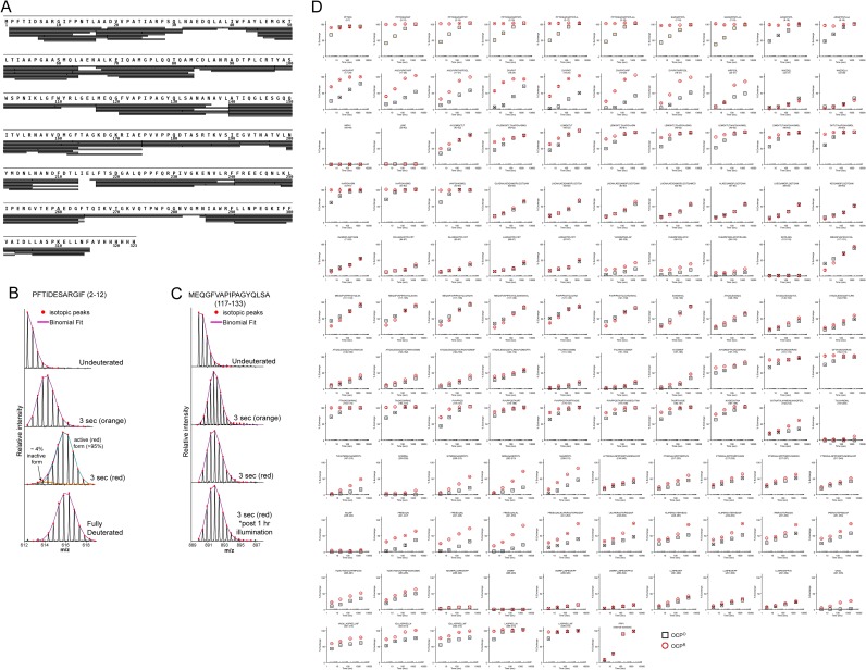
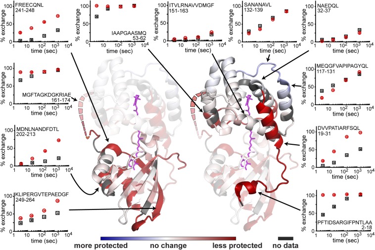
HDX-MS, in contrast, uses the natural exchange of protons at backbone amide positions to probe amide proton accessibility (14, 15). Amides involved in hydrogen bonds through stable secondary structure, or that are substantially solvent-occluded, exchange much slower than accessible ones. Pepsin digestion yielded 116 observable fragments, resulting in a net sequence coverage of 98% (Fig. S6A). Differences in amide exchange between OCPo and OCPR are summarized in Fig. 3. Additional controls were performed to ensure that the light-activated OCP measured by HDX-MS was fully activated OCPR and that the level of continuous illumination did not damage the protein (Fig. S6 B and C).
Fig. S6.
(A) Peptic coverage map of OCP. Each observable peptic peptide is shown as a gray bar under the primary sequence of OCP. The figure was made using HXMS tools (www.HXMS.com/mstools). (B) Mass spectra for residues 2–12 after 3 s of deuteration in the orange and red states. Binomial distributions used to fit the mass envelopes are shown in purple. For the OCPR dataset, there is a minor subpopulation of nonactivated OCPO. Based on fitting of two binomial distributions (orange and blue), this minor population corresponds to ∼4% of the total OCP. (C) Mass spectra for residues 117–133 after 3 s of deuterium exchange in the dark (“orange”), in the presence of blue light, and with 30 s of preillumination (“red”), or in the presence of blue light after 1 h of constant illumination (“post 1 hr illumination”). The identical spectra in the last two panels indicate that even extensive illumination did not disrupt the protein structure. (D) HDX-MS for all observable peptides. The uptake plots for all peptides of OCPO (orange squares) and OCPR (red circles) are shown. The average (mean) percent deuterium exchange is shown relative to the fully deuterated and “zero” standards (SI Methods). Error bars show SD between duplicate measurements. The PPPI internal standard showing identical exchange conditions between the two samples is the last plot at the bottom.
Fig. 3.
Local structural changes in the OCP upon light activation as measured by HDX-MS. The changes in solvent accessibility upon light activation are plotted on the OCP crystal structure (PDB ID code 3MG1) (5) showing regions becoming less protected from exchange (red) and more protected from exchange (blue). The carotenoid is shown in magenta sticks. Residues 164–170, not modeled in the crystal structure, are shown as dashed lines. Individual exchange plots for peptides of interest in OCPO (black squares) and OCPR (red circles) are shown on the periphery with their corresponding location in the structure indicated by arrows. Exchange data reflect the average from duplicate measurements with error bars showing the SD. Exchange plots for all observed peptides are shown in Fig. S6D.
We observed large increases in solvent accessibility by XF-MS in two regions of the protein: first, at the major interface between the NTD-CTD and, second, at the minor interface of the N-terminal helix and CTD (Fig. 2) (5). At the major interface, the largest increase (10-fold) was observed at the conserved NTD residue R155, which forms a salt bridge with E244 on the CTD in the OCPO (4, 5). We also observed 2- and 1.5-fold increases at absolutely conserved residues P276-W277-Y278 and the moderately conserved M284, respectively. All of these residues are involved in the stabilization of the carotenoid in the major interface (4, 5, 9). In addition, residue W277 H-bonds with N104 on the NTD in the OCPO (5, 10). Upon photoactivation to OCPR, the NTD peptide 97–106 showed a cluster of modifications at Y98-W101-P103 that resulted in an overall twofold increase in accessibility. By HDX-MS, residues 241–248 proximal to the major interface showed increased accessibility in OCPR. In contrast, the NTD helix αI, containing R155, remained protected from exchange upon photoactivation (Fig. 3) and the peptides within NTD helices αF and αG, which include N104, and the CTD β3 sheet, which includes W277, showed only minor changes in the deuterium exchange kinetics. Together, these results suggest that a disruption of the side-chain interactions at the major interface, but without measurable local unfolding of secondary structure (helices), leads to formation of OCPR.
At the minor interface, NTD residues A8–R9 and 13–22 on the loop joining αA to αB showed a significant increase in accessibility in OCPR relative to OCPO by XF-MS, commensurate with the corresponding residues V301-A302-I303 on the surface of the β-sheet facing helix αA. Previous chemical footprinting experiments have also shown increased access to the minor interface, suggesting helix dissociation (11). By HDX-MS, large changes were also observed within the N-terminal helix (residues 2–18), which became completely exchanged within 3 s in OCPR. This rapid exchange shows that the N-terminal helix (αA) not only dissociates from the CTD but becomes disordered upon photoactivation (Fig. 3 and Fig. S6D). The unfolding of this helix is consistent with the slight loss of helical structure predicted by comparison of the far-UV CD spectrum of OCPR versus that of OCPO (Table S3). Interestingly, even in OCPO, this helix exchanges considerably faster than other helical segments, suggesting that this region is somewhat dynamic and only loosely associated with the CTD in the OCPO. Additionally, HDX-MS also showed that residues 19–31 adjacent to the N-terminal helix become less protected from exchange in OCPR (Fig. 3). The combination of CD and HDX-MS demonstrates that activation of the OCP results in disruption of the minor interface, the interaction between the N-terminal α-helix and the solvent-exposed β-sheet of the CTD, via the unfolding of the αA (the N-terminal helix). The critical role of the αA helix in OCP photochemistry and function is consistent with a recent mutational analysis that implicated changes in this helix as important to the interaction of the OCP with the PB and the FRP (24). A similar disruption of a helix:domain interaction upon activation was observed in other photoactive proteins, including PYP, BLUF, and LOV (5, 10). BLUF and LOV domain β-scaffolds are well known for their involvement in both intra- and interprotein interactions (25), and we note that previous in silico docking experiments between the CTD of the OCP and the FRP implicated β-scaffold residues (including V232, D220, and F299) in the CTD–FRP interaction (8). Accordingly, disruption of the minor interface seems to mediate both an intraprotein (domain dissociation) response and a putative interprotein interaction (with the FRP) that collectively control OCP’s photoprotective function.
Local Structural Changes in the Carotenoid Pocket in the OCP upon Light Activation.
The recently reported crystal structure of a constitutively active NTD [Synechocystis “red carotenoid protein” (RCP) expressed in and isolated from Escherichia coli] revealed a 12-Å translocation of the carotenoid into a previously unknown binding configuration in the NTD (9). Additional evidence for light-dependent carotenoid translocation was provided by mutational analysis and XF-MS analysis of carotenoid binding residues in E. coli-expressed OCP (binding a mixture of ECN and CAN). Our results support carotenoid translocation in Synechocystis-expressed OCP-ECN after photoactivation (Fig. 2): a greater than twofold decrease in solvent accessibility at residue cluster W41-Y42-Y44 in the NTD was observed concomitantly with a greater than twofold increase in solvent accessibility in CTD residue cluster P276-W277-F278, consistent with previously reported results (9). In this study, complementary HDX-MS data on OCP-ECN showed that, whereas some regions are moderately less protected from exchange in OCPR, the local secondary structure remains largely intact near these carotenoid-binding residues. The combined XF- and HDX-MS data thus solidify the assignment of reciprocal solvent accessibility changes at these sidechains to a reconfiguration of carotenoid-sidechain interactions. Similar reciprocal changes are observed in the light-dependent XF-MS data for carotenoid binding residues in the NTD of OCP-ECN (less than twofold accessibility decrease at M47, M74-P76-M83, and M117) and CTD (less than twofold accessibility increase at M284 and F290). Notably, the OCPO carotenoid-binding residue M202, which is buried in the hydrophobic core of the CTD, showed no light dependent change in accessibility.
We obtained previously unidentified evidence supporting carotenoid translocation at the linker region (residues 120–131), which connects the two four-helix bundles of the NTD between helices αG and αH. XF-MS showed the largest decrease (greater than fivefold) in accessibility at the NTD residue cluster F121-P124-P126 after photoactivation. HDX-MS data were consistent with these results because the area around the αG-αH was the only region that was more protected from exchange in OCPR (Fig. 3). Based on the structure of the isolated NTD (9), the increased protection is likely attributable to the ligand shifting into direct contact with the loop and thereby stabilizing the local structure in this region (Fig. 2D).
We observed reciprocal solvent accessibility changes in two structurally proximal regions at the major interface: first, in the loop joining β1 and αM in the CTD (P225-P226-F227 & P230; 1.7-fold decrease) and adjacent residues in the NTD (R9, P22, and N156; twofold increase) (Fig. 2C); second, in the loop joining αC and αD, which joins the two four-helix bundles in NTD (A54-A55-P56; 1.3-fold decrease), and the adjacent residue in CTD (W277; twofold increase) (Fig. 2D). Similar reciprocal changes were previously described for a membrane-embedded helix and proposed to involve helix reorientation as well as movement of internal water (26). In the Synechocystis OCP, the residues involved in this reciprocal interaction are highly conserved and may play an important role in structural stabilization of the hydrophobic core of the CTD after the carotenoid translocation as well as its stabilization in NTD.
In contrast, relatively few changes upon illumination were observed in the accessibility of residues surrounding the carotenoid in the CTD. As noted above, HDX-MS showed that, with the exception of the αA-helix, the secondary structure remains largely intact within OCPR. Side-chains located on helices αK and αL, which project toward the core of the CTD (peptide 192–215), showed detectable modification in both OCPO and OCPR but did not undergo any change in accessibility. Residues Y201 and W288 (no modification) were absolutely conserved and participated in H-bonding to the keto group of the carotenoid in OCPO (5). Residue F240, projecting toward the β-ionone ring of the carotenoid, and F241, projecting toward the core of the CTD, showed no modification in either OCPO or OCPR. Collectively, these results suggest that no major structural changes occur within the carotenoid binding pocket of the CTD upon photoactivation. The translocation of the carotenoid into the NTD upon photoactivation presumably generates a void space in the CTD, and the protection from hydroxyl radical-mediated modification may be explained by the loop joining β1 and αM, limiting the accessibility of the CTD core. The decrease in the accessibility of the loop residues (P225-P226-F227 and P230) is consistent with its capping of the CTD in OCPR.
The Role of Bound Water and H-Bonding Networks in Carotenoid-to-Protein Signal Propagation.
It is known that bound water molecules are important for protein structural stabilization, ligand binding, and conformational changes (27–29). XF-MS generates hydroxyl radicals by activating bulk as well as protein-bound water, which can react directly with proximal side-chains (13, 30). In contrast to bulk water, protein-bound water (identified by X-ray crystallography) shows a longer residence time in solution and facilitates a significantly higher yield of hydroxyl radical modifications at reactive residues (23, 31). XF-MS has been used to probe the positions of internal water molecules, changes in the H-bonding networks involving bound water, and the dynamic role of internal water in hydrophobic cavities inside protein cores (23, 26, 31–33). In addition to carotenoid translocation (9), our data demonstrate two other structural changes upon photoactivation of the OCP: unfolding and release of the N-terminal helix from the CTD surface and complete dissociation of the NTD and CTD. Our XF-MS analysis detects associated solvent accessibility changes at the functionally important side-chains in these regions that are in close proximity to the bound water. Accordingly, it is plausible that signal propagation occurs from the light-activated carotenoid to the protein surface via internal hydrogen bonding networks and involves bound water molecules.
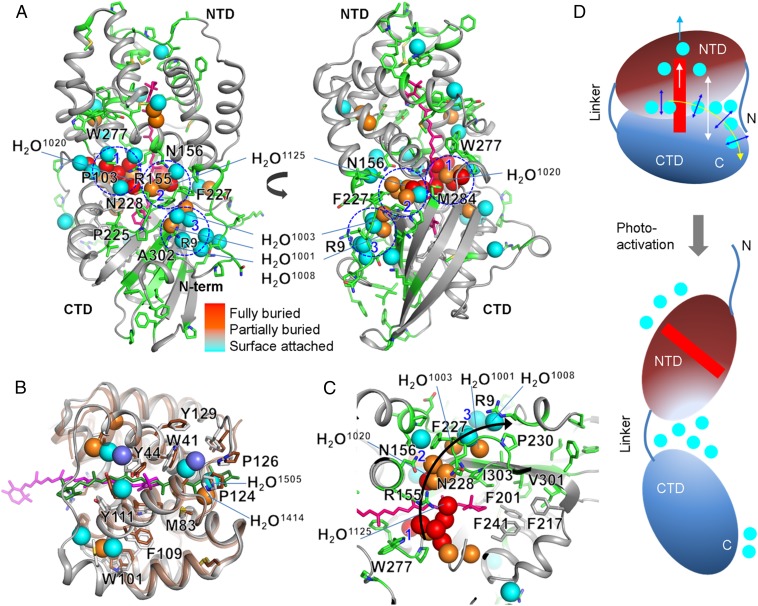
Comparison of the crystal structures of Synechocystis and Arthrospira maxima OCPO reveals three clusters of water molecules in both structures at the major and minor interfaces (Fig. 4); these waters are conserved and/or are within 0.5 Å of each other in both crystal structures (Fig. S1 and Table S6). Waters HOH1020 and 1125 are at the center of two clusters of water molecules (labeled 1 and 2) at the major interface within 3.9 Å of the carotenoid (Fig. 4 A and B, Fig. S1, and Table S6). R155, which participates in a salt bridge between the NTD and CTD, and which plays an important role in interaction with the phycobilisome during energy dissipation (12), is associated with water cluster 1, which includes HOH1020 (Fig. 4 and Fig. S1F). These water molecules participate in a constellation of residue–water interactions: R155-HOH1151-W277, R155-HOH1200-HOH1044-S102/P103, W277-N104-HOH1044/HOH1200-R155, and T275/P276-HOH1020. As noted above, most of these conserved residues showed large increases in accessibility upon photoactivation, implying a concomitant change in the water–protein network after domain separation. Also at the major interface, water cluster 2 connects M284, P226, F227, and N156, all of which undergo changes in side-chain accessibility upon photoactivation. Water cluster 3, at the minor interface, consists of structurally conserved and partially buried water molecules (HOH1001, HOH1003, and HOH1008) and connects A302-I303 of β5 to R9 of the N-terminal helix αA, and F227-P226 of the loop joining β1 and αM (Fig. 4C) facing the minor interface. In addition, residues F227–P226 and the adjacent Q228 of the same loop are directly connected to water cluster 2 at the major interface. These water clusters and adjacent residues may form a pathway (water cluster 1 > R155-N156 of NTD > water cluster 2 > F227/P226 of CTD > water cluster 3) (Fig. 4C) and may even provide a means of communication from the carotenoid binding pocket to the surface of the protein, while concomitantly playing a role in domain dissociation. The alternate and reciprocal (increase-decrease-increase) accessibility along the major interface, the loop joining β1 and αM, and the minor interface indicate a continuous movement/displacement of water molecules, as well as reorganization of the H-bonding network that drives domain dissociation after the carotenoid translocation.
Fig. 4.
Water network for carotenoid signaling in OCP. (A) Modified residues and the carotenoid are represented by green and pink sticks, respectively, on the X-ray crystal structure of OCPO from Synechocystis (PDB ID code 3MG1). The spheres represent water molecules, which are either conserved (labeled) or located at approximately the same position (within 0.5 Å) in both the crystal structure of A. maxima (4) and Synechocystis OCP (5). The coloring of the waters indicates their depth from the surface of the OCP (see Fig. S1 C–E for additional details). Three major water clusters, at the minor and major interfaces, are marked by blue dashed lines and numbered. These waters potentially form internal H-bonding contacts with side chains, amide backbone, and other water molecules (Fig. S1F and Table S6). Residues that undergo the highest degree of solvent accessibility change and are important for the H-bonding network at the major and minor interface are labeled. (B) Superimposed structure of the constitutively active RCP (protein in brown, carotenoid in green, buried water as violet spheres) (9) and Synechocystis OCPO (gray, carotenoid in pink, conserved and buried water colored as in A) (5). Residues that are proposed to stabilize the carotenoid in the activated state are labeled. (C) Proposed signal propagation pathway from the carotenoid through the water-side chain H-bonding network to the protein surface that facilitates carotenoid shift, dissociation of NTD-CTD, and detachment of the N-terminal helix from CTD. (D) Schematic of the OCPO and OCPR showing regions with the largest conformational rearrangement associated with the change in H-bonding network and water rearrangements.
Table S6.
List of water molecules that are either conserved or found to be at the same location (within 0.5 Å) in the crystal structures of A. maxima and Synechocystis OCP
| Water name in PDB ID code 3MG1 | B Factor | Nearest residue(s) that can form H bond | Distance, Å | Corresponding H2O in PDB ID code 1M98 | Location and water cluster (WC) |
| HOH 1001* | 19.6 | I303-O† | 2.8 | HOH451 | Minor interface /WC3 |
| HOH1042-O | 2.7 | ||||
| HOH 1003* | 21.4 | HOH1042-O | 3.8 | HOH420 | Minor interface /WC3 |
| L105-O | 3.7 | ||||
| L105-N | 2.8 | ||||
| HOH 1008* | 21.3 | R9-NH2 | 4.0 | HOH490 | Minor interface /WC3 |
| R9-O | 2.8 | ||||
| HOH 1019 | 6.8 | ‡ | ‡ | HOH526 | CTD |
| HOH 1020* | 16.4 | T275-OG1† | 2.9 | Cl403 | Major interface /WC1 |
| HOH 1023 | 8.9 | ‡ | ‡ | HOH500 | CTD |
| HOH 1042 | 22.2 | F227-N† | 2.9 | HOH442 | Minor interface /WC3 |
| HOH1001 | 2.7 | ||||
| P225-O† | 3.8 | ||||
| HOH1003-O | 3.7 | ||||
| HOH 1044 | 19.0 | N104-ND2† | 3.8 | HOH519 | Major interface /WC1 |
| N104-N† | 3.0 | ||||
| HOH1200-O | 2.8 | ||||
| E243-O | 2.9 | ||||
| HOH 1046 | 17.6 | S102-OG§ | 2.7 | HOH506 | Major interface /WC1 |
| E243-O | 3.0 | ||||
| HOH 1060* | 25.1 | Y44-OH | 2.7 | HOH432 | NTD |
| HOH 1061 | 1.0 | ‡ | ‡ | HOH447 | CTD |
| HOH 1070 | 22.5 | ‡ | ‡ | HOH614 | NTD |
| HOH 1081 | 0.1 | ‡ | ‡ | HOH447 | CTD |
| HOH 1084 | 20.9 | T15-O† | 2.9 | HOH663 | Minor interface /WC3 |
| D304-OD2† | 3.8 | ||||
| HOH1395-O | 2.7 | ||||
| HOH 1109 | 34.0 | ‡ | ‡ | HOH699 | NTD |
| HOH 1125* | 20.0 | M284-SD | 3.6 | HOH410 | Major interface /WT2 |
| HOH1342-O | 2.7 | ||||
| HOH 1128 | 21.9 | N227-NE2 | 2.7 | HOH676 | Major interface /WC2 |
| HOH 1142 | 15.9 | E311-OE1† | 2.8 | HO438 | Near minor interface /WC3 |
| K268-NZ† | 2.8 | ||||
| HOH 1147 | 24.9 | Y101-O | 2.8 | HOH169 | Near P103 at the major interface and water cluster 1 |
| HOH 1151 | 19.1 | N104-OD1† | 2.9 | HOH509 | Major interface /WC1 |
| R155-NE† | 3.2 | ||||
| W277-NE1† | 3.7 | ||||
| HOH 1164 | 22.3 | ‡ | ‡ | HOH558 | NTD |
| HOH 1200 | 18.1 | R155-NH2† | 3.5 | HOH460 | Major interface /WC2 |
| N104-OD1† | 3.3 | ||||
| HOH 1203 | 18.7 | ‡ | ‡ | HOH408 | Others |
| HOH 1218 | 16.7 | HOH1671-O | 2.6 | HOH412 | Major interface /WC1 |
| HOH1020-O | 3.2 | ||||
| HOH1200-O | 3.6 | ||||
| E244-O | 2.9 | ||||
| HOH 1259 | 21.3 | HOH1268-O | 2.9 | HOH512 | Near P103 at the major interface and water cluster 1 |
| HOH 1285 | 36.2 | ‡ | ‡ | HOH640 | NTD |
| HOH 1290 | 21.6 | D304-OD1† | 2.7 | HOH478 | Minor interface /WC3 |
| HOH1003-O | 2.9 | ||||
| HOH1042-O | 2.8 | ||||
| HOH 1311 | 5.8 | ‡ | ‡ | HOH590 | Near P179 of the linker |
| HOH 1313 | 14.5 | E244-O | 3.0 | HOH473 | Major interface /WC1 |
| HOH 1317 | 20.2 | ‡ | ‡ | HOH501 | Others |
| HOH 1342 | 20.8 | HOH1128-O | 3.4 | Major interface /WC2 | |
| HOH1391-O | 3.0 | ||||
| HOH 1391 | 23.5 | N156-ND2† | 3.0 | HOH685 | Major interface /WC2 |
| N156-OD1† | 3.5 | HOH474 | |||
| HOH 1395 | 19.1 | HOH1084-O | 2.7 | HOH507 | Minor interface /WC3 |
| T15-OG1† | 3.9 | ||||
| I11–O† | 3.3 | ||||
| HOH 1448 | 39.6 | ‡ | ‡ | HOH550 | NTD |
| HOH 1467 | 27.7 | N156-ND2† | 2.9 | HOH695 | Major interface /WC2 |
| F227†, P22† | |||||
| HOH 1491 | 23.3 | ‡ | ‡ | HOH469 | NTD |
| HOH 1610 | 22.3 | N228-OE1 | 3.0 | HOH583 | Major interface /WC2 |
| HOH 1628 | 22.3 | HOH1259-O | 2.9 | HOH520 | Near P103 at the major interface and water cluster 1 |
| HOH 1664 | 35.3 | ‡ | ‡ | HOH558 | NTD |
| HOH 1671 | 18.6 | R155-NH2† | 3.1 | HOH629 | Major interface /WC1 |
The nearest neighboring atom on the amino acid residue that can form H bonding with these water molecules, is determined from the crystal structure using PyMOL v1.2r3pre (Schrödinger, LLC).
Conserved water reported in OCP structures.
Absolutely conserved amino acid residue.
The nearest residue that can form H bond are not shown, and the distance is not measured. The water molecule is located either at the opposite side of major interface or NTD. This water molecule is not linked to the WC, and can play a role in structural stabilization.
Moderately conserved.
To summarize, the combination of structural MS methods with CD and SAXS provides both local and global structural insight into the structure of the OCPR, which has remained recalcitrant to crystallization. We have defined the specific changes in interactions at the major NTD–CTD interface, and, critically, SAXS results have confirmed that complete separation of the NTD and CTD occurs in the OCPR form. Domain separation is predicated on carotenoid translocation, as well as unfolding of the loosely associated N-terminal helix and its dissociation from the CTD (Fig. 4D). We also demonstrate that a change in the oligomeric state (i.e., dimerization) is not required for photoactivation and that the secondary structural content in each individual domain remains largely intact during the photoactivation process, with the exception of the α1-helix. Collectively, these results offer insight that will aid efforts to investigate the interactions of OCPR with its binding partners in the course of regulating photoprotection, such as cross-linking studies (18) and in silico modeling of protein–protein interactions (8, 9, 18). These findings will also enable more accurate modeling of the OCP–PB complex, a major step in delineating the mechanisms of the complete photoprotective system.
SI Methods
Protein Purification.
OCP was purified from an OCP overexpressing mutant strain of Synechocystis PCC 6803 (a gift of Diana Kirilovsky, Commissariat à l'Energie Atomique, Institut de Biologie et Technologies de Saclay, Gif-sur-Yvette, France) using a procedure similar to that described previously (7). In the Synechocystis PCC 6803 strain used for protein expression, the CrtR gene (responsible for carotenoid hydroxylation) is interrupted by an antibiotic resistance cassette, and its OCP is known to bind predominantly ECN (34). Modifications to the previously described OCP purification procedure included the following: (i) lysis by bead-beating (9 × 20-s cycles at 0 °C), (ii) intermediate purification by ion-exchange chromatography (IEC) on a HiPrep Q-Sepharose HP 16/10 column (GE Healthcare), and (iii) a final gel filtration purification step after IEC using a Superdex 75 16/60 SEC column and 50 mM Tris⋅HCl (pH 7.5), 200 mM NaCl running buffer. Sample purity was qualified by SDS/PAGE and the UV-visible absorbance spectrum of the purified holoprotein. OCP was buffer exchanged by gel filtration before XF-MS and HDX experiments using a Superdex 75 10/300 GL column (GE Healthcare) preequilibrated with 20 mM potassium phosphate, 100 mM NaCl (pH 7.4).
Analytical SEC.
Analytical SEC was performed on an AKTA Pure M2 chromatography system (GE Healthcare) with triple wavelength absorbance detection. A set of globular standards (cat. no. 151-1901; Bio-Rad) was used to calibrate the column for MW estimations by determining the best linear fit to a plot of partition coefficient versus log(MW) for three globular standards in the linear fractionation range of the Superdex 200 10/300 GL column. Identical running conditions (100-μL injection volume, 0.75 mL/min flow rate, 4 °C run temperature) were used for both OCP samples and standards in all buffers, and a separate standard calibration curve was calculated in each buffer. For SEC runs of OCPR, OCP was preilluminated for 5 min on ice with a blue LED lamp (∼470 nm λmax, three LEDs per lamp, Luxeon Rebel LXML-PB01-0030 LED; Lumileds).
Circular Dichroism.
Measurement of far-UV CD spectra.
OCP samples were exchanged into 20 mM potassium phosphate, 100 mM NaF (pH 7.4) by gel filtration on a Superdex 75 10/300 GL column before CD measurements. Sample concentrations ranged from 0.10 to 0.17 mg/mL for four independent replicates, and concentrations were determined using the maximum extinction coefficient of ECN (118,000 M-1⋅cm−1) (35) and the known 1:1 binding stoichiometry of ECN:OCP. OCP CD spectra in the far-UV range were collected in a 1-mm pathlength quartz cuvette using a Chirascan-plus spectropolarimeter (Applied Photophysics). Individual scans were collected in the 260- to 185-nm range with 0.5-nm steps, 0.5 s per point, and 1-nm spectral bandwidth. Smoothed spectra were obtained by averaging eight scans, subtracting a smoothed background spectrum, and the application of a Savitsky–Golay filter to the averaged data. Units of ellipticity (in millidegrees) were converted to mean-residue-ellipticity as described in ref. 36. The OCPR CD spectrum was collected after illumination (5 min with a blue LED at 0 °C) of the same sample/cuvette used for the OCPO spectrum acquisition. UV-visible absorption spectra collected before and after collection of the OCPR CD dataset confirmed that the sample remained predominantly in the OCPR state during the time necessary to make the CD measurement (Fig. S2E). After OCPR CD measurements, the sample was allowed to thermally revert to OCPO at 24 °C. UV-visible absorption (Fig. S2E) and far-UV CD spectra collected after thermal reversion confirmed that the light-induced changes observed by CD were largely reversible.
Secondary structure estimation.
Estimations of protein secondary structure were performed using CDSSTR, CONTINLL, and SELCON3 packages in the CDPro program suite (17) using five different reference sets of soluble protein CD spectra. Analyses were performed on averaged and smoothed CD spectra (OCPR and OCPO) from four independent experiments.
SAXS Data Collection and Analysis.
SAXS measurements were conducted on beamline 4-2 at the Stanford Synchrotron Radiation Lightsource. The focused 11-keV X-ray beam irradiated a thin-wall quartz capillary cell, placed at 1.7 m upstream of the Rayonix MX 225HE detector. One hundred microliters of OCP (1.3 mg/mL) were injected onto a high resolution Sepharose 200 column (GE Healthcare) with a flow rate of 50 μL/min in 20 mM Na3PO4 (pH 7.4), 150 mM NaCl, 0.02% NaN3, 1 mM EDTA. The flow from the column passed through a UV detector cell, followed by 15 cm of clear tubing leading to the inlet of the quartz capillary cell. As the flow passed the quartz capillary, 1-s exposures were collected every 5 s throughout the run, with a circulating water bath maintaining the capillary cell temperature at 8 °C. The detector pixel numbers were converted to the momentum transfer q = 4p*sinq/l, where 2*q is the scattering angle and l the X-ray wavelength of 1.127 A°, using a silver behenate powder standard placed at the capillary position (37). A background scattering curve was obtained from the first 100 exposures (before the void volume), which was subtracted from all subsequent exposures during the elution profile. Rg and I(0) for each frame were batch-analyzed using autoRg (38), and frames with stable Rg values were merged in PRIMUS (39) for the final scattering curve.
The inactive OCP dataset was collected by wrapping all clear lines from the column in aluminum foil in the absence of any background light in the hutch. An additional dataset was collected bypassing the UV detector to ensure that the UV cell was not causing any activation of OCP (Fig. S3D). Activated OCP was measured by placing one blue LED to illuminate the clear tubing leading to the capillary and a second blue LED to illuminate the capillary just before the beam path. The duration of illumination with this setup was ∼20–25 seconds, and there was only 1–2 s between illumination and X-ray exposure. The half-life of reversion of OCPR to OCPO is ∼30 s at 22 °C and much slower at lower temperatures (7); therefore, no significant reversion should occur before X-ray exposure. The LEDs were placed either 20 cm away (weak illumination), 7 cm away (medium illumination), or 2 cm away (strong illumination) from the lines and capillary. The LEDs were on for collection of the background scattering signal and turned on 5 min before elution of OCP from the column to minimize the heating of the lines that can occur over longer illumination times.
The particle distance distribution function [P(r)] plot was calculated from the merged scattering data using GNOM (40). The transformed data were subsequently used for generating ensembles of ab initio shape reconstructions using DAMMIN v5.3 (21) or GASBOR22i (20). The 12 calculated bead models were very consistent with the scattering data as reflected by low χ values (DAMMIN, 1.14 ± 0.01, 1.25 ± 0.02; GASBOR, 1.15 ± 0.02, 1.31 ± 0.03, for OCPO and OCPR, respectively). The bead models were aligned using SUPCOMB13 with enantiomers considered (41), and spatially filtered using DAMFILT (42). The normalized spatial discrepancy (NSD) value for each ensemble was below 2, indicating good consistency between the individual bead models (DAMMIN, 0.64 ± 0.02, 0.55 ± 0.01; GASBOR, 1.23 ± 0.03, 1.38 ± 0.07, for OCPO and OCPR, respectively). Domains of OCP were manually docked into the reconstructed shape volumes using Chimera (43).
Coral (ATSAS package) was used for rigid body modeling of the SAXS data for OCPR. The structure of the isolated N-terminal domain (9) (residues 18–165) and C-terminal domains (residues 185–306 from PDB ID code 3MG1) were treated as rigid bodies. The N termini (2–17), C terminus with the His tag (307–322), and linker region (166–184) were treated as flexible portions. Ten calculations were performed with the default Coral settings to sample various domain and linker orientations.
X-Ray Radiolysis and Mass Spectrometry.
OCPECN (expressed in Escherichia coli) was exchanged into 20 mM potassium phosphate (pH 7.4), 100 mM NaCl by SEC on a Superdex-75 10/300 GL column before XF-MS experiments. Before X-ray irradiation of OCPR, the sample syringe was cooled with an ice pack and illuminated with a blue LED array (470 nm Luxeon Rebel; Philips Lumileds). Protein samples were irradiated in the microsecond time range at beamline 5.3.1 at the Advanced Light Source as reported previously (30). All samples, including the control (no X-ray irradiation), were subjected to cys-alkylation and de-salting before overnight trypsin and endoproteinase GluC digestions at pH 8 and 37 °C. Proteolyzed samples were analyzed in an Agilent 6550 iFunnel Q-TOF mass spectrometer (Agilent Technologies) coupled to an Agilent 1290 LC system (Agilent) using Sigma-Aldrich Ascentis Peptides ES-C18 reverse phase column (2.1 mm × 100 mm, 2.7-μm particle size; Sigma-Aldrich). The concentrations of the components in the gradient are measured as vol/vol. Approximately 10 pmol of samples were loaded onto the column via an Infinity Autosampler (Agilent) with buffer A (2% acetonitrile, 0.1% formic acid) flowing at 0.400 mL/min. The peptides were separated and eluted into the mass spectrometer via a gradient with initial condition of 5% buffer B (98% acetonitrile, 0.1% formic acid) increasing to 70% B over 15 min. Subsequently, B was increased to 90% over 1 min and held for 3 min at a flow rate of 0.6 mL/min, followed by a ramp back down to 5% over 1 min, where it was held for minutes to reequilibrate the column to the original condition. Peptides were introduced to the mass spectrometer from the LC using a Jet Stream source (Agilent) operating in positive-ion mode (3,500 V). The data were acquired with MassHunter B.05.00 operating in Auto MS/MS mode whereby the three most intense ions (charge states 2–5) within 300 m/z to 1,400 m/z mass range above a threshold of 1,000 counts were selected for MS/MS analysis. MS/MS spectra were collected with the quadrupole set to “Narrow” resolution and collision energy to optimize fragmentation. MS/MS spectra were scanned from m/z 100–1,700 and were collected until 40,000 total counts were collected or for a maximum accumulation time of 333 ms. Parent ions were excluded for 0.1 min after MS/MS acquisition. MS/MS data of native and modified peptide fragments were interpreted by Mascot MS/MS Ions Search, as well as verified manually. The abundance of native and modified peptides at any irradiation time point area were measured (peak area) from their respective extracted ion chromatogram using Agilent Mass Hunter v 2.0.
The peak area from the extracted ion chromatograms of a specific peptide fragment with a particular mass-to-charge ratio and associated +16-, +32-, or +48-Da side-chain modifications was used to quantify the amount of modification at a given irradiation time. Increasing irradiation progressively reduces the fraction of unmodified products and provides a site-specific dose–response plot (as in Fig. S5B). The hydroxyl radical reactivity rate (k), which depends on both intrinsic reactivity and solvent accessibility, was obtained by fitting the dose–response to a single exponential decay (based on a pseudo-first order reaction scheme using Origin 7.5 (OriginLabs). The ratio (R) of the measured reactivity of the side chains residues between OCPO and OCPR (R = kOCPO/kOCPR) gave information on solvent accessibility changes independent of the intrinsic reactivity (Table S5).
Hydrogen–Deuterium Exchange and Mass Spectrometry.
Ten microliters of the OCP stock (0.5 mg/mL in PBS: 10 mM NaPO4, pH 7.0, 150 mM NaCl, 10 mM DTT) was exposed to the blue LED (described in Analytical SEC) at 25 °C at a distance of 7 cm for 30 s, at which point 90 μL of buffered D2O (PBS in 95% D2O) was added. The sample remained illuminated through the course of deuterium exchange, after which the sample was transferred to a tube on ice containing 100 μL of 0.4% formic acid with 0.15 mg/mL pepsin for a final pH of 2.5. The samples were digested on ice for 5 min, flash frozen in liquid nitrogen, and stored at −80 °C until LC-MS analysis. Sample heating from the LED at this distance was less than 1 °C over a 30-min time course. Undeuterated samples were prepared identically substituting D2O for optima LC-MS grade water. A fully deuterated sample was prepared by denaturing a solution of OCP at 0.5 mg/mL in 3 M GndHCl, heating at 85 °C for 30 min, diluting into D2O just as for the other samples, incubating for 1 h at 60 °C, then quenching just as for the other samples. A “zero” time point to correct for “IN exchange” was prepared by adding 10 μL of the OCP stock to prequenched D2O and digesting just as for the other samples. An internal tetrapeptide (PPPI) was included in all samples to ensure that both OCPO and OCPR were exposed to the same deuterium exchange conditions (last plot in Fig. S3) (44).
Deuterated samples were thawed and rapidly analyzed by LC-MS on a Waters Synapt Q-TOF mass spectrometer. Peptides were captured and desalted on a trap column for 3 min (Vanguard BEH, 1.7 μ 1 × 17 mm; Waters) and resolved on a 1 × 50-mm 2.1 μ C18 Hypersil gold Thermo using a linear gradient of 5–40% B in 8 min (A, 0.05% TFA, 5% acetonitrile; B, 0.05% TFA 80% acetonitrile). The injection loop, columns, and lines were kept under melting ice to minimize back-exchange. For peptide identification, data-dependent MS/MS scans of the peptic digests were acquired on an Orbitrap LTQ (Thermo). MS/MS data along with exact mass information were used to identify peptic fragments with the aid of protein prospector (prospector.ucsf.edu). Deuterium levels were analyzed using HX-Express v2 (45), and percent exchange for each time point (Dx) was calculated relative to the fully deuterated (DT) and zero (D0) standards [%D = (Dx − D0)/(DT − D0)].
Dynamic and Static Light Scattering.
All experiments were performed on a Dynapro (Wyatt Technologies) with 30 acquisitions of 10 s each at 4 °C, either in the absence of light (OCPO), or after 1 min of illumination on ice (OCPR). The cuvette and tubes were left on ice before measurements to maintain the sample at low temperatures to minimize reversion back to the dark state, which occurs only with a half-life of >45 min at this temperature (7).
Methods
OCP was purified from an OCP-overexpressing mutant strain of Synechocystis PCC 6803 (a gift of Diana Kirilovsky, Commissariat à l'Energie Atomique, Institut de Biologie et Technologies de Saclay, Gif-sur-Yvette, France) using a procedure described previously (5, 7) with modifications described in SI Methods. SAXS measurements were conducted on beamline 4-2 at the Stanford Synchrotron Radiation Laboratory, with an online SEC-SAXS setup with varying levels of blue LED illumination immediately before X-ray exposure. OCPO was converted to OCPR by blue LED illumination on ice before XF-MS measurements. Deuterium exchange reactions were conducted either in the absence of light or under constant illumination of the blue LED. XF-MS (13) measurements were made at the Advanced Light Source beamline 5.3.1 (30). Extensive details of the data collection for all methods are provided in SI Methods.
Acknowledgments
We thank Tsutomu Matsui and the support staff at the Stanford Synchrotron Radiation Lightsource (SSRL) for assistance with SAXS data collection, as well as Rich Celestre for help with data collection on Advanced Light Source beamline 5.3.1. This work was supported by National Institutes of Health (NIH) Grants F32-GM097805 and R01-GM099989 (to K.K.L. and M.G.) and Office of Science of the US Department of Energy (DOE) Grant DE-FG02-91ER20021 (to C.A.K., E.G.P., and R.L.L.). This work was supported in part by NIH Grant P30EB009998. The Advanced Light Source at Lawrence Berkeley National Laboratory is supported by the Director, Office of Science, Office of Basic Energy Sciences, US DOE under Contract DE-AC02-05CH11231. The SSRL Structural Molecular Biology Program is supported by the Department of Energy Office of Biological and Environmental Research and by NIH National Institute of General Medical Sciences Grant P41GM103393 and National Center for Research Resources Grant P41RR001209. This research used resources of the Joint BioEnergy Institute supported by the Office of Science, Office of Biological and Environmental Research, US DOE under contract DE-AC02-05CH11231.
Footnotes
The authors declare no conflict of interest.
This article is a PNAS Direct Submission.
See Commentary on page 12547.
This article contains supporting information online at www.pnas.org/lookup/suppl/doi:10.1073/pnas.1512240112/-/DCSupplemental.
References
- 1.Müller P, Li XP, Niyogi KK. Non-photochemical quenching: A response to excess light energy. Plant Physiol. 2001;125(4):1558–1566. doi: 10.1104/pp.125.4.1558. [DOI] [PMC free article] [PubMed] [Google Scholar]
- 2.Pascal AA, et al. Molecular basis of photoprotection and control of photosynthetic light-harvesting. Nature. 2005;436(7047):134–137. doi: 10.1038/nature03795. [DOI] [PubMed] [Google Scholar]
- 3.Niyogi KK, Truong TB. Evolution of flexible non-photochemical quenching mechanisms that regulate light harvesting in oxygenic photosynthesis. Curr Opin Plant Biol. 2013;16(3):307–314. doi: 10.1016/j.pbi.2013.03.011. [DOI] [PubMed] [Google Scholar]
- 4.Kerfeld CA, et al. The crystal structure of a cyanobacterial water-soluble carotenoid binding protein. Structure. 2003;11(1):55–65. doi: 10.1016/s0969-2126(02)00936-x. [DOI] [PubMed] [Google Scholar]
- 5.Wilson A, et al. Structural determinants underlying photoprotection in the photoactive orange carotenoid protein of cyanobacteria. J Biol Chem. 2010;285(24):18364–18375. doi: 10.1074/jbc.M110.115709. [DOI] [PMC free article] [PubMed] [Google Scholar]
- 6.Leverenz RL, et al. Structural and functional modularity of the orange carotenoid protein: Distinct roles for the N- and C-terminal domains in cyanobacterial photoprotection. Plant Cell. 2014;26(1):426–437. doi: 10.1105/tpc.113.118588. [DOI] [PMC free article] [PubMed] [Google Scholar]
- 7.Wilson A, et al. A photoactive carotenoid protein acting as light intensity sensor. Proc Natl Acad Sci USA. 2008;105(33):12075–12080. doi: 10.1073/pnas.0804636105. [DOI] [PMC free article] [PubMed] [Google Scholar]
- 8.Sutter M, et al. Crystal structure of the FRP and identification of the active site for modulation of OCP-mediated photoprotection in cyanobacteria. Proc Natl Acad Sci USA. 2013;110(24):10022–10027. doi: 10.1073/pnas.1303673110. [DOI] [PMC free article] [PubMed] [Google Scholar]
- 9.Leverenz RL, et al. A 12 Å carotenoid translocation in a photoswitch associated with cyanobacterial photoprotection. Science. 2015;348(6242):1463–1466. doi: 10.1126/science.aaa7234. [DOI] [PubMed] [Google Scholar]
- 10.Kirilovsky D, Kerfeld CA. The Orange Carotenoid Protein: A blue-green light photoactive protein. Photochem Photobiol Sci. 2013;12(7):1135–1143. doi: 10.1039/c3pp25406b. [DOI] [PubMed] [Google Scholar]
- 11.Liu H, et al. Mass spectrometry footprinting reveals the structural rearrangements of cyanobacterial orange carotenoid protein upon light activation. Biochim Biophys Acta. 2014;1837(12):1955–1963. doi: 10.1016/j.bbabio.2014.09.004. [DOI] [PubMed] [Google Scholar]
- 12.Wilson A, et al. The essential role of the N-terminal domain of the orange carotenoid protein in cyanobacterial photoprotection: Importance of a positive charge for phycobilisome binding. Plant Cell. 2012;24(5):1972–1983. doi: 10.1105/tpc.112.096909. [DOI] [PMC free article] [PubMed] [Google Scholar]
- 13.Xu G, Chance MR. Hydroxyl radical-mediated modification of proteins as probes for structural proteomics. Chem Rev. 2007;107(8):3514–3543. doi: 10.1021/cr0682047. [DOI] [PubMed] [Google Scholar]
- 14.Konermann L, Pan J, Liu YH. Hydrogen exchange mass spectrometry for studying protein structure and dynamics. Chem Soc Rev. 2011;40(3):1224–1234. doi: 10.1039/c0cs00113a. [DOI] [PubMed] [Google Scholar]
- 15.Marcsisin SR, Engen JR. Hydrogen exchange mass spectrometry: What is it and what can it tell us? Anal Bioanal Chem. 2010;397(3):967–972. doi: 10.1007/s00216-010-3556-4. [DOI] [PMC free article] [PubMed] [Google Scholar]
- 16.Putnam CD, Hammel M, Hura GL, Tainer JA. X-ray solution scattering (SAXS) combined with crystallography and computation: Defining accurate macromolecular structures, conformations and assemblies in solution. Q Rev Biophys. 2007;40(3):191–285. doi: 10.1017/S0033583507004635. [DOI] [PubMed] [Google Scholar]
- 17.Sreerama N, Woody RW. Estimation of protein secondary structure from circular dichroism spectra: Comparison of CONTIN, SELCON, and CDSSTR methods with an expanded reference set. Anal Biochem. 2000;287(2):252–260. doi: 10.1006/abio.2000.4880. [DOI] [PubMed] [Google Scholar]
- 18.Zhang H, et al. Molecular mechanism of photoactivation and structural location of the cyanobacterial orange carotenoid protein. Biochemistry. 2014;53(1):13–19. doi: 10.1021/bi401539w. [DOI] [PMC free article] [PubMed] [Google Scholar]
- 19.Fischer H, de Oliveira Neto M, Napolitano HB, Craievich AF, Polikarpov I. The molecular weight of proteins in solution can be determined from a single SAXS measurement on a relative scale. J Appl Cryst. 2010;43:101–109. [Google Scholar]
- 20.Svergun DI, Petoukhov MV, Koch MH. Determination of domain structure of proteins from X-ray solution scattering. Biophys J. 2001;80(6):2946–2953. doi: 10.1016/S0006-3495(01)76260-1. [DOI] [PMC free article] [PubMed] [Google Scholar]
- 21.Svergun DI. Restoring low resolution structure of biological macromolecules from solution scattering using simulated annealing. Biophys J. 1999;76(6):2879–2886. doi: 10.1016/S0006-3495(99)77443-6. [DOI] [PMC free article] [PubMed] [Google Scholar]
- 22.Petoukhov MV, et al. New developments in the ATSAS program package for small-angle scattering data analysis. J Appl Cryst. 2012;45(Pt 2):342–350. doi: 10.1107/S0021889812007662. [DOI] [PMC free article] [PubMed] [Google Scholar]
- 23.Gupta S, D’Mello R, Chance MR. Structure and dynamics of protein waters revealed by radiolysis and mass spectrometry. Proc Natl Acad Sci USA. 2012;109(37):14882–14887. doi: 10.1073/pnas.1209060109. [DOI] [PMC free article] [PubMed] [Google Scholar]
- 24.Thurotte A, et al. Regulation of Orange Carotenoid Protein activity in cyanobacterial photoprotection. Plant Physiol. 2015;169(1):737–747. doi: 10.1104/pp.15.00843. [DOI] [PMC free article] [PubMed] [Google Scholar]
- 25.Zoltowski BD, Gardner KH. Tripping the light fantastic: Blue-light photoreceptors as examples of environmentally modulated protein-protein interactions. Biochemistry. 2011;50(1):4–16. doi: 10.1021/bi101665s. [DOI] [PMC free article] [PubMed] [Google Scholar]
- 26.Gupta S, et al. Visualizing the kinetic power stroke that drives proton-coupled zinc(II) transport. Nature. 2014;512(7512):101–104. doi: 10.1038/nature13382. [DOI] [PMC free article] [PubMed] [Google Scholar]
- 27.Breiten B, et al. Water networks contribute to enthalpy/entropy compensation in protein-ligand binding. J Am Chem Soc. 2013;135(41):15579–15584. doi: 10.1021/ja4075776. [DOI] [PubMed] [Google Scholar]
- 28.Bhat TN, et al. Bound water molecules and conformational stabilization help mediate an antigen-antibody association. Proc Natl Acad Sci USA. 1994;91(3):1089–1093. doi: 10.1073/pnas.91.3.1089. [DOI] [PMC free article] [PubMed] [Google Scholar]
- 29.Ball P. Water as an active constituent in cell biology. Chem Rev. 2008;108(1):74–108. doi: 10.1021/cr068037a. [DOI] [PubMed] [Google Scholar]
- 30.Gupta S, Celestre R, Petzold CJ, Chance MR, Ralston C. Development of a microsecond X-ray protein footprinting facility at the Advanced Light Source. J Synchrotron Radiat. 2014;21(Pt 4):690–699. doi: 10.1107/S1600577514007000. [DOI] [PMC free article] [PubMed] [Google Scholar]
- 31.Angel TE, Gupta S, Jastrzebska B, Palczewski K, Chance MR. Structural waters define a functional channel mediating activation of the GPCR, rhodopsin. Proc Natl Acad Sci USA. 2009;106(34):14367–14372. doi: 10.1073/pnas.0901074106. [DOI] [PMC free article] [PubMed] [Google Scholar]
- 32.Gupta S, et al. Conformational changes during the gating of a potassium channel revealed by structural mass spectrometry. Structure. 2010;18(7):839–846. doi: 10.1016/j.str.2010.04.012. [DOI] [PMC free article] [PubMed] [Google Scholar]
- 33.Frankel LK, et al. Radiolytic mapping of solvent-contact surfaces in Photosystem II of higher plants: Experimental identification of putative water channels within the photosystem. J Biol Chem. 2013;288(32):23565–23572. doi: 10.1074/jbc.M113.487033. [DOI] [PMC free article] [PubMed] [Google Scholar]
- 34.Wilson A, Punginelli C, Couturier M, Perreau F, Kirilovsky D. Essential role of two tyrosines and two tryptophans on the photoprotection activity of the Orange Carotenoid Protein. Biochim Biophys Acta. 2011;1807(3):293–301. doi: 10.1016/j.bbabio.2010.12.009. [DOI] [PubMed] [Google Scholar]
- 35.Britton G, Liaaen-Jensen S, Pfander H, editors. Carotenoids Handbook. Birkhäuser; Basel: 2004. [Google Scholar]
- 36.Greenfield NJ. Using circular dichroism spectra to estimate protein secondary structure. Nat Protoc. 2006;1(6):2876–2890. doi: 10.1038/nprot.2006.202. [DOI] [PMC free article] [PubMed] [Google Scholar]
- 37.Smolsky IL, et al. Biological small-angle X-ray scattering facility at the Stanford Synchrotron Radiation Laboratory. J Appl Cryst. 2007;40:s453–s458. [Google Scholar]
- 38.Petoukhov MV, Konarev PV, Kikhney AG, Svergun DI. ATSAS 2.1: Towards automated and web-supported small-angle scattering data analysis. J Appl Cryst. 2007;40:s223–s228. [Google Scholar]
- 39.Konarev PV, Volkov VV, Sokolova AV, Koch MHJ, Svergun DI. PRIMUS: A Windows PC-based system for small-angle scattering data analysis. J Appl Cryst. 2003;36(5):1277–1282. [Google Scholar]
- 40.Svergun DI. Determination of the regularization parameter in indirect-transform methods using perceptual criteria. J Appl Cryst. 1992;25:495–503. [Google Scholar]
- 41.Kozin MB, Svergun DI. Automated matching of high- and low-resolution structural models. J Appl Cryst. 2001;34:33–41. [Google Scholar]
- 42.Volkov VV, Svergun DI. Uniqueness of ab-initio shape determination in small-angle scattering. J Appl Cryst. 2003;36:860–864. doi: 10.1107/S0021889809000338. [DOI] [PMC free article] [PubMed] [Google Scholar]
- 43.Pettersen EF, et al. UCSF Chimera: A visualization system for exploratory research and analysis. J Comput Chem. 2004;25(13):1605–1612. doi: 10.1002/jcc.20084. [DOI] [PubMed] [Google Scholar]
- 44.Zhang Z, Zhang A, Xiao G. Improved protein hydrogen/deuterium exchange mass spectrometry platform with fully automated data processing. Anal Chem. 2012;84(11):4942–4949. doi: 10.1021/ac300535r. [DOI] [PubMed] [Google Scholar]
- 45.Guttman M, Weis DD, Engen JR, Lee KK. Analysis of overlapped and noisy hydrogen/deuterium exchange mass spectra. J Am Soc Mass Spectrom. 2013;24(12):1906–1912. doi: 10.1007/s13361-013-0727-5. [DOI] [PMC free article] [PubMed] [Google Scholar]


