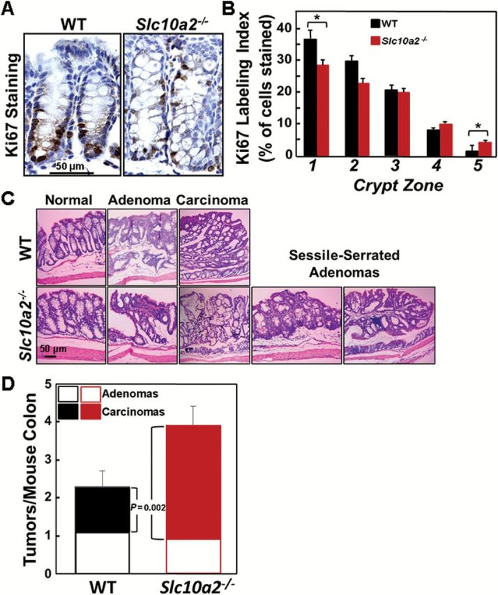
Figure 2.

Asbt deficiency promotes colonocyte proliferation and advanced colon neoplasia. (A) Representative images of Ki67 staining in colonic crypts from WT and Slc10a2 −/− mice. (B) Effect of Asbt deficiency on proliferation of normal colonocytes. The Ki67 labeling index was measured in normal crypts from the distal half of the colon. Crypts were divided into five equal zones, starting from the base (zone 1) and extending to the luminal surface (zone 5), and a minimum of 1000 crypt colonocytes was assessed in the colon from each mouse (N = 20 WT and 12 Slc10a2 −/− mice). Note increased Ki67 staining in upper portions of crypts in normal colons from Asbt-deficient mice. (*P = 0.05 compared to crypts from WT mice). (C) Representative hematoxylin and eosin-stained sections of normal colon, adenomas, adenocarcinomas and sessile-serrated adenomas from WT and Slc10a2 −/− mice. Sessile-serrated adenomas were observed only in colons from AOM/DSS-treated Slc10a2 −/− mice. (D) Asbt deficiency promotes progression from adenoma to adenocarcinoma. Bar graph shows distribution of colon adenomas and adenocarcinomas in WT and Slc10a2 −/− mice treated with AOM plus DSS. There was no difference in the numbers of adenomas between the two groups but the number of adenocarcinomas was significantly greater in Slc10a2 −/− compared to WT mice (P = 0.002). Error bars denote SEM.
