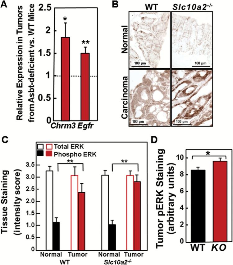
Figure 4.

Increased muscarinic and EGF receptor expression, and activation of post-receptor signaling in tumors from Slc10a2 −/− mice. (A) qRT-PCR of genes for type 3 muscarinic (Chrm3) and epidermal growth factor (Egfr) receptors. Expression of Chrm3 and Egfr was significantly increased in tumors from Slc10a2 −/− mice compared to those from WT mice (*P < 0.05, **P < 0.01; n = 9–11). (B) Representative images of phospho-ERK staining in normal colon tissue and adenocarcinomas from WT and Slc10a2 −/− mice. (C) Total ERK staining is unchanged but phospho-ERK staining is significantly increased in tumors compared to normal colon epithelium from WT and Asbt-deficient mice. Bars represent mean ± SEM for staining intensity score measured in 0.5 increments by a senior pathologist on a scale from 0, no staining to 3, maximal staining (**P < 0.01; n = 10–13). (D) Phospho-ERK (pERK) staining was significantly increased in tumors from Asbt-deficient (KO) mice compared to those from WT mice. Bars represent mean ± SEM for staining intensity expressed in arbitrary units (*P < 0.05; n = 10–13).
