


Abstract
It is widely acknowledged that cardiovascular heart disease (CHD) has a genetic influence. Several studies have investigated the role of inflammatory markers like C-reactive protein (CRP) and tumor necrosis factor α (TNF-α) in the causation of cardiovascular diseases. Although there have been several positive studies associating CRP and TNF-α genes with CHD, the evidence is not entirely consistent. Therefore, we performed a meta-analysis to gain a better understanding into this issue. The meta-analysis was conducted with 22 articles of genetic association studies of CRP (G1059C rs1800947, C1444T rs1130864, C717T rs2794521 and G3872A rs1205) and TNF-α (C857T rs1799724, C863A rs1800630 and T1031C rs1799964) genes. To analyze the association of these variants with CHD we used the following models: allelic, additive, dominant and recessive. In addition, we performed a sub-group analysis by Caucasian population using the same four models. CRP and TNF-α gene polymorphisms showed a positive significant association with CHD. This study provides evidence that rs2794521 of the CRP gene and rs1799724, rs1800630 and rs1799964 of the TNF-α gene polymorphisms may be risk factors to manifest CHD. The analysis of rs1800947 and rs1205 of the CRP gene yielded a protective effect in the pathogenesis of this disease. Only the analysis of the rs1130864 polymorphism showed a lack of association with CHD. To have conclusive outcomes it is necessary to integrate more studies to confirm our findings.
Keywords: Cardiovascular disease, polymorphisms, C-reactive protein, TNF-α, meta-analysis and systematic review
Introduction
The global burden of cardiovascular heart disease (CHD) represents the highest cause of mortality and one of the highest causes of morbidity around the world [1]. Several reports have explored a contribution in the pathogenesis of CHD considering environmental and intrinsic variables such as smoking, infections, age, obesity, lipid levels, blood pressure, among other causes, but nowadays there is growing evidence indicating a substantial genetic component [2,3]. Several studies have investigated the role of inflammatory markers like C-reactive protein (CRP) and tumor necrosis factor α (TNF-α) in the causation of cardiovascular diseases [4,5]. The human CRP gene is located on chromosome 1q21-1q23. It spans approximately 1,900 base pairs and contains 2 exons [6,7]. There are more than 30 single nucleotide polymorphisms (SNPs) of the human CRP gene listed (accessed on September 2014) in the National Center for Biotechnology Information (NCBI) SNP Database (http://www.ncbi.nlm.nih.gov/SNP). The most studied variant of the CRP gene is rs1800947 in which a change of guanine to cytosine at position 1059 has occurred. CRP polymorphisms have been related to several diseases [8-11]. The other CRP variant associated with CHD is rs1130864, where the change of cytosine to thymine at position 1444 has taken place [12-14]. With regard to polymorphisms rs2794521 and rs1205, these have been less studied in association studies with CHD, but similarly to the above mentioned polymorphisms they have yielded interesting outcomes [14-16]. The genetic change of rs2794521 occurs at position 717, where a cytosine is replaced by thymine; as for rs1205 a guanine is substituted by adenine at position 3872 [11,17]. With regard to the gene encoding TNF-α, it is located on chromosome 6p21 [18,19]. Some studies have reported that rs1799964, a specific TNF-α gene polymorphism, is associated with increased TNF-α secretion (Bennet et al., 2006; Cui et al., 2012; Ghazouani et al., 2009); in this variant T is substituted by C in the promoter region at position 1031. Other studies have shown that the TNF-α gene promoter region bears other polymorphisms, including C857T rs1799724 and C863A rs1800630, which are associated with CAD. Several studies have investigated whether the TNF-α gene polymorphisms that affect TNF-α expression or activity influences the susceptibility of cardiovascular disorders (Asifa et al., 2013; Bennet et al., 2006; Cho et al., 2013; Koch et al., 2001).
Although numerous studies have been conducted, this issue has not been settled conclusively. Some of these studies did not find an association between CRP and TNF-α with CHD, whereas other reports presented some evidence in favor of this hypothesis [20,21]. It is necessary to conduct larger and more detailed analyses to get more reliable assessment for the possibility of any moderate causal role. For this reason our aim was to perform a meta-analysis and updated systematic review with the following polymorphisms: G1059C rs1800947, C1444T rs1130864, C717T rs2794521 and G3872A rs1205 of the CRP gene, and C857T rs1799724, C863A rs1800630 and T1031C rs1799964 of the TNF-α gene to get a better understanding of the role these polymorphisms play in the pathogenesis of cardiovascular disease.
Materials and methods
The meta-analysis and systematic review were performed by following the Preferred Reporting Items for Systematic Reviews and Meta-Analyses (PRISMA) criteria [22,23].
Identification and selection of publications
The literature search was conducted using PubMed and EBSCO databases, using the following keywords: “C-reactive protein AND polymorphisms AND cardiovascular heart disease”, “C-reactive protein AND polymorphisms AND coronary heart disease”, “CRP AND polymorphisms AND CVD”, “CRP AND polymorphisms AND CHD (PubMed: 474; EBSCO: 486) and “TNF-α AND polymorphisms AND cardiovascular heart disease”, “TNF-α AND polymorphisms AND coronary heart disease”, “TNF-α AND polymorphisms AND CVD”, “TNF-α AND polymorphisms AND CHD (PubMed: 316; EBSCO: 310). These words were combined to retrieve the summaries. The search also implicated the review of the bibliography cited at the end of the various research articles.
Inclusion criteria
Two researchers (González-Castro and Juárez-Rojop) working independently screened each of the titles, abstracts and full texts to determine inclusion. When the researchers were in disagreement a third researcher (Tovilla-Zárate) was consulted. Eligible studies fulfilled the following criteria: (1) to be published in peer-reviewed journals, (2) to contain independent data, (3) to be association studies in which the frequencies of three genotypes were clearly stated or could be calculated, (3) diagnosis of cardiovascular disease in the patient study group, and (4) the articles had to be written in English. The following polymorphisms were studied: G1059C rs1800947, C1444T rs1130864, C717T rs2794521 and G3872A rs1205 of the CRP gene, and C857T rs1799724, C863A rs1800630 and T1031C rs1799964 of the TNF-α gene
Data extraction
The same authors mentioned previously extracted the information from all the included reports. These researchers worked independently and in agreement with the inclusion criteria listed above. The following data were obtained from each of the studies: authors, year of publication, location, ethnic group, number of cases and/or controls, age, gender, cardiovascular diagnosis of the participants and CRP serum levels. If these data were not available in the studies, we contacted the respective authors to ask for the lacking information. The outcomes of the meta-analysis were built by taking into consideration the categories reported in previously published studies [25-28].
Statistical analysis
For the meta-analysis the odds ratio (OR) and 95% confidence interval (CI) were estimated and used to evaluate the strength of the association of CRP and TNF-α variants with CHD risk. Pooled ORs were calculated respectively for each of the models used, viz.: allelic (Example: C vs. A), additive (e.g. C/C vs A/A), dominant (e.g. C/C+C/A vs A/A) and recessive (e.g. C/C vs C/A+A/A); OR and 95% CI values were estimated for each study. Also, we analyzed these CRP variants by populations. For all these procedures we used the EPIDAT 3.1 program (http://dxsp.sergas.es). This software is freely available for epidemiologic analysis of tabulated data. Data were analyzed with the random-effects model following the reports in the literature [29-31]. On the other hand, to explore the problem of publication bias, the Egger’s test and funnel plots were calculated with the same software. This last approach standardizes the effect of each of the published studies on the vertical axis and its correspondent precision on the horizontal axis. Sample heterogeneity was analyzed with the Dersimonian and Laird’s Q test. Q test results were complemented with graphs to help the visualization of those studies favoring heterogeneity. Next, a chi-squared (χ2) analysis was used to calculate the Hardy-Weinberg equilibrium to evaluate genotype distribution.
In order to strengthen the analysis we evaluated publication bias using the GRADE approach and assessed the risk of bias. The Newcastle-Ottawa Assessment Scale (NOS) was used for inclusion in the systematic review by scoring the methodological quality. We established a score of six or more as a cut-off point to distinguish high from low quality studies (see Additional file).
Results
Figure 1 shows the stages of the meta-analysis and systematic review. The combined search included 1586 potentially relevant articles (790 in PubMed and 796 in EBSCO). In the end, 22 articles were obtained after discarding overlapping references that did not agree with the inclusion criteria [8-17,32-41,44,47]. The total data were gathered from 17 521 CHD patients and 13 089 controls with respect to CRP gene variants, and 12 268 CHD patients and 12 162 controls for TNF-α gene variants. We divided the included studies into Caucasian populations and the sample sizes were: 15 077 CHD patients and 10 853 controls for CRP gene variants and 5844 CHD patients and 5824 controls for TNF-α gene variants. Also, we measured the Hardy-Weinberg equilibrium (HWE) in both groups (cases and controls) for all the samples of CRP and TNF-α gene variants and additionally we explored all populations in a combined way (Table 1). To develop the analysis further we present descriptive characteristics of the studies in Table 2.
Figure 1.
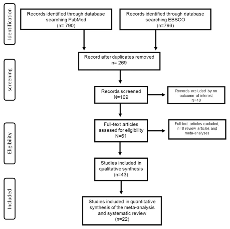
Flow-chart showing the search strategy and inclusion/exclusion criteria used in the meta-analysis and systematic review.
Table 1.
Genotype and allele distribution in association studies on CRP (rs1800947, rs1130864, rs2794521 and rs1205) and TNF-α (rs1799724, rs1800630 and rs1799964) gene variants with CHD
| Reference | Genotype cases | Genotype controls | Allele cases | Allele controls | HWE cases | HWE controls | ||||||
|---|---|---|---|---|---|---|---|---|---|---|---|---|
| CRP gene variants | ||||||||||||
| G1059C rs1800947 | GG | GC | CC | GG | GC | CC | G | C | G | C | p value | p value |
| Zee R.Y. [8] | 142 | 81 | 3 | 638 | 86 | 2 | 1365 | 87 | 1362 | 90 | 0.614 | 0.02* |
| Brull D.J. [9] | 163 | 231 | 1 | 209 | 23 | 1 | 349 | 25 | 441 | 25 | 0.670 | 0.00* |
| Miller D.T. [14] | 568 | 75 | 2 | 615 | 79 | 2 | 1209 | 77 | 1309 | 83 | 0.748 | 0.774 |
| Balistreri C.R. [32] | 84 | 18 | 4 | 112 | 7 | 1 | 186 | 26 | 231 | 9 | 0.03* | 0.02* |
| Dai D.F. [10] | 303 | 55 | 7 | 138 | 29 | 4 | 661 | 69 | 305 | 37 | 0.113 | 0.02* |
| Pai J.K. [16] | 414 | 80 | 5 | 89 | 100 | 4 | 908 | 90 | 1882 | 108 | 0.00* | 0.607 |
| Grammer T.B. [12] | 2238 | 305 | 12 | 620 | 72 | 1 | 4781 | 329 | 1312 | 74 | 0.463 | 0.643 |
| Pasalic D. [33] | 184 | 20 | 2 | 101 | 23 | 1 | 388 | 24 | 225 | 25 | 0.803 | 0.098 |
| Akbarzadeh-Najar R. [11] | 845 | 92 | 13 | 834 | 105 | 11 | 1782 | 118 | 1773 | 127 | 0.00* | 0.00* |
| Ghattas M.H. [34] | 128 | 21 | 1 | 121 | 27 | 2 | 277 | 123 | 269 | 3 | 0.725 | 0.891 |
| Singh P. [13] | 140 | 37 | 3 | 142 | 31 | 2 | 317 | 43 | 315 | 35 | 0.833 | 0.759 |
| Total | 5209 | 1015 | 53 | 3619 | 582 | 31 | 12223 | 1011 | 9424 | 616 | 0.155 | 0.646 |
| C144T rs1130864 | CC | CT | TT | CC | CT | TT | C | T | C | T | p value | p value |
| Brull D.J. [9] | 86 | 78 | 22 | 122 | 92 | 13 | 250 | 122 | 336 | 118 | 0.42 | 0.50 |
| Miller D.T. [14] | 315 | 270 | 58 | 331 | 298 | 67 | 900 | 386 | 960 | 432 | 0.99 | 0.98 |
| Grammer T.B. [12] | 1150 | 1124 | 281 | 305 | 316 | 76 | 3424 | 1686 | 926 | 468 | 0.66 | 0.78 |
| Singh P. [13] | 90 | 75 | 15 | 113 | 55 | 7 | 255 | 105 | 281 | 69 | 0.92 | 0.91 |
| Total | 1892 | 1739 | 418 | 1344 | 1144 | 266 | 5523 | 2575 | 3832 | 1676 | 0.32 | 0.53 |
| C717T rs2794521 | CC | CT | TT | CC | CT | TT | C | T | C | T | p value | p value |
| Brull D.J. [9] | 98 | 73 | 14 | 117 | 88 | 17 | 269 | 101 | 322 | 122 | 0.93 | 0.93 |
| Chen J. [17] | 452 | 163 | 4 | 439 | 158 | 18 | 1067 | 179 | 1067 | 179 | 0.41 | 0.00* |
| Miller D.T. [14] | 352 | 247 | 44 | 361 | 281 | 54 | 951 | 335 | 1003 | 389 | 0.94 | 0.94 |
| Grammer T.B. [12] | 1395 | 981 | 179 | 351 | 294 | 52 | 3771 | 1339 | 996 | 398 | 0.37 | 0.71 |
| Akbarzadeh-Najar R. [11] | 683 | 252 | 15 | 648 | 249 | 53 | 1618 | 282 | 1545 | 355 | 0.00* | 0.12 |
| Singh P. [13] | 112 | 60 | 8 | 137 | 35 | 3 | 284 | 76 | 309 | 41 | 0.66 | 0.99 |
| Total | 3092 | 1776 | 264 | 2053 | 1105 | 197 | 7960 | 2312 | 5242 | 1484 | 0.06 | 0.66 |
| G3872A rs1205 | CC | CT | TT | CC | CT | TT | C | T | C | T | p value | p value |
| Miller D.T. [14] | 248 | 303 | 92 | 313 | 308 | 75 | 799 | 487 | 934 | 458 | 0.95 | 0.97 |
| Mathew J.P. [15] | 153 | 132 | 10 | 61 | 73 | 14 | 438 | 152 | 195 | 101 | 0.23 | 0.00* |
| Pai J.K. [16] | 218 | 221 | 56 | 447 | 447 | 89 | 657 | 1333 | 1341 | 625 | 0.12 | 0.99 |
| Total | 619 | 656 | 158 | 821 | 828 | 178 | 1894 | 1972 | 2470 | 1184 | 0.13 | 0.42 |
| TNF-α gene variants | ||||||||||||
| C857T rs1799724 | CC | CT | TT | CC | CT | TT | C | T | C | T | p value | p value |
| Herrmann, S.M.[35] | 153 | 41 | 1 | 125 | 45 | 1 | 348 | 43 | 295 | 47 | 0.47 | 0.20 |
| Herrmann, S.M.[35] | 302 | 122 | 10 | 340 | 155 | 10 | 726 | 142 | 835 | 175 | 0.72 | 0.12 |
| Bennet, A.M. [36] | 959 | 161 | 16 | 1265 | 210 | 21 | 2079 | 193 | 2740 | 252 | 0.00* | 0.00* |
| Cui, G. [37] | 907 | 459 | 22 | 700 | 299 | 28 | 2273 | 503 | 1699 | 355 | 0.00* | 0.66 |
| Cui, G. [37] | 642 | 278 | 41 | 529 | 262 | 30 | 1562 | 360 | 1320 | 322 | 0.13 | 0.82 |
| Cho, H.C. [38] | 133 | 54 | 10 | 145 | 30 | 53 | 320 | 74 | 672 | 136 | 0.16 | 0.00* |
| Total | 3096 | 1115 | 100 | 3104 | 1001 | 143 | 7308 | 1315 | 7561 | 1287 | 0.97 | 0.30 |
| C863A rs1800630 | CC | CA | AA | CC | CA | AA | C | A | C | A | p value | p value |
| Koch, W. [39] | 716 | 261 | 21 | 242 | 90 | 8 | 1693 | 303 | 574 | 106 | 0.71 | 1.00 |
| Koch, W. [39] | 546 | 227 | 20 | 242 | 90 | 8 | 1319 | 267 | 574 | 106 | 0.61 | 1.00 |
| Bennet, A.M. [36] | 792 | 314 | 37 | 1011 | 438 | 43 | 1898 | 388 | 2460 | 524 | 0.40 | 0.65 |
| Asifa, G.Z. [40] | 92 | 215 | 3 | 201 | 101 | 8 | 399 | 221 | 503 | 117 | 0.00* | 0.35 |
| Cho, H.C. [38] | 141 | 54 | 2 | 313 | 51 | 40 | 336 | 58 | 677 | 131 | 0.26 | 0.00* |
| Total | 2287 | 1071 | 83 | 2009 | 770 | 107 | 5645 | 1237 | 4788 | 984 | 0.07 | 0.06 |
| T1031C rs1799964 | TT | TC | CC | TT | TC | CC | T | C | T | C | p value | p value |
| Bennet, A.M. [36] | 726 | 364 | 55 | 889 | 523 | 68 | 1816 | 474 | 2301 | 659 | 0.00* | 0.24 |
| Ghazouani, L. [41] | 270 | 134 | 14 | 284 | 111 | 11 | 674 | 162 | 679 | 133 | 0.00* | 1.00 |
| Cui, G. [37] | 650 | 281 | 30 | 557 | 239 | 25 | 1581 | 341 | 1353 | 289 | 0.89 | 1.00 |
| Asifa, G.Z. [40] | 178 | 106 | 26 | 182 | 210 | 18 | 462 | 158 | 474 | 146 | 0.97 | 0.98 |
| Total | 2696 | 1360 | 166 | 2511 | 1464 | 169 | 6752 | 1692 | 6386 | 1702 | 0.88 | 0.99 |
p value: statistical significance.
Table 2.
Descriptive characteristics of the studies about variants of the CRP and TNF-α genes included in the meta-analysis and systematic review
| Reference | Date of publication | Cases | Controls | Mean age cases | Mean age controls | Location | Origin | Diagnosis or procedure | CRP serum levels in cases/controls |
|
| |||||||||
| CRP gene variants | |||||||||
| G1059C rs1800947 | |||||||||
| Zee R.Y. [8] | May 2002 | 726 | 726 | 60 | 60 | USA | Caucasians | Arterial thrombosis | 1.43 mg/L/1.23 mg/L |
| Brull D.J. [9] | July 2003 | 187 | 233 | 63 | 63 | UK | Caucasians | Patients with coronary artery bypass | 1.39 mg/L/1.19 mg/L |
| Miller D.T. [14] | Jun 2005 | 643 | 696 | 60 | 60 | USA | Caucasians | Myocardial infarction and ischemic stroke | 1.6 mg/L/1.2 mg/L |
| Balistreri C.R. [32] | May 2006 | 106 | 120 | 41 | 39 | Italy | Caucasians | Acute myocardial infarction | - |
| Mathew J.P. [15] | Jan 2007 | 295 | 148 | 61 | 60 | USA | Caucasians | Patients with coronary artery bypass | - |
| Dai D.F. [10] | May 2007 | 365 | 171 | 60 | 62 | China | Asians | Coronary angiography | 3.3 mg/L/- |
| Pai J.K. [16] | Jan 2008 | 499 | 995 | 60 | 60 | USA | Caucasians | Acute myocardial infarction | 1.02 mg/L/- |
| Grammer T.B. [12] | Jan 2009 | 2555 | 693 | 64 | 58 | Germany | Caucasians | Coronary angiography | 4.2 mg/L/2.8 mg/L |
| Pasalic D. [33] | April 2009 | 206 | 125 | 61 | 58 | Croatia | Caucasians | Coronary angiography | 2.8 mg/L/2.8 mg/L |
| Akbarzadeh-Najar R. [11] | April 2012 | 950 | 950 | 53 | 50 | Iran | Asians | Acute myocardial infarction | 2.5 mg/L/2.5 mg/L |
| Ghattas M.H. [34] | Nov 2012 | 150 | 150 | - | - | Egypt | Africans | Acute myocardial infarction | - |
| Singh P. [13] | Jun 2014 | 180 | 175 | 55 | 54 | Punyab | Indians | Coronary angiography | 2.3 mg/L/1.4 mg/L |
| C144T rs1130864 | |||||||||
| Brull D.J. [9] | July 2003 | 227 | 186 | 63 | 63 | UK | Caucasians | Myocardial infarction and ischemic stroke | 1.6 mg/L/1.2 mg/L |
| Miller D.T. [14] | Jun 2005 | 643 | 696 | 60 | 60 | USA | Caucasians | Myocardial infarction and ischemic stroke | 1.6 mg/L/1.2 mg/L |
| Pai J.K. [16] | Jan 2008 | 485 | 959 | 60 | 60 | USA | Caucasians | Acute myocardial infarction | 1.02 mg/L/- |
| Grammer T.B. [12] | Jan 2009 | 2555 | 697 | 64 | 58 | Germany | Caucasians | Coronary angiography | 4.2 mg/L/2.8 mg/L |
| C717T rs2794521 | |||||||||
| Brull D.J. [9] | July 2003 | 185 | 222 | 63 | 63 | UK | Caucasians | Patients with coronary artery bypass | 1.39 mg/L/1.19 mg/L |
| Chen J. [17] | Jan 2005 | 619 | 615 | 53 | 53 | China | Asians | Acute mycocardial infarction | 2.38 mg/L-1.82 mg/L |
| Miller D.T. [14] | Jun 2005 | 643 | 696 | 60 | 60 | USA | Caucasians | Myocardial infarction and ischemic stroke | 1.6 mg/L/1.2 mg/L |
| Grammer T.B. [12] | Jan 2009 | 2555 | 697 | 64 | 58 | Germany | Caucasians | Coronary angiography | 4.2 mg/L/2.8 mg/L |
| Akbarzadeh-Najar R. [11] | April 2012 | 950 | 950 | 53 | 50 | Iran | Asians | Acute myocardial infarction | 2.5 mg/L/2.5 mg/L |
| Singh P. [13] | Jun 2014 | 180 | 175 | 55 | 54 | Punyab | Indians | Coronary angiography | 2.3 mg/L/1.4 mg/L |
| G3872A rs1205 | |||||||||
| Miller D.T. [14] | Jun 2005 | 643 | 696 | 60 | 60 | USA | Caucasians | Myocardial infarction and ischemic stroke | 1.6 mg/L/1.2 mg/L |
| Mathew J.P. [15] | Jan 2007 | 295 | 148 | 61 | 60 | USA | Caucasians | Patients with coronary artery bypass | - |
|
| |||||||||
| Reference | Date of publication | Cases | Controls | Mean age cases | Mean age controls | Location | Origin | Diagnosis | CRP serum levels in cases/controls |
|
| |||||||||
| TNF-α gene variants | |||||||||
| C857T rs1799724 | |||||||||
| Herrmann, S.M. [35] | Jan 1998 | 195 | 171 | - | - | UK | Caucasians | Myocardial infarction or angina pectoris | - |
| Herrmann, S.M. [35] | Jan 1998 | 434 | 505 | - | - | France | Caucasians | Myocardial infarction or angina pectoris | - |
| Bennet, A.M. [36] | Jan 2006 | 1136 | 1496 | 61 | 61 | Sweden | Caucasians | Myocardial infarction | 1.8 mg/L/1.2 mg/L |
| Cui, G. [37] | Oct 2012 | 1338 | 1027 | - | - | China | Asians | Ischemic stroke | - |
| Cui, G. [37] | Oct 2012 | 961 | 821 | - | - | China | Asians | Ischemic stroke | - |
| Cho, H.C. [38] | March 2013 | 394 | 808 | 61 | 62 | Korea | Asians | Myocardial infarction | 1.9 mg/L/- |
| C863A rs1800630 | |||||||||
| Koch, W. [39] | Jan 2001 | 998 | 340 | 64 | 63 | Germany | Caucasians | Coronary stenosis | - |
| Koch, W. [39] | Jan 2001 | 793 | 340 | 63 | 63 | Germany | Caucasians | Myocardial infarction | - |
| Bennet, A.M. [36] | Jan 2006 | 1143 | 1492 | 61 | 61 | Sweden | Caucasians | Myocardial infarction | 1.8 mg/L/1.2 mg/L |
| Asifa, G.Z. [40] | Sep 2013 | 310 | 310 | 54 | 53 | Pakistan | Oriental | Coronary angiography | - |
| Cho, H. C. [38] | March 2013 | 394 | 808 | 61 | 62 | Korea | Asians | Myocardial infarction | 1.3 mg/L/- |
| T1031C rs1799964 | |||||||||
| Bennet, A.M. [36] | Jan 2006 | 1145 | 1480 | 61 | 61 | Sweden | Caucasians | Myocardial infarction | 1.8 mg/L/1.2 mg/L |
| Ghazouani, L. [41] | July 2009 | 418 | 406 | 58 | 57 | Tunisia | African | Myocardial infarction | - |
| Cui, G. [37] | Oct 2012 | 1338 | 1027 | 0 | 0 | China | Asians | Ischemic stroke | - |
| Cui, G. [37] | Oct 2012 | 961 | 821 | 0 | 0 | China | Asians | Ischemic stroke | - |
| Asifa, G.Z. [40] | Sep 2013 | 310 | 310 | 54 | 53 | Pakistan | Oriental | Ischemic stroke | - |
PCR gene variants
G1059C rs1800947 polymorphism
In the analysis of rs1800947, we included eleven association studies with CHD. When we explored the role of this variant in all populations in the allelic and recessive models with heterogeneity we found a slight association [fixed: OR 0.86; CI 95%: (0.77-0.96); Q test: <0.0001; Egger’s test: 0.12; and fixed: OR 0.85; (CI 95%: 0.75-0.96); Q test: <0.0001; Egger’s test: 0.74, respectively (Figures 2 and 3)]. With regard to the rest of the models, the analysis of all populations in the study yielded a lack of association. Moreover, we performed a sub-analysis in Caucasian populations to get a more comprehensive understanding of this common variant and we could confirm the association in the same allelic and recessive models with heterogeneity [fixed: OR 0.83; CI 95%: (0.72-0.95); Q test: 0.0007; Egger’s test: 0.87 and fixed: OR 0.75; (CI 95%: 0.65-0.87); Q test: <0.0001; Egger’s test: 0.72] (see Additional file). The rest of the outcomes as well as the results excluding heterogeneity and bias in the publication are shown in Table 3.
Figure 2.

Odds ratios and forest plots of the PCR rs1800947 variant in overall studies, without heterogeneity, using the following models: A. Allelic; B. Additive; C. Dominant, and D. Recessive.
Figure 3.
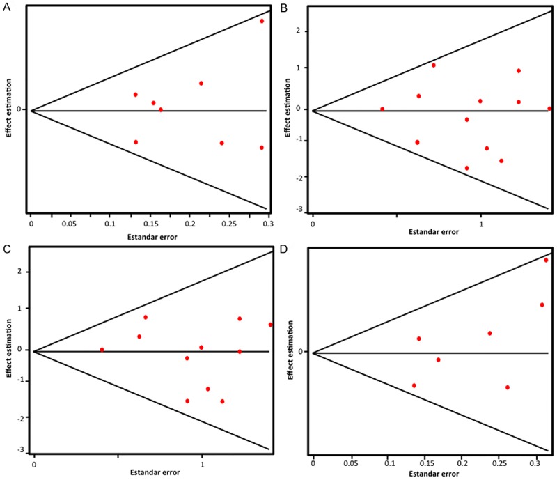
Egger’s funnel plots in overall studies indicating publication bias in studies on CHD and PCR rs1800947 variant, without heterogeneity, using the following models: A. Allelic; B. Additive; C. Dominant, and D. Recessive.
Table 3.
Analysis of association studies on CRP (rs1800947, rs1130864, rs2794521 and rs1205) and TNF-α (rs1799724, rs1800630 and rs1799964) gene variants with CHD in Caucasians and all populations in the study
| Model analysis | Caucasians | P value of Q test | P value of Egger’s test | All populations | P value of Q test | P value of Egger’s test | |||
|---|---|---|---|---|---|---|---|---|---|
|
|
|
||||||||
| Random effects OR (CI 95%) | Fixed effects OR (CI 95%) | Fixed effects OR (CI 95%) | Fixed effects OR (CI 95%) | ||||||
| PCR gene variants | |||||||||
| G1059C rs1800947 | |||||||||
| Allelic | With heterogeneity | 0.82 (0.61-1.10) | 0.83 (0.72-0.95) | 0.0007 | 0.87 | 0.74 (0.55-1.01) | 0.86 (0.77-0.96) | <0.0001 | 0.12 |
| Without heterogeneity | 0.91 (0.78-1.07) | 0.91 (0.78-1.07) | 0.60 | 0.90 | 0.99 (0.86-1.14) | 0.99 (0.87-1.12) | 0.30 | 0.50 | |
| Additive | With heterogeneity | ||||||||
| Without heterogeneity | 0.62 (0.22-1.74) | 0.74 (0.35-1.54) | 0.08 | 0.20 | 0.83 (0.48-1.45) | 0.86 (0.54-1.38) | 0.27 | 0.42 | |
| Dominant | With heterogeneity | ||||||||
| Without heterogeneity | 0.68 (0.31-1.48) | 0.70 (0.34-1.46) | 0.35 | 0.39 | 0.84 (0.52-1.34) | 0.84 (0.52-1.34) | 0.53 | 0.65 | |
| Recessive | With heterogeneity | 0.65 (0.23-1.78) | 0.75 (0.65-0.87) | <0.0001 | 0.72 | 0.79 (0.41-1.50) | 0.85 (0.75-0.96) | <0.0001 | 0.74 |
| Without heterogeneity | 1.06 (0.72-1.57) | 0.95 (0.78-1.16) | 0.06 | 0.08 | 1.05 (0.87-1.26) | 1.02 (0.88-1.18) | 0.16 | 0.15 | |
| C1444T rs1130864 | |||||||||
| Allelic | With heterogeneity | 0.92 (0.77-1.09) | 0.99 (0.91-1.07) | 0.004 | 0.06 | ||||
| Without heterogeneity | 1.00 (0.88-1.14) | 1.02 (0.94-1.11) | 0.09 | 0.25 | 1.00 (0.88-1.14) | 1.02 (0.94-1.11) | 0.09 | 0.25 | |
| Additive | With heterogeneity | 0.87 (0.61-1.25) | 0.99 (0.82-1.20) | 0.02 | 0.08 | ||||
| Without heterogeneity | 0.98 (0.71-1.35) | 1.03 (0.85-1.25) | 0.06 | 0.30 | 1.02 (0.76-1.37) | 1.05 (0.87-1.28) | 0.11 | 0.28 | |
| Dominant | With heterogeneity | 0.91 (0.67-1.23) | 0.98 (0.82-1.18) | 0.05 | 0.12 | ||||
| Without heterogeneity | 0.98 (0.73-1.30) | 1.01 (0.84-1.22) | 0.09 | 0.35 | 1.03 (0.81-1.30) | 1.04 (0.86-1.25) | 0.23 | 0.38 | |
| Recessive | With heterogeneity | 0.93 (0.76-1.13) | 0.99 (0.89-1.10) | 0.02 | 0.06 | ||||
| Without heterogeneity | 1.03 (0.92-1.16) | 1.03 (0.93-1.15) | 0.35 | 0.15 | 1.03 (0.92-1.16) | 1.03 (0.93-1.15) | 0.35 | 0.15 | |
| C717T rs2794521 | |||||||||
| Allelic | With heterogeneity | 1.02 (0.86-1.21) | 1.10 (1.01-1.19) | 0.001 | 0.08 | ||||
| Without heterogeneity | 1.10 (1.00-1.21) | 1.10 (1.00-1.21) | 0.81 | 0.29 | 1.13 (1.03-1.23) | 1.13 (1.04-1.22) | 0.30 | 0.47 | |
| Additive | With heterogeneity | 1.47 (0.86-2.54) | 1.39 (1.11-1.73) | <0.0001 | 0.79 | ||||
| Without heterogeneity | 1.15 (0.90-1.47) | 1.15 (0.90-1.47) | 0.93 | 0.45 | 1.08 (0.80-1.45) | 1.10 (0.86-1.40) | 0.29 | 0.12 | |
| Dominant | With heterogeneity | 3.60 (1.83-7.09) | 2.79 (2.24-3.47) | <0.0001 | 0.37 | ||||
| Without heterogeneity | 2.05 (1.61-2.62) | 2.05 (1.61-2.62) | 0.93 | 0.98 | 2.04 (1.60-2.60) | 2.04 (1.60-2.60) | 0.97 | 0.53 | |
| Recessive | With heterogeneity | 1.03 (0.86-1.24) | 1.10 (1.00-1.21) | 0.009 | 0.37 | ||||
| Without heterogeneity | 1.14 (1.01-1.29) | 1.14 (1.01-1.29) | 0.74 | 0.12 | 1.14 (1.04-1.26) | 1.14 (1.04-1.26) | 0.92 | 0.28 | |
| G3872A rs1205 | |||||||||
| Allelic | With heterogeneity | 0.64 (0.22-1.88) | 0.44 (0.40-0.48) | <0.0001 | 0.45 | 0.64 (0.22-1.88) | 0.44 (0.40-0.48) | <0.0001 | 0.45 |
| Without heterogeneity | |||||||||
| Additive | With heterogeneity | 1.05 (0.53-2.09) | 0.79 (0.62-1.01) | 0.001 | 0.07 | 1.05 (0.53-2.09) | 0.79 (0.62-1.01) | 0.001 | 0.07 |
| Without heterogeneity | 0.70 (0.54-0.90) | 0.70 (0.54-0.90) | 0.48 | 1.00 | 0.70 (0.54-0.90) | 0.70 (0.54-0.90) | 0.48 | 1.00 | |
| Dominant | With heterogeneity | 1.02 (0.58-1.81) | 0.83 (0.66-1.04) | 0.007 | 0.07 | 1.02 (0.58-1.81) | 0.83 (0.66-1.04) | 0.007 | 0.07 |
| Without heterogeneity | 0.74 (0.58-0.95) | 0.74 (0.58-0.95) | 0.7567 | 1.00 | 0.74 (0.58-0.95) | 0.74 (0.58-0.95) | 0.75 | 1.00 | |
| Recessive | With heterogeneity | 0.99 (0.71-1.37) | 0.91 (0.79-1.06) | 0.01 | 0.29 | 0.99 (0.71-1.37) | 0.91 (0.79-1.06) | 0.01 | 0.29 |
| Without heterogeneity | 0.85 (0.69-1.04) | 0.85 (0.73-0.99) | 0.19 | 1.00 | 0.85 (0.69-1.04) | 0.85 (0.73-0.99) | 0.19 | 1.00 | |
| TNF-α gene variants | |||||||||
| C857T rs1799724 | |||||||||
| Allelic | With heterogeneity | ||||||||
| Without heterogeneity | 1.04 (0.90-1.20) | 1.04 (0.90-1.20) | 0.65 | 0.10 | 1.00 (0.92-1.09) | 1.00 (0.92-1.09) | 0.65 | 0.45 | |
| Additive | With heterogeneity | 1.42 (0.08-2.51) | 1.35 (1.03-1.79) | 0.003 | 0.80 | ||||
| Without heterogeneity | 0.96 (0.57-1.61) | 0.96 (0.57-1.61) | 0.96 | 0.73 | 1.08 (0.80-1.46) | 1.08 (0.80-1.46) | 0.65 | 0.99 | |
| Dominant | With heterogeneity | 1.46 (1.07-2.75) | 1.38 (1.05-1.82) | 0.0006 | 0.82 | ||||
| Without heterogeneity | 0.95 (0.56-1.59) | 0.95 (0.56-1.59) | 0.95 | 0.89 | 1.07 (0.79-1.45) | 1.07 (0.80-1.45) | 0.40 | 0.98 | |
| Recessive | With heterogeneity | ||||||||
| Without heterogeneity | 1.06 (1.00-1.24) | 1.06 (0.90-1.24) | 0.48 | 0.13 | 1.02 (1.00-1.14) | 1.01 (0.92-1.12) | 0.32 | 0.08 | |
| C863A rs1800630 | |||||||||
| Allelic | With heterogeneity | 1.16 (0.84-1.61) | 1.10 (1.00-1.22) | <0.0001 | 0.58 | ||||
| Without heterogeneity | 0.98 (0.88-1.10) | 0.98 (0.88-1.10) | 0.64 | 0.52 | 0.97 (0.88-1.08) | 0.97 (0.88-1.08) | 0.75 | 0.94 | |
| Additive | With heterogeneity | ||||||||
| Without heterogeneity | 1.05 (1.04-1.51) | 1.05 (1.04-1.51) | 0.89 | 0.60 | 0.77 (0.42-1.39) | 0.92 (0.65-1.28) | 0.06 | 0.19 | |
| Dominant | With heterogeneity | 1.41 (0.86-2.34) | 1.18 (1.06-1.33) | <0.0001 | 0.26 | ||||
| Without heterogeneity | 0.97 (0.86-1.10) | 0.97 (0.86-1.10) | 0.54 | 0.41 | 1.02 (0.88-1.18) | 1.00 (0.89-1.13) | 0.28 | 0.09 | |
| Recessive | With heterogeneity | 0.64 (0.32-1.28) | 0.87 (0.62-1.22) | 0.01 | 0.08 | ||||
| Without heterogeneity | 1.06 (0.75-1.52) | 1.06 (0.75-1.52) | 0.88 | 0.44 | 0.99 (0.70-1.40) | 0.99 (0.70-1.40) | 0.47 | 0.14 | |
| T1031C rs1799964 | |||||||||
| Allelic | With heterogeneity | 0.99 (0.70-1.40) | 0.99 (0.70-1.40) | 0.47 | 0.14 | ||||
| Without heterogeneity | 0.97 (0.86-1.10) | 0.94 (0.87-1.02) | 0.06 | 0.06 | |||||
| Additive | With heterogeneity | ||||||||
| Without heterogeneity | 0.97 (0.71-1.32) | 0.93 (0.74-1.17) | 0.13 | 0.32 | |||||
| Dominant | With heterogeneity | 0.88 (0.73-1.06) | 0.88 (0.80-0.96) | 0.003 | 0.92 | ||||
| Without heterogeneity | 0.94 (0.81-1.11) | 0.92 (0.83-1.01) | 0.06 | 0.06 | |||||
| Recessive | With heterogeneity | ||||||||
| Without heterogeneity | 1.05 (1.00-1.51) | 1.00 (0.80-1.25) | 0.06 | 0.39 | |||||
C1444T rs1130864 polymorphism
When the whole sample was explored with respect to this variant gene, the analysis revealed no significant association of this polymorphism in relation to CHD using the following models: allelic [random: OR 1.00; CI 95% (0.88-1.14); Q test: 0.09; Egger’s test: 0.25], additive [random: OR 1.02; CI 95% (0.76-1.37); Q test: 0.11; Egger’s test: 0.28], dominant [random: OR 1.03; CI 95% (0.81-1.30); Q test: 0.23; Egger’s test: 0.38], and recessive [random: OR 1.03; CI 95% (0.92-1.16); Q test: 0.35; Egger’s test: 0.15] (Figures 4 and 5). Due to these results, we explored the role of this variant gene in the Caucasian population only (see Additional file), and even though the results did not reveal heterogeneity we confirmed the same results of the first analysis, using the same models: allelic [random: OR 1.00; CI 95% (0.88-1.14); Q test: 0.09; Egger’s test: 0.25], additive [random: OR 0.98; CI 95% (0.71-1.35); Q test: 0.06; Egger’s test: 0.30], dominant [random: OR 0.98; CI 95% (0.73-1.30); Q test: 0.09; Egger’s test: 0.35], and recessive [random: OR 1.03; CI 95% (0.92-1.16); Q test: 0.35; Egger’s test: 0.15]. Additional data can be found in Table 3.
Figure 4.
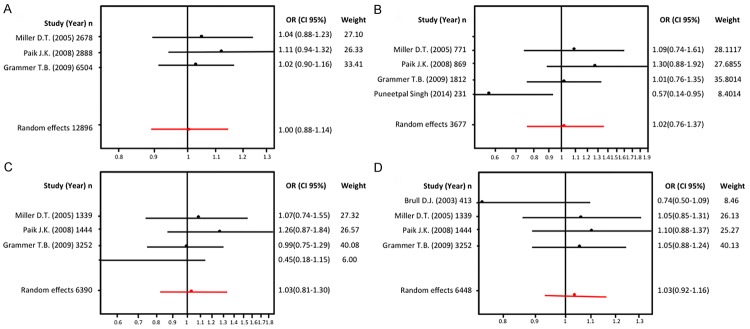
Odds ratios and forest plots of PCR rs1130864 variant in overall studies, without heterogeneity, using the following models: A. Allelic; B. Additive; C. Dominant, and D. Recessive.
Figure 5.
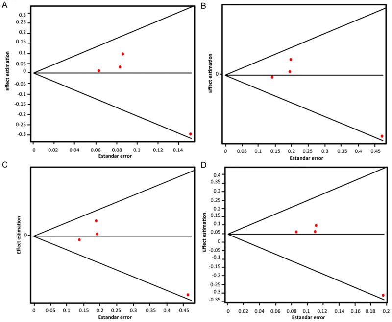
Egger’s funnel plots in overall studies indicating publication bias in studies on CHD and PCR rs1130864 variant, without heterogeneity, using the following models: A. Allelic; B. Additive; C. Dominant, and D. Recessive.
C717T rs2794521 polymorphism
The analysis of all the populations as a whole showed that the rs2794521 polymorphism in the four models analyzed yielded a significant relation to present CHD clinically in the following models: allelic [random: OR 1.13; CI 95% (1.04-1.22); Q test: 0.30; Egger’s test: 0.47; without heterogeneity], dominant [random: OR 2.04; CI 95% (1.60-2.60); Q test 0.97; Egger’s test: 0.53; without heterogeneity] and recessive [random: OR 1.14; CI 95% (1.04-1.26); Q test: 0.92; Egger’s test: 0.28; without heterogeneity] (Figures 6 and 7). Furthermore, a more careful analysis of this gene variant was performed reporting Caucasian population only and we still observed the same results showing a strong relationship of this polymorphism and CHD: allelic [random: OR 1.10; CI 95% (1.00-1.21); Q test: 0.81; Egger’s test: 0.29], dominant [random: OR 2.05; CI 95% (1.61-2.62); Q test: 0.93; Egger’s test: 0.98] and recessive [random: OR 1.14; CI 95% (1.01-1.29); Q test: 0.74; Egger’s test: 0.12]. These data did not reveal heterogeneity (Table 3).
Figure 6.

Odds ratios and forest plots of the PCR rs2794521 variant in overall studies, without heterogeneity, using the following models: A. Allelic; B. Additive; C. Dominant, and D. Recessive.
Figure 7.
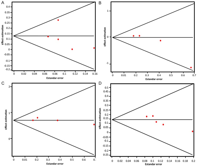
Egger’s funnel plots in overall studies indicating publication bias in studies on CHD and PCR rs2794521 variant, without heterogeneity, using the following models: A. Allelic; B. Additive; C. Dominant, and D. Recessive.
G3872A rs1205 polymorphism
In this gene variant we only included reports with Caucasian populations (see Additional file). We conducted only one analysis taking the whole population as sample and found a significant association of this polymorphism with CHD in the following models: additive [random: OR 0.70; CI 95% (0.54-0.90); Q test: 0.48; Egger’s test: 1.00] and dominant [random: OR 0.74; CI 95% (0.58-0.95); Egger’s test: 0.75; Egger’s test: 1.00] (Figures 8 and 9). In the analysis with the allelic model we could not exclude heterogeneity (Table 3).
Figure 8.
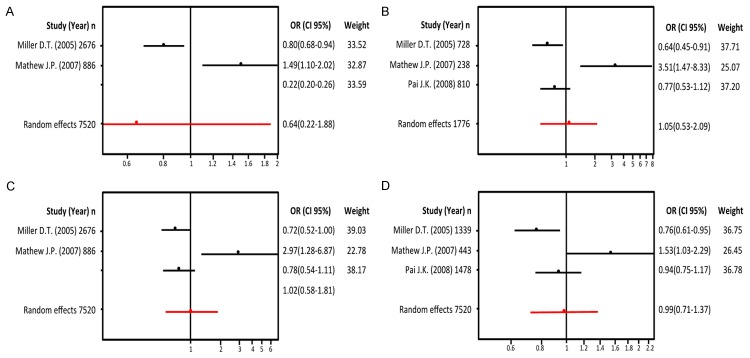
Odds ratios and forest plots of the PCR rs1205 variant in overall studies, without heterogeneity, using the following models: A. Allelic; B. Additive; C. Dominant, and D. Recessive.
Figure 9.
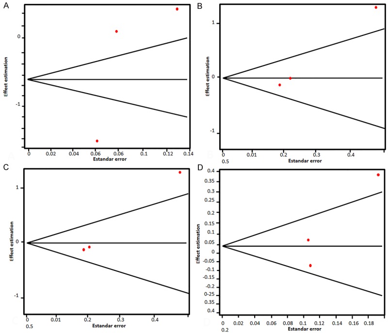
Egger’s funnel plots in overall studies indicating publication bias in studies on CHD and PCR rs1205 variant, without heterogeneity, using the following models: A. Allelic; B. Additive; C. Dominant, and D. Recessive.
TNF-α gene variants
C857T rs1799724 polymorphism
The analysis of this variant included the same models proposed in the CRP polymorphisms: allelic, additive, dominant and recessive with and without heterogeneity. In this way we could observe in the recessive model without heterogeneity a significant association of allele T of the rs1799724 polymorphism in whole CHD population [random: OR 1.02; CI 95% (1.00-1.14); Q test: 0.32; Egger’s test: 0.08] (Figures 10 and 11). This result was confirmed when we explored this same variant but only in Caucasians, using the recessive model without heterogeneity [random: OR 1.06; CI 95% (1.00-1.24); Q test: 0.48; Egger’s test: 0.13] (see Additional file). In addition, the findings in the others models did not show C857T polymorphisms as a predisposing factor in CHD (Table 3).
Figure 10.

Odds ratios and forest plots of the TNF-α rs1799724 variant in overall studies, without heterogeneity, using the following models: A. Allelic; B. Additive; C. Dominant, and D. Recessive.
Figure 11.

Egger’s funnel plots in overall studies indicating publication bias in studies on CHD and TNF-α rs1799724 variant, without heterogeneity, using the following models: A. Allelic; B. Additive; C. Dominant, and D. Recessive.
C863A rs1800630 polymorphism
First, the interaction of this variant in CHD was analyzed; the outcomes did not show a significant association in the population when heterogeneity was discarded. Subsequently, we explored this association only in Caucasians (see Additional file) and we could observe in the additive model without heterogeneity a slight 5% increase in the risk to present CHD [random: OR 1.05; CI 95% (1.04-1.51); Q test: 0.89; Egger’s test: 0.60] (Figures 12 and 13).
Figure 12.

Odds ratios and forest plots of the TNF-α rs1800630 variant in overall studies, without heterogeneity, using the following models: A. Allelic; B. Additive; C. Dominant, and D. Recessive.
Figure 13.

Egger’s funnel plots in overall studies indicating publication bias in studies on CHD and TNF-α rs1800630 variant, without heterogeneity, using the following models: A. Allelic; B. Additive; C. Dominant, and D. Recessive.
T1031C rs1799964 polymorphism
The findings of this variant in the dominant model with heterogeneity revealed a probability that this SNP could have a protective effect in the CHD for allele T [random: OR 0.88; CI 95% (0.80-0.96); Q test: 0.003; Egger’s test: 0.92]. Although the outcome in the recessive model without heterogeneity [random: OR 1.05; CI 95% (1.00-1.51); Q test: 0.06; Egger’s test: 0.39] showed that allele C of the variant T1031C could be considered a risk factor (Figures 14 and 15). An analysis by ethnicity was not performed.
Figure 14.

Odds ratios and forest plots of the TNF-α rs1799964 variant in overall studies, without heterogeneity, using the following models: A. Allelic; B. Additive; C. Dominant, and D. Recessive.
Figure 15.

Egger’s funnel plots in overall studies indicating publication bias in studies on CHD and TNF-α rs1799964 variant, without heterogeneity, using the following models: A. Allelic; B. Additive; C. Dominant, and D. Recessive.
Discussion
It is well known that CHD is a complex disease where multiple genetic markers interact and inflammatory markers play a vital role in its pathogenesis. Due to this, the present study aimed to explore the involvement of TNF-α and CRP genetic variants with CHD in a meta-analysis and systematic review to get a more comprehensive understanding of this pathology. To start, we chose the TNF-α and CRP genes because there is strong evidence that they are powerful predictors of incident cardiovascular events. As a result, the most common variants studied in association with CHD were the following polymorphisms: G1059C rs1800947, C1444T rs1130864, C717T rs2794521 and G3872A rs1205 of the CRP gene, and C857T rs1799724, C863A rs1800630 and T1031C rs1799964 of the TNF-α gene with interesting results.
First, we explored the rs1800947 polymorphism and found that in all the studies concerning this variant, the pooled ORs suggested a possible protective role to present CHD clinically. The allelic model was used to analyze all the populations as a whole [fixed: OR 0.86; CI 95% (0.77-0.96)], and also Caucasian populations only [fixed: OR 0.83; CI 95% (0.72-0.95); with heterogeneity] (Table 3). This polymorphism is the most studied CRP gene in relation to CHD. We found that our results are in agreement with other reports on the subject [33,34]. However, there are also reports not showing such association [9,10,14]. We encourage researchers to take into consideration that the fixed model is not as conservative as the random model, because the latter assumes the existence of normal distribution effects and parameterizes inter-studies. Therefore, the outcomes from the random effects model are more reliable when heterogeneity is present.
Next, we analyzed the rs1130864 variant in association studies with CHD, but we could not find any significant association in all the analyses performed (Table 3); these outcomes are in agreement with other studies [9,13]. The lack of association could be explained by the scarce number of studies, fewer than for rs1800947. Hence, it is necessary to increase the number of studies to have a definitive outcome. In the next analysis we studied the role of rs2794521 in CHD and we encountered that pooled ORs in almost all the models used in this variant showed a tendency from 13-260% to be a risk factor to present CHD (Table 3). Even though this variant has not been as studied as rs1800947, our data could be helpful in the search for genetic markers in this pathology. Researchers should take this information with caution because the sample size limits the outcomes obtained. Finally, the last CRP variant addressed was rs1205. Even though this polymorphism has been even less studied than the rest, the outcomes showed the same results as for the rs180097 polymorphism (Table 3), i.e., this variant may have a protective effect in CHD. However, we cannot take this result as conclusive -even with the agreement encountered with other studies [15]- given that when the meta-analysis was performed we could not exclude heterogeneity in all the models and this may have an effect on population variation and stratification. Moreover, we could not perform an analysis by ethnic group, since we thought useful to explore the interaction of the gene depending on the origin of the subjects.
With regard to the TNF-α gene, we selected polymorphisms C857T rs1799724, C863A rs1800630 and T1031C rs1799964 for this study. The first approach was with the rs1799724 variant where we obtained a probability range of 2-46% for this variant to be a marker of risk in CHD in the general population. But when we analyzed this role in Caucasians we only observed a 6% risk. This same tendency was seen in the analysis of rs1800630 where in the case of Caucasians we got a 5% risk to manifest CHD. Moreover, in the analysis of all populations with the random effects model, no percentage of risk was obtained. This may be so because in some association studies the genotype distribution may depend on the ethnicity of the subjects studied and probably in other populations the interaction of a hypothetical gene is necessary to manifest CHD [36,40]. In the final variant, rs1799964, the statistical analysis yielded a slight association with CHD in the recessive model (CC+TC vs TT) (Table 3). Therefore, there is a remote possibility of a protective effect with allele T when using the dominant form with heterogeneity in the fixed effects model. But as was explained previously, we did not take into consideration these results given the limitations present with the fixed effects model when heterogeneity is present.
To our knowledge the present study is the first meta-analysis and systematic review with several gene variants of inflammatory markers in the analysis. We also used different models for the analyses (allelic, additive, dominant and recessive; Caucasian population only) to gain a better comprehension of CHD. On the other hand, there are some limitations in our study that must be taken into consideration. First, our findings can be ambiguous because the case-control design of the studies can be affected by population stratification, where systematic differences in the ancestry can lead to spurious findings, even though we tried to address this problem by conducting an analysis which only included Caucasians. Second, although we analyzed several TNF-α and CRP gene variants, some of the studies used a small sample size, which may not provide sufficient power to detect small effects of the gene. Alternatively, we did not conduct an analysis of all TNF-α and CRP gene variants in the Asian population or an analysis by gender, because there were not enough studies with available data. In addition, although we evaluated heterogeneity in the studies, we could not exclude it from the sample as a whole.
In conclusion the present investigation demonstrates that polymorphisms rs2794521 of CRP gene, and rs1799724, rs1800630 and rs1799964 of TNF-α gene are possible risk markers to present CHD. Conversely, the analysis of rs1800947 and rs1205 of the CRP gene could exert a protective effect on the pathogenesis of this disease. Only the analysis of polymorphism rs1130864 shows a lack association with CHD, but it is necessary to integrate more studies to obtain more robust conclusions.
Disclosure of conflict of interest
None.
References
- 1.Fan AZ, Yesupriya A, Chang MH, House M, Fang J, Ned R, Hayes D, Dowling NF, Mokdad AH. Gene polymorphisms in association with emerging cardiovascular risk markers in adult women. BMC Med Genet. 2010;11:6. doi: 10.1186/1471-2350-11-6. [DOI] [PMC free article] [PubMed] [Google Scholar]
- 2.Koenig W. High-sensitivity C-reactive protein and atherosclerotic disease: from improved risk prediction to risk-guided therapy. Int J Cardiol. 2013;168:5126–5134. doi: 10.1016/j.ijcard.2013.07.113. [DOI] [PubMed] [Google Scholar]
- 3.Kovacs A, Green F, Hansson LO, Lundman P, Samnegard A, Boquist S, Ericsson CG, Watkins H, Hamsten A, Tornvall P. A novel common single nucleotide polymorphism in the promoter region of the C-reactive protein gene associated with the plasma concentration of C-reactive protein. Atherosclerosis. 2005;178:193–198. doi: 10.1016/j.atherosclerosis.2004.08.018. [DOI] [PubMed] [Google Scholar]
- 4.Li CS, Guo BR, Guo Z, Yang J, Zheng HF, Wang AL. Association between C-reactive protein gene +1059 G/C polymorphism and the risk of coronary heart disease: a meta-analysis. Chin Med J (Engl) 2013;126:4780–4785. [PubMed] [Google Scholar]
- 5.López NJ, Quintero A, Llancaqueo M, Jara L. Effects of periodontal therapy on markers of systemic inflammation in patients with coronary heart disease risk. Rev Med Chil. 2009;137:1315–1322. [PubMed] [Google Scholar]
- 6.Abd El-Aziz TA, Mohamed RH. Human C-reactive protein gene polymorphism and metabolic syndrome are associated with premature coronary artery disease. Gene. 2013;532:216–221. doi: 10.1016/j.gene.2013.09.042. [DOI] [PubMed] [Google Scholar]
- 7.Bahramali E, Firouzabadi N, Jonaidi-Jafari N, Shafiei M. Renin-angiotensin system genetic polymorphisms: Lack of association with CRP levels in patients with coronary artery disease. J Renin Angiotensin Aldosterone Syst. 2013;15:559–65. doi: 10.1177/1470320312474051. [DOI] [PubMed] [Google Scholar]
- 8.Zee RY, Ridker PM. Polymorphism in the human C-reactive protein (CRP) gene, plasma concentrations of CRP, and the risk of future arterial thrombosis. Atherosclerosis. 2002;162:217–219. doi: 10.1016/s0021-9150(01)00703-1. [DOI] [PubMed] [Google Scholar]
- 9.Brull DJ, Serrano N, Zito F, Jones L, Montgomery HE, Rumley A, Sharma P, Lowe GD, World MJ, Humphries SE, Hingorani AD. Human CRP gene polymorphism influences CRP levels: implications for the prediction and pathogenesis of coronary heart disease. Arterioscler Thromb Vasc Biol. 2003;23:2063–2069. doi: 10.1161/01.ATV.0000084640.21712.9C. [DOI] [PubMed] [Google Scholar]
- 10.Dai DF, Chiang FT, Lin JL, Huang LY, Chen CL, Chang CJ, Lai LP, Hsu KL, Tseng CD, Tseng YZ, Hwang JJ. Human C-reactive protein (CRP) gene 1059G>C polymorphism is associated with plasma CRP concentration in patients receiving coronary angiography. J Formos Med Assoc. 2007;106:347–354. doi: 10.1016/S0929-6646(09)60319-3. [DOI] [PubMed] [Google Scholar]
- 11.Akbarzadeh Najar R, Ghaderian SM, Tabatabaei Panah AS. C-reactive protein (CRP) gene polymorphisms: implication in CRP plasma levels and susceptibility to acute myocardial infarction. Mol Biol Rep. 2012;39:3705–3712. doi: 10.1007/s11033-011-1145-z. [DOI] [PubMed] [Google Scholar]
- 12.Grammer TB, Marz W, Renner W, Bohm BO, Hoffmann MM. C-reactive protein genotypes associated with circulating C-reactive protein but not with angiographic coronary artery disease: the LURIC study. Eur Heart J. 2009;30:170–182. doi: 10.1093/eurheartj/ehn191. [DOI] [PubMed] [Google Scholar]
- 13.Singh P, Singh M, Nagpal HS, Kaur T, Khullar S, Kaur G, Dhillon H, Di Napoli M, Mastana S. A novel haplotype within C-reactive protein gene influences CRP levels and coronary heart disease risk in Northwest Indians. Mol Biol Rep. 2014;41:5851–5862. doi: 10.1007/s11033-014-3459-0. [DOI] [PubMed] [Google Scholar]
- 14.Miller DT, Zee RY, Suk Danik J, Kozlowski P, Chasman DI, Lazarus R, Cook NR, Ridker PM, Kwiatkowski DJ. Association of common CRP gene variants with CRP levels and cardiovascular events. Ann Hum Genet. 2005;69:623–638. doi: 10.1111/j.1529-8817.2005.00210.x. [DOI] [PubMed] [Google Scholar]
- 15.Mathew JP, Podgoreanu MV, Grocott HP, White WD, Morris RW, Stafford-Smith M, Mackensen GB, Rinder CS, Blumenthal JA, Schwinn DA, Newman MF. Genetic variants in P-selectin and C-reactive protein influence susceptibility to cognitive decline after cardiac surgery. J Am Coll Cardiol. 2007;49:1934–1942. doi: 10.1016/j.jacc.2007.01.080. [DOI] [PubMed] [Google Scholar]
- 16.Pai JK, Mukamal KJ, Rexrode KM, Rimm EB. C-reactive protein (CRP) gene polymorphisms, CRP levels, and risk of incident coronary heart disease in two nested case-control studies. PLoS One. 2008;3:e1395. doi: 10.1371/journal.pone.0001395. [DOI] [PMC free article] [PubMed] [Google Scholar]
- 17.Chen J, Zhao J, Huang J, Su S, Qiang B, Gu D. -717A>G polymorphism of human C-reactive protein gene associated with coronary heart disease in ethnic Han Chinese: the Beijing atherosclerosis study. J Mol Med (Berl) 2005;83:72–78. doi: 10.1007/s00109-004-0585-5. [DOI] [PubMed] [Google Scholar]
- 18.Shimokata K, Yamada Y, Kondo T, Ichihara S, Izawa H, Nagata K, Murohara T, Ohno M, Yokota M. Association of gene polymorphisms with coronary artery disease in individuals with or without nonfamilial hypercholesterolemia. Atherosclerosis. 2004;172:167–173. doi: 10.1016/j.atherosclerosis.2003.09.019. [DOI] [PubMed] [Google Scholar]
- 19.Koch W, Tiroch K, von Beckerath N, Schomig A, Kastrati A. Tumor necrosis factor-alpha, lymphotoxin-alpha, and interleukin-10 gene polymorphisms and restenosis after coronary artery stenting. Cytokine. 2003;24:161–171. doi: 10.1016/j.cyto.2003.08.004. [DOI] [PubMed] [Google Scholar]
- 20.Hermann M, Fischer D, Hoffmann MM, Gasser T, Quitzau K, Meinertz T, Munzel T, Luscher TF. CRP and CD14 polymorphisms correlate with coronary plaque volume in patients with coronary artery disease--IVUS substudy of the ENCORE trials. Atherosclerosis. 2012;220:172–176. doi: 10.1016/j.atherosclerosis.2011.10.019. [DOI] [PubMed] [Google Scholar]
- 21.Tiong WN, Sim EU, Fong AY, Ong TK. Early detection of C-reactive protein and von Willebrand factor levels in Malaysian patients with acute coronary syndrome. J Cardiovasc Dis Res. 2013;4:3–6. doi: 10.1016/j.jcdr.2012.08.004. [DOI] [PMC free article] [PubMed] [Google Scholar]
- 22.Swartz MK. The PRISMA statement: a guideline for systematic reviews and meta-analyses. J Pediatr Health Care. 2011;25:1–2. doi: 10.1016/j.pedhc.2010.09.006. [DOI] [PubMed] [Google Scholar]
- 23.Moher D, Liberati A, Tetzlaff J, Altman DG. Preferred reporting items for systematic reviews and meta-analyses: the PRISMA Statement. Open Med. 2009;3:e123–130. [PMC free article] [PubMed] [Google Scholar]
- 24.Little J, Higgins JP, Ioannidis JP, Moher D, Gagnon F, von Elm E, Khoury MJ, Cohen B, Davey-Smith G, Grimshaw J, Scheet P, Gwinn M, Williamson RE, Zou GY, Hutchings K, Johnson CY, Tait V, Wiens M, Golding J, van Duijn C, McLaughlin J, Paterson A, Wells G, Fortier I, Freedman M, Zecevic M, King R, Infante-Rivard C, Stewart A, Birkett N. STrengthening the REporting of Genetic Association studies (STREGA)--an extension of the STROBE statement. Eur J Clin Invest. 2009;39:247–266. doi: 10.1111/j.1365-2362.2009.02125.x. [DOI] [PMC free article] [PubMed] [Google Scholar]
- 25.Gonzalez-Castro TB, Tovilla-Zarate CA, Juarez-Rojop I, Pool Garcia S, Genis A, Nicolini H, Lopez Narvaez L. Association of 5HTR1A gene variants with suicidal behavior: Case-control study and updated meta-analysis. J Psychiatr Res. 2013;30:00130–00131. doi: 10.1016/j.jpsychires.2013.04.011. [DOI] [PubMed] [Google Scholar]
- 26.Tovilla-Zárate C, Juárez-Rojop I, Ramón-Frías T, Villar-Soto M, Pool-García S, Medellín BC, Genis Mendoza AD, Narváez LL, Humberto N. No association between COMT val158met polymorphism and suicidal behavior: meta-analysis and new data. BMC Psychiatry. 2011;11:151. doi: 10.1186/1471-244X-11-151. [DOI] [PMC free article] [PubMed] [Google Scholar]
- 27.González-Castro TB, Nicolini H, Lanzagorta N, López-Narváez L, Genis A, Pool García S, Tovilla-Zárate CA. The role of brain-derived neurotrophic factor (BDNF) Val66Met genetic polymorphism in bipolar disorder: a case-control study, comorbidities, and meta-analysis of 16,786 subjects. Bipolar Disord. 2014;17:27–38. doi: 10.1111/bdi.12227. [DOI] [PubMed] [Google Scholar]
- 28.González-Castro TB, Juárez-Rojop I, López-Narváez ML, Tovilla-Zárate CA. Association of TPH-1 and TPH-2 gene polymorphisms with suicidal behavior: a systematic review and meta-analysis. BMC Psychiatry. 2014;14:196. doi: 10.1186/1471-244X-14-196. [DOI] [PMC free article] [PubMed] [Google Scholar]
- 29.Kavvoura FK, Ioannidis JP. Methods for meta-analysis in genetic association studies: a review of their potential and pitfalls. Hum Genet. 2008;123:1–14. doi: 10.1007/s00439-007-0445-9. [DOI] [PubMed] [Google Scholar]
- 30.Angles MR, Ocaña DB, Medellín BC, Tovilla-Zárate C. No association between the HTR1A gene and suicidal behavior: a meta-analysis. Rev Bras Psiquiatr. 2012;34:38–42. doi: 10.1590/s1516-44462012000100008. [DOI] [PubMed] [Google Scholar]
- 31.González-Castro TB, Tovilla-Zárate C, Juárez-Rojop I, Pool García S, Velázquez-Sánchez MP, Genis A, Nicolini H, López Narváez L. Association of the 5HTR2A gene with suicidal behavior: case-control study and updated meta-analysis. BMC Psychiatry. 2013;13:13–25. doi: 10.1186/1471-244X-13-25. [DOI] [PMC free article] [PubMed] [Google Scholar]
- 32.Balistreri CR, Vasto S, Listi F, Grimaldi MP, Lio D, Colonna-Romano G, Caruso M, Caimi G, Hoffmann E, Caruso C, Candore G. Association between +1059G/C CRP polymorphism and acute myocardial infarction in a cohort of patients from Sicily: a pilot study. Ann N Y Acad Sci. 2006;1067:276–281. doi: 10.1196/annals.1354.036. [DOI] [PubMed] [Google Scholar]
- 33.Pasalic D, Marinkovic N, Grskovic B, Ferencak G, Bernat R, Stavljenic-Rukavina A. C-reactive protein gene polymorphisms affect plasma CRP and homocysteine concentrations in subjects with and without angiographically confirmed coronary artery disease. Mol Biol Rep. 2009;36:775–780. doi: 10.1007/s11033-008-9244-1. [DOI] [PubMed] [Google Scholar]
- 34.Ghattas MH, Abo-Elmatty DM, El-Eraki AZ. C-reactive protein 1059G/C gene polymorphism, C-reactive protein levels and acute myocardial infarction. J Cardiovasc Med (Hagerstown) 2012;13:716–719. doi: 10.2459/JCM.0b013e3283577170. [DOI] [PubMed] [Google Scholar]
- 35.Herrmann SM, Ricard S, Nicaud V, Mallet C, Arveiler D, Evans A, Ruidavets JB, Luc G, Bara L, Parra HJ, Poirier O, Cambien F. Polymorphisms of the tumour necrosis factor-alpha gene, coronary heart disease and obesity. Eur J Clin Invest. 1998;28:59–66. doi: 10.1046/j.1365-2362.1998.00244.x. [DOI] [PubMed] [Google Scholar]
- 36.Bennet AM, van Maarle MC, Hallqvist J, Morgenstern R, Frostegard J, Wiman B, Prince JA, de Faire U. Association of TNF-alpha serum levels and TNFA promoter polymorphisms with risk of myocardial infarction. Atherosclerosis. 2006;187:408–414. doi: 10.1016/j.atherosclerosis.2005.09.022. [DOI] [PubMed] [Google Scholar]
- 37.Cui G, Wang H, Li R, Zhang L, Li Z, Wang Y, Hui R, Ding H, Wang DW. Polymorphism of tumor necrosis factor alpha (TNF-alpha) gene promoter, circulating TNF-alpha level, and cardiovascular risk factor for ischemic stroke. J Neuroinflammation. 2012;9:1742–2094. doi: 10.1186/1742-2094-9-235. [DOI] [PMC free article] [PubMed] [Google Scholar]
- 38.Cho HC, Yu G, Lee MY, Kim HS, Shin DH, Kim YN. TNF-alpha polymorphisms and coronary artery disease: association study in the Korean population. Cytokine. 2013;62:104–109. doi: 10.1016/j.cyto.2013.02.008. [DOI] [PubMed] [Google Scholar]
- 39.Koch W, Kastrati A, Bottiger C, Mehilli J, von Beckerath N, Schomig A. Interleukin-10 and tumor necrosis factor gene polymorphisms and risk of coronary artery disease and myocardial infarction. Atherosclerosis. 2001;159:137–144. doi: 10.1016/s0021-9150(01)00467-1. [DOI] [PubMed] [Google Scholar]
- 40.Asifa GZ, Liaquat A, Murtaza I, Kazmi SA, Javed Q. Tumor necrosis factor-alpha gene promoter region polymorphism and the risk of coronary heart disease. ScientificWorldJournal. 2013;5:1–5. doi: 10.1155/2013/203492. [DOI] [PMC free article] [PubMed] [Google Scholar]
- 41.Ghazouani L, Khalifa SB, Abboud N, Addad F, Khalfallah AB, Brahim N, Mediouni M, Almawi WY, Mahjoub T. -308G>A and -1031T>C tumor necrosis factor gene polymorphisms in Tunisian patients with coronary artery disease. Clin Chem Lab Med. 2009;47:1247–1251. doi: 10.1515/CCLM.2009.287. [DOI] [PubMed] [Google Scholar]
- 42.Cao H, Hegele RA. Human C-reactive protein (CRP) 1059G/C polymorphism. J Hum Genet. 2000;45:100–101. doi: 10.1007/s100380050022. [DOI] [PubMed] [Google Scholar]
- 43.Kathiresan S, Larson MG, Vasan RS, Guo CY, Gona P, Keaney JF Jr, Wilson PW, Newton-Cheh C, Musone SL, Camargo AL, Drake JA, Levy D, O’Donnell CJ, Hirschhorn JN, Benjamin EJ. Contribution of clinical correlates and 13 C-reactive protein gene polymorphisms to interindividual variability in serum C-reactive protein level. Circulation. 2006;113:1415–1423. doi: 10.1161/CIRCULATIONAHA.105.591271. [DOI] [PubMed] [Google Scholar]
- 44.Suk Danik J, Chasman DI, Cannon CP, Miller DT, Zee RY, Kozlowski P, Kwiatkowski DJ, Ridker PM. Influence of genetic variation in the C-reactive protein gene on the inflammatory response during and after acute coronary ischemia. Ann Hum Genet. 2006;70:705–716. doi: 10.1111/j.1469-1809.2006.00272.x. [DOI] [PubMed] [Google Scholar]
- 45.Lange LA, Carlson CS, Hindorff LA, Lange EM, Walston J, Durda JP, Cushman M, Bis JC, Zeng D, Lin D, Kuller LH, Nickerson DA, Psaty BM, Tracy RP, Reiner AP. Association of polymorphisms in the CRP gene with circulating C-reactive protein levels and cardiovascular events. JAMA. 2006;296:2703–2711. doi: 10.1001/jama.296.22.2703. [DOI] [PubMed] [Google Scholar]
- 46.Lin JW, Dai DF, Chou YH, Huang LY, Hwang JJ. Exploring the effects of C-reactive protein (CRP) and interleukin-1 beta single nucleotide polymorphisms on CRP concentration in patients with established coronary artery disease. Classification tree approach. Int Heart J. 2007;48:463–475. doi: 10.1536/ihj.48.463. [DOI] [PubMed] [Google Scholar]
- 47.Wang Q, Hunt SC, Xu Q, Chen YE, Province MA, Eckfeldt JH, Pankow JS, Song Q. Association study of CRP gene polymorphisms with serum CRP level and cardiovascular risk in the NHLBI Family Heart Study. Am J Physiol Heart Circ Physiol. 2006;291:H2752–2757. doi: 10.1152/ajpheart.01164.2005. [DOI] [PubMed] [Google Scholar]