

Abstract
Myocardial ischemia/reperfusion injury is a serious problem involved in cardiovascular diseases. There data which indicate that some steroids induce cardioprotective effects on myocardial ischemia-reperfusion injury; however their activity and the molecular mechanism involved on myocardial ischemia-reperfusion injury are very confusing. Therefore, in this study some estrogen derivatives (compound 3 to 7) were synthesized with the objective of evaluating its activity on myocardial ischemia/reperfusion injury using an isolated heart model. Additionally, molecular mechanism involved in the activity exerted by the compounds 3 to 7 on perfusion pressure and coronary resistance was evaluated by measuring left ventricular pressure in absence or presence of following compounds; prazosin, metoprolol, indomethacin and nifedipine. The results showed that 7 reduce infarct size compared with the estrone and other estrogen derivatives (compounds 3, 4, 5, and 6). Other results showed that 7 significantly increase the perfusion pressure and coronary resistance in isolated heart in comparison with estrone, 3, 4, 5, and 6. Finally, other data indicate that 7 increased the left ventricular pressure in a dose-dependent manner; however, this phenomenon was significantly inhibited by nifedipine. In conclusion, all these data suggest that 7 exert a cardioprotective effect through calcium channels activation and consequently induce changes in the left ventricular pressure levels. This phenomenon results in decrease of myocardial necrosis after ischemia and reperfusion.
Keywords: Estrogen, ischemia/reperfusion injury left ventricular pressure, nifedipine
Introduction
Several clinical and laboratory data indicate that myocardial infarction is a main cause of death worldwide [1,2]. In addition, there are studies which indicate that myocardial infarction can be induced by prolonged ischemia which consequently brings in cell viability, and ultimately cardiac function. Acute myocardial infarction can produce alterations in the topography of both the infarcted and non-infarcted regions of the ventricle [3]. There are some reports which indicate the most effective method of limiting necrosis is restoration of blood flow; however, the effects of reperfusion itself may also be associated with tissue injury [4]. In the search of therapeutic alternatives to reduce the ischemia-reperfusion injury, some drugs have been evaluated; for example, there is evidence that methylene blue decreases ischemia-reperfusion injury by inhibiting xanthine oxidase, which could bring as a consequence changes on superoxide concentration using an in vitro model of ischemia and reoxygenation [5]. Other data indicate that the compound 5-(N,N-dimethyl) amiloride induced protective effects on ischemia-reperfusion injury in rat hearts via inhibition of the Na+-H+ exchanger [6]. In addition, a study showed the protective effect of magnolol on ischemia-reperfusion injury attributed to its antioxidant and anti-inflammatory activity using a hind limb ischemic-reperfusion animal model [7].
Other studies showed that glibenclamide reduces myocardial damage caused by ischemia/reperfusion in Guinea pig heart via activation of ATP-regulated K+ channels [8]. Also, other data indicates that Cyclosporin A can reduce the ischemia/reperfusion-injury in isolated rat hearts through changes in cAMP levels [9]. In addition, some naphthalene derivatives have been developed to evaluate their biological activity in several animal models; for example, a study showed that the naphthalene derivative ((±)-(R,S)-5,6-dihydroxy-2-methylamino-1,2,3,4-tetrahydro-naphthalenehydro-chloride) can induces protective effects on ischemia-reperfusion injury in isolated heart rat via decrease of norepinephrine [10].
On the other hand, also the steroid induces cardioprotective activity on ischemia-reperfusion injury; for example, there are studies which show that 17β-estradiol, but not 17α-estradiol, reduces myocardial necrosis in rabbits after ischemia and reperfusion [11]. Other studies suggest that 17β-Estradiol prevents dysfunction of canine coronary endothelium and myocardium and reperfusion arrhythmias after brief ischemia/reperfusion [12]. In addition, progesterone has been used in conjunction with estrogen in an ischemia/reperfusion model, resulting in a significantly decreased myocardial injury; the protective effect could be mediated by attenuation of inflammation and its possible interaction with endogenous estrogen [13]. Additionally, other reports indicate that effect of 17β-estradiol have a cardioprotective effect after myocardial ischemia and reperfusion by activation of the mitochondrial and sarcolemma ATP-sensitive K+ channels [14]; this effect was independent of estrogen receptor. Nevertheless, some data indicate that estrogen receptor plays a role in the protective effects of estrogen following global, warm ischemia-reperfusion of the isolated mouse heart [15]. Here, it is important to mention that some studies indicate that interaction of 17β-estradiol with receptor estrogen depend on the functional groups involved in its chemical structure [16]. However, other data indicate that some estrogen derivatives induce their effect via L-type calcium channel at cardiovascular level [17]. Therefore, differences in the chemical structure of estrogens may be in part responsible of their activity on myocardial infarction-reperfusion injury. To test this information, the present study was designed to investigate the effects induced by an estrogen derivative in a myocardial infarction-reperfusion model. In addition, to evaluate the molecular mechanism involved in the activity of the estrogen derivative on left ventricular pressure which were used as pharmacological tools for blocking various biological systems; tamoxifen (estrogen receptor antagonist) [18], prazosin (α1 adrenoreceptor antagonist) [19], metoprolol (selective β1 receptor blocker) [20], indomethacin (prostaglandin synthesis inhibitor) [21] and nifedipine (antagonist calcium channel type L) [22].
Material and methods
Chemical synthesis
The compound 17-(2-Amino-ethylimino)-13-methyl-7,8,9,11,12,13,14,15,16,17-deca hydro-6H-cyclopenta[a]phenanthren-3-ol was synthetized using previously method reported [23]. The other compounds evaluated in this study were purchased from Sigma-Aldrich Co., Ltd. The melting point for compounds was determined on an Electrothermal (900 model). Infrared spectra (IR) were recorded using KBr pellets on a Perkin Elmer Lambda 40 spectrometer. 1H and 13C NMR (nuclear magnetic resonance) spectra were recorded on a Varian VXR-300/5 FT NMR spectrometer at 300 and 75.4 MHz (megahertz) in CDCl3 (deuterated chloform) using TMS (tetramethylsilane) as internal standard. EIMS (electron impact mass spectroscopy) spectra were obtained with a Finnigan Trace Gas Chromatography Polaris Q Spectrometer. Elementary analysis data were acquired from a Perkin Elmer Ser. II CHNS/0 2400 elemental analyzer.
Synthesis of 3-(13-Methyl-17-oxo-7,8,9,11,12,13,14,15,16,17-decahydro-6H-cyclopenta[a]phenanthren-3-yloxy)-5-nitro-benzoic acid (3)
A solution of estrone (100 mg, 0.37 mmol) and 3,5-dinitro benzoic acid (80 µl, 0.37 mmol) potassium carbonate anhydrous (50 mg, 0.36 mmol) in 5 ml of Dimethyl sulfoxyde was stirring for 72 h to room temperature. The reaction mixture was evaporated to dryness under reduced pressure. The obtained solid was washed with water, yielding 68% of product, m.p. 242-244°C; IR Vmax = 1720, 1702 and 1150 cm-1; 1H NMR (300 MHz, CDCl3) δH: 0.98 (s, 3H), 1.22-1.50 (m, 5H), 1.80-2.00 (m, 3H), 2.10-2.30-2.46 (m, 4H), 2.70-3.00 (m, 3H), 7.06-7.20 (m, 3H), 7.50-8.46 (m, 3H), 11.60 (broad, 1H) ppm. 13C NMR (75.4 Hz, CDCl3) δC: 13.60, 21.00, 25.12, 26.30, 29.34, 30.02, 35.62, 37.54, 44.70, 44.00, 48.60, 112.34, 112.60, 114.08, 117.40, 119.10, 124.58, 132.56, 134.38, 139.56, 144.64, 160.20, 164.08, 164.52, 219.78 ppm. EI-MS m/z: 435.16 (M+11). Anal. Calcd. for C25H25NO6: C, 68.95; H, 5.79; N, 3.22; O, 22.04. Found: C, 68.90; H, 5.70.
Synthesis of 3-(Naphtalen-2-yloxy)-5-(17-oxo-7,8,9,11,12,13,14,15,16,17-decahydro-6H-cyclopenta[a]phenanthren-3-yloxy)benzoic acid (4)
A solution of compound 3 (150 mg, 0.35 mmol), β-naphtol (50 mg, 0.35 mmol) and potassium carbonate anhydrous (50 mg, 0.36 mmol) in 5 ml of Dimethyl sulfoxyde was stirring for 72 h to room temperature. The reaction mixture was evaporated to dryness under reduced pressure. The obtained solid was washed with water, yielding 76% of product, m.p. 198-200°C; IR Vmax = 1724, 1700 and 1152 cm-1; 1H NMR (300 MHz, CDCl3) δH: 0.98 (s, 3H), 1.22-1.50 (m, 5H), 1.76-2.00 (m, 3H), 2.12-2.20 (m, 3H), 2.44-3.00 (m, 4H), 5.70 (broad, 1H), 686 (m, 1H), 7.04-7.18 (m, 3H), 7.34-7.36 (m, 2H), 7.50-7.90 (m, 7H) ppm. 13C NMR (75.4 Hz, CDCl3) δC: 13.60, 21.84, 25.73, 26.30, 29.34, 30.90, 35.62, 37.42, 44.73, 48.00, 48.70, 104.26, 106.08, 107.28, 110.64, 111.00, 112.15, 116.00, 120.79, 123.00, 124.10, 124.60, 125.92, 127.64, 130.28, 131.00, 132.56, 137.10, 139.56, 153.18, 156.08, 167.50, 168.00, 168.40, 219.78 ppm. EI-MS m/z: 532.22 (M+10). Anal. Calcd. for C35H32O5: C, 78.92; H, 6.06; O, 15.02. Found: C, 78.86; H, 6.00.
Synthesis of 3-[17-(2-Amino-ethylimino)-13-methyl-7,8,9,11,12,13,14,15,16,17-decahydro-6H-cyclopenta[a]phenanthren-3-yloxy]-5-nitro-benzoic acid (6)
A solution of compound 5 (100 mg, 0.34 mmol) and 3,5-dinitro benzoic acid (72 mg, 0.34 mmol) and potassium carbonate anhydrous (50 mg, 0.36 mmol) in 5 ml of Dimethyl sulfoxyde was stirring for 72 h to room temperature. The reaction mixture was evaporated to dryness under reduced pressure. The obtained solid was washed with water, yielding 76% of product, m.p. 210-212°C; IR Vmax = 3380, 1702 and 1150 cm-1; 1H NMR (300 MHz, CDCl3) δH: 1.00 (s, 3H), 1.22-1.80 (m, 4H), 1.84-1.88 (m, 3H), 2.10-2.26 (m, 4H), 2.60-2.80 (m, 4H), 3.10-3.50 (m, 4H), 6.70 (broad, 3H), 7.10-7.30 (m, 3H), 7.50-8.50 (m, 3H) ppm. 13C NMR (75.4 Hz, CDCl3) δC: 16.09, 21.93, 25.74, 26.00, 27.48, 29.75, 32.40, 37.56, 41.00, 43.33, 46.04, 54.14, 54.24, 112.31, 112.66, 114.09, 117.38, 119.10, 125.88, 134.05, 134.40, 140.84, 144.68, 160.20, 164.16, 164.52, 166.29 ppm. EI-MS m/z: 477.22 (M+12). Anal. Calcd. for C27H31N3O5: C, 67.91; H, 6.54; N, 8.80; O, 16.75. Found: C, 67.86; H, 6.50.
Synthesis of 3-[17-(2-Amino-ethylimino)-13-methyl-7,8,9,11,12,13,14,15,16,17-decahydro-6H-cyclopenta[a]phenanthren-3-yloxy]-5-(naphtalen-2-yloxy)-benzoic acid (7)
A solution of compound 6 (200 mg, 0.42 mmol) and β-naphtol (60 µl, 0.42 mmol) in 5 ml of Dimethyl sulfoxide was stirring for 72 h to room temperature. The reaction mixture was evaporated to dryness under reduced pressure. The obtained solid was washed with water, yielding 40% of product, m.p. 148-150°C; IR Vmax = 3384, 1700 and 1152 cm-1; 1H NMR (300 MHz, CDCl3) δH: 0.97 (s, 3H), 1.20 (m, 1H), 1.74-1.90 (m, 6H), 2.04-2.26 (m, 4H), 2.60-2.80 (m, 4H), 3.04-3.50 (m, 4H), 4.80 (broad, 3H), 6.86 (m, 1H), 7.04-7.30 (m, 3H), 7.34-7.36 (m, 2H), 7.52-7.90 (m, 7H) ppm. 13C NMR (75.4 Hz, CDCl3) δC: 16.10, 21.94, 25.76, 26.00, 27.50, 29.72, 32.38, 37.56, 41.00, 43.30, 46.04, 54.10, 54.20, 104.22, 106.10, 107.34, 110.64, 111.02, 112.20, 116.00, 120.78, 123.00, 124.10, 125.90, 125.98, 127.62, 130.36, 131.02, 134.04, 137.12, 140.80, 153.20, 156.08, 166.34, 167.50, 168.00, 168.43 ppm. EI-MS m/z: 574.28 (M+9). Anal. Calcd. for C37H38N2O4: C, 77.33; H, 6.66; N, 4.87; O, 11.14. Found: C, 77.28; H, 6.60.
Physicochemical parameters evaluation
In study, physicochemical descriptors such as Log P, π, Rm, Vm, Pc and St were evaluated using previously methods reported [24].
Biological method
All experimental procedures and protocols used in this investigation were reviewed and approved by the Animal care and use Committee of University Autonomous of Campeche (No. PI-420/12) and were in accordance with the guide for the care and use of laboratory animals [25]. Male Wistar rats; weighing 200-250 g were obtained from University Autonomous of Campeche.
Reagents
All drugs were dissolved in methanol and different dilutions were obtained using Krebs-Henseleit solution (≤ 0.01%, v/v).
Experimental design
Briefly, the male rat (200-250 g) was anesthetized by injecting them with pentobarbital at a dose rate of 50 mg/Kg body weight. Then the chest was opened, and a loose ligature passed through the ascending aorta. The heart was then rapidly removed and immersed in ice cold physiologic saline solution. The heart was trimmed of non-cardiac tissue and retrograde perfused via a non-circulating perfusion system at a constant flow rate. The perfusion medium was the Krebs-Henseleit solution (pH = 7.4, 37°C) composed of (mmol); 117.8 NaCl; 6 KCl; 1.75 CaCl2; 1.2 NaH2PO4; 1.2 MgSO4; 24.2 NaHCO3; 5 glucose and 5 sodium pyruvate. The solution was actively bubbled with a mixture of O2/CO2 (95: 5/5%). The coronary flow was adjusted with a variable speed peristaltic pump. An initial perfusion rate of 15 ml/min for 5 min was followed by a 15 min equilibration period at a perfusion rate of 10 ml/min. All experimental measurements were done after this equilibration period.
Perfusion pressure
Evaluation of measurements of perfusion pressure changes induced by drugs administration in this study were assessed using a pressure transducer connected to the chamber where the hearts were mounted and the results entered into a computerized data capture system (Biopac).
Evaluation of left ventricular pressure
Contractile function was assessed by measuring left ventricular developed pressure (LV/dP), using a saline-filled latex balloon (0.01 mm, diameter) inserted into the left ventricle via the left atrium [26]. The latex balloon was bound to cannula which was linked to pressure transducer that was connected with the MP100 data acquisition system.
First stage
Ischemia/reperfusion model
After of 15-minute equilibration time, the hearts were subjected to ischemia for 30 minutes by turning off the perfusion system [27]. After this period, the system was restarted and the hearts were reperfused by 30 minutes with Krebs-Henseleit solution. The hearts were randomly divided into 2 major treatment groups with n = 9:
Group I. Hearts were subjected to ischemia/reperfusion but received vehicle only (Krebs-Henseleit solution).
Group II. Hearts were subjected to ischemia/reperfusion and treated with estrone and the estrogen derivatives (compounds 3 to 7; 0.001 nM) before ischemia period (for 10 minutes) and during the entire period of reperfusion. At the end of each experiment, the perfusion pump was stopped, and 0.5 ml of fluorescein solution (0.10%) was injected slowly through a sidearm port connected to the aortic cannula. The dye was passed through the heart for 10 sec to ensure its uniform tissue distribution. The presence of fluorescein was used to demarcate the tissue that was not subjected to regional ischemia, as opposed to the risk region. The heart was removed from the perfusion apparatus and cut into two transverse sections at right angles to the vertical axis. The right ventricle, apex, and atrial tissue were discarded. The areas of the normal left ventricle non risk region, area at risk, and infarct region were determined using methods previously reported [27]. Total area at risk was expressed as the percentage of the left ventricle.
Second stage
Effect induced by the estrone and compounds 3 to 7 on perfusion pressure
Changes in perfusion pressure as a consequence of increases in time (3 to 18 min) in absence (control) or presence of estrone and compounds 3 to 7 at a concentration of 0.001 nM were determined. The effects were obtained in isolated hearts perfused at a constant-flow rate of 10 ml/min.
Evaluation of effects exerted by estrone and the compounds 3 to 7 on coronary resistance
The coronary resistance in absence (control) or presence of estrone and the compounds 3 to 7 at a concentration of 0.001 nM was evaluated. The effects were obtained in isolated hearts perfused at a constant flow rate of 10 ml/min. Since a constant flow was used changes in coronary pressure reflected the changes in coronary resistance.
Third stage
Effects induced by the compound 7 on left ventricular pressure through estrogen receptors
Intracoronary boluses (50 µl) of compound 7 (0.001 to 100 nM) were administered and the corresponding effect on the left ventricular pressure was determined. The dose-response curve (control) was repeated in the presence of tamoxifen at a concentration of 1 nM (duration of preincubation with tamoxifen was by a 10 min equilibration period).
Effects induced by the compound 7 on left ventricular pressure through α1- adrenergic receptor
Intracoronary boluses (50 μl) of the compound 7 (0.001 to 100 nM) were administered and the corresponding effect on the left ventricular pressure was determined. The dose-response curve (control) was repeated in the presence of prazosin at a concentration of 1 nM (duration of preincubation with prazosin was by a 10 min equilibration period).
Effects induced by the compound 7 on left ventricular pressure through β1- adrenergic receptor
Intracoronary boluses (50 μl) of the compound 7 (0.001 to 100 nM) were administered and the corresponding effect on the left ventricular pressure was determined. The dose-response curve (control) was repeated in the presence of metoprolol at a concentration of 1 nM (duration of preincubation with metoprolol was by a 10 min equilibration period).
Effect exerted by the compound 7 on left ventricular pressure in the presence of indomethacin
The boluses (50 μl) of the compound 7 [0.001 to 100 nM] were administered and the corresponding effect on the left ventricular pressure was evaluated. The bolus injection administered was done in the point of cannulation. The dose response curve (control) was repeated in the presence of indomethacin at a concentration of 1 nM (duration of the pre-incubation with indomethacin was for a period of 10 min).
Effects of the compound 7 on left ventricular pressure through the calcium channel
Intracoronary boluses (50 μl) of the compound 7 [0.001 to 100 nM] were administered and the corresponding effect on the left ventricular pressure was evaluated. The dose-response curve (control) was repeated in the presence of nifedipine at a concentration of 1 nM (duration of the pre-incubation with nifedipine was for a period of 10 min).
Statistical analysis
The obtained values are expressed as average ± SE, using each heart (n = 9) as its own control. The data obtained were put under Analysis of Variance (ANOVA) with the Bonferroni correction factor using the SPSS 12.0 program [28]. The differences were considered significant when P was equal or smaller than 0.05.
Results and discussion
Chemical analyses
Since several years ago, several procedures for the synthesis of estrogen derivatives have been used [29-31]; nevertheless, expensive reagents and special conditions are required; therefore, in this study a new estrogens derivative was synthesized using some chemical tools. The first stage was achieved by the synthesis of the compound 3 via displacement of nitro group from 3,5-dinitrobenzoic acid. It is important to mention that there are several methods for displacement of nitro groups using dipolar aprotic solvent [32]; in general, dipolar solvents are used to attain high yield of ether groups. Therefore, in this study, the compound 7 was synthetized by the reaction of 3,5-dinitro benzoic acid with estrone in presence of Dimetyhyl sulfoxide at mild conditions (Figure 1). The structure of 7 was confirmed using IR and NMR spectroscopy. The 1H NMR spectrum of 3 shows signals at 0.98 ppm for methyl group; at 1.22-7.20 ppm for steroid moiety; at 7.50-8.46 ppm for phenyl group bound to both nitro and ether groups; at 11.60 ppm for carboxyl group. The 13C NMR spectra displays chemical shifts at 13.60 ppm for methyl group; at 21.00-112.60, 124.58-132.56, 139.56 and 160.20 ppm for steroid moiety; at 114.08-119.10, 134.38, 144.68, 149.68 and 164.08 ppm for phenyl group bound to both nitro and ether groups; at 164.52 ppm for carboxyl group; at 219.78 ppm for ketone group. Finally, the presence of 3 was further confirmed from mass spectrum which showed a molecular ion at m/z 435.16.
Figure 1.

Synthesis of an estrogen derivative (4). The first stage involve the reaction of estrone (1) with 3,5-dinitrobenzoic acid (2) to form 3. The second stage was achieved by the reaction of 3 with β-naphtol to form 4. i and ii = Dimethyl sulfoxide (DMSO)/K2CO3/rt.
The second stage was achieved by the reaction between the compound 3 and β-naphtol to form a new ether group involved in the compound 4 using the method above mentioned for displacement of nitro groups. The 1H NMR spectrum of 4 shows signals at 0.98 ppm for methyl group; at 1.22-3.00 and 7.04-7.18 ppm for steroid moiety; at 5.70 ppm for carboxyl group; at 6.86 and 7.34-7.36 ppm for phenyl group bound to both ether and carboxyl groups; at 7.50-7.90 ppm for naphthalene rings. The 13C NMR spectra displays chemical shifts at 13.60 ppm for methyl group; at 21.84-48.70, 110.64-11.00, 124.60, 132.56, 139.56 and 156.08 ppm for steroid moiety; at 104.26-106.08, 112.15, 137.19, 167.50 and 168.40 ppm for phenyl group bound to both ether and carboxyl groups; at 107.28, 116.00-124.10, 125.92-131.00 and 153.18 ppm naphthalene rings; at 168.00 for carboxyl group; at 219.78 ppm for ketone group. In addition, the presence of the compound 4 was further confirmed from mass spectrum which showed a molecular ion at m/z 532.22.
The third stage involved the synthesis of 6 by the reaction of 5 with 3,5-dinitro benzoic acid (Figure 2). The 1H NMR spectrum of 6 shows signals at 1.00 ppm for methyl group; at 1.22-2.80 and 7.10-7.30 ppm for steroid moiety; at 3.10-3.50 ppm for methylene groups bound to both amino and steroid nucleus; at 7.50-7.80 ppm for phenyl group; at 6.70 ppm for carboxyl group. The 13C NMR spectra displays chemical shifts at 16.09 ppm for methyl group; at 21.93-37.56, 43.33-46.04, 54.24-112.66, 125.88-134.05, 140.84 and 160.20 ppm for steroid moiety; at 41.00 and 54.14 for methylene groups bound to both steroid nucleus and amine group; at 114.09-119.10, 134.40, 144.68 and 164.16 ppm for phenyl group bound to both carboxyl and nitro groups; at 164.52 ppm for carboxyl group; at 166.29 ppm for imino group. Finally, the presence of 6 was further confirmed from mass spectrum which showed a molecular ion at m/z 477.22.
Figure 2.

Synthesis de an estrogen derivative (7). The first stage was achieved by reaction of 5 with 3,5-dinitrobenzoic acid (2) to form 6. The second stage involved the preparation of 7 by the reaction of 6 with β-naphtol. i and ii = DMSO/K2CO3/rt.
The fourth stage was achieved by synthesis of 7 by the reaction of 6 with β-naphtol in Dimetyhyl sulfoxide at mild conditions (Figure 2). The 1H NMR spectrum of 7 shows signals at 0.97 ppm for methyl group; at 1.20-2.80 and 7.04-7.30 ppm for steroid moiety; at 3.04-3.50 ppm for methylene groups bound to steroid nucleus and amino groups; at 4.80 ppm for both carboxyl and amino groups; at 6.86 and 7.34-7.36 ppm for phenyl group bound to both carboxyl and ether groups; at 7.52-7.90 ppm for naphthalene rings. The 13C NMR spectra displays chemical shifts at 16.10 ppm for methyl group; at 21.94-37.56, 43.30-46.04,54.20, 110.64-111.02, 125.90, 134.04, 142.80 and 156.08 ppm for steroid moiety; at 41.00 and 54.10 ppm for methylene groups bound to both steroid nucleus and amino group; at 104.22-106.10, 112.20, 137.12, 167.50 and 168.43 ppm for phenyl group bound to both ether and carboxyl groups; at 107.34, 116.00-124.10, 125.98-131.02 and 153.20 ppm for naphthalene rings; at 166.34 ppm for imino group; at 168.00 ppm for carboxyl group. Additionally, the presence of 7 was further confirmed from mass spectrum which showed a molecular ion at m/z 574.28.
Physicochemical parameters
In order to delineate the structural chemical requirements involved in the degree of lipophilicity of the compounds 3, 4, 5, 6, 7 and estrone (compound 1), some parameters such as the descriptors [33] log P and π were calculated. Is important to mention that, the descriptor log P estimates the logarithmic octanol-water partition coefficient; therefore, log P represents the lipophilic effects of a molecule which includes the sum of the lipophilic contributions of the parent molecule and its substituent [34]. The difference between the substituted and unsubstituted log P values is conditioned by the π value for a particular substituent. Several years ago, Hammett showed that π values measure the free energy change caused by particular substituent to relate to biological activity [35]. Therefore, in this study, the log P and π parameters were calculated by previously methods reported24. The results (Tables 1 and 2) showed an increase in log P and π values in the compound 7 with respect to the compounds 1, 3, 4, 5 and 6; this phenomenon is conditioned mainly, by the contribution of all substituent atoms involved in the chemical structure of compounds. All these results suggest that different functional groups involved in the chemical structure of 7 induce changes in the higher degree of lipophilicity in comparison with the compounds 3, 4 and 6; however, there are other studies which indicate that other type of physicochemical parameters such as the molar volume (Vm) and molar refractivity (Rm) that are steric constant may induce changes in some biological activities. These physicochemical parameters are useful tool for the correlation of different properties that depend on characteristics of substituents attached to a constant reaction center [36,37]. Therefore in this study, both Vm and Rm descriptors (Table 3) were calculated using ACD/Chem Sketch algorithms [24]. The results showed an increase in both Rm and Vm values for 7 in comparison with the compounds 1, 3, 4, 5 and 6. These results indicate that steric impediment, conformational preferences and internal rotation of 7 could influence some biological activity exerted by 7 in comparison with the compounds 1, 3, 4, 5 and 6.
Table 1.
Physicochemical parameters [log P (log Kow), and π] of compounds 3, 4 and 5
| Compound | Functional group/Log Kow and π | Values | |
|---|---|---|---|
| 1 (C18H22O2) | -CH3 | [aliphatic carbon] | 0.5473 |
| -CH2- | [aliphatic carbon] | 2.9466 | |
| -CH | [aliphatic carbon] | 1.0842 | |
| Aromatic Carbon | 1.7640 | ||
| -OH | [hydroxy, aromatic attach] | -0.4802 | |
| -C(= O)- | [carbonyl, aliphatic attach] | -1.5586 | |
| -tert Carbon | [3 or more carbon attach] | 0.2676 | |
| Fused aliphatic ring unit correction | -1.3684 | ||
| Equation Constant | 0.2290 | ||
| π | -0.5085 | ||
| Log Kow | 3.4315 | ||
| 3 (C25H25N1O6) | -CH3 | [aliphatic carbon] | 0.5473 |
| -CH2- | [aliphatic carbon] | 2.9466 | |
| -CH | [aliphatic carbon] | 1.0842 | |
| Aromatic Carbon | 3.5280 | ||
| -O- | [aliphatic O, two aromatic attach] | 0.2923 | |
| -C(= O)- | [carbonyl, aliphatic attach] | -1.5586 | |
| -NO2 | [nitro, aromatic attach] | -0.1823 | |
| -COOH | [acid, aromatic attach] | -0.1186 | |
| -tert Carbon | [3 or more carbon attach] | 0.2676 | |
| Fused aliphatic ring unit correction | -1.3684 | ||
| Equation Constant | 0.2290 | ||
| π | 2.2356 | ||
| Log Kow | 5.6671 | ||
| 4 (C35H32O5) | -CH3 | [aliphatic carbon] | 0.5473 |
| -CH2- | [aliphatic carbon] | 3.9288 | |
| -CH | [aliphatic carbon] | 1.0842 | |
| -C | [aliphatic carbon - No H, not tert] | 0.9723 | |
| -NH2 | [aliphatic attach] | -1.4148 | |
| Aromatic Carbon | 1.7640 | ||
| -OH | [hydroxy, aromatic attach] | -0.4802 | |
| -tert Carbon | [3 or more carbon attach] | 0.2676 | |
| -N = C | [aliphatic attach] | -0.0010 | |
| Fused aliphatic ring unit correction | -1.3684 | ||
| >C = N-C | [cyclic-type imine, ali carbon att] | -1.5500 | |
| Equation Constant | 0.2290 | ||
| π | -1.6883 | ||
| Log Kow | 3.9788 | ||
Table 2.
Physicochemical parameters [log P (log Kow), and π] of compounds 6 and 7
| Compound | Functional group/Log Kow and π | Values | |
|---|---|---|---|
| 5 (C20H28N2O) | -CH3 | [aliphatic carbon] | 0.5473 |
| -CH2- | [aliphatic carbon] | 3.9288 | |
| -CH | [aliphatic carbon] | 1.0842 | |
| C | [aliphatic carbon - No H, not tert] | 0.9723 | |
| -NH2 | [aliphatic attach] | -1.4148 | |
| Aromatic Carbon | 1.7640 | ||
| -OH | [hydroxy, aromatic attach] | -0.4802 | |
| -tert Carbon | [3 or more carbon attach] | 0.2676 | |
| -N = C | [aliphatic attach] | -0.0010 | |
| >C = N-C | [cyclic-type imine, ali carbon att] | -1.5500 | |
| Fused aliphatic ring unit correction | -1.3684 | ||
| Equation Constant | 0.2290 | ||
| π | 0.5473 | ||
| Log Kow | 3.9788 | ||
| 6 (C27H31N3O5) | -CH3 | [aliphatic carbon] | 0.5473 |
| -CH2- | [aliphatic carbon] | 3.9288 | |
| -CH | [aliphatic carbon] | 1.0842 | |
| C | [aliphatic carbon - No H, not tert] | 0.9723 | |
| -NH2 | [aliphatic attach] | -1.4148 | |
| Aromatic Carbon | 3.5280 | ||
| -O- | [aliphatic O, two aromatic attach] | 0.2923 | |
| -NO2 | [nitro, aromatic attach] | -0.1823 | |
| -COOH | [acid, aromatic attach] | -0.1186 | |
| -tert Carbon | [3 or more carbon attach] | 0.2676 | |
| -N = C | [aliphatic attach] | -0.0010 | |
| >C = N-C | [cyclic-type imine, ali carbon att] | -1.5500 | |
| Fused aliphatic ring unit correction | -1.3684 | ||
| Equation Constant | 0.2290 | ||
| π | 2.2356 | ||
| Log Kow | 6.2144 | ||
| 7 (C37H38N2O4) | -CH3 | [aliphatic carbon] | 0.5473 |
| -CH2- | [aliphatic carbon] | 3.9288 | |
| -CH | [aliphatic carbon] | 1.0842 | |
| C | [aliphatic carbon - No H, not tert] | 0.9723 | |
| -NH2 | [aliphatic attach] | -1.4148 | |
| Aromatic Carbon | 6.4680 | ||
| -O- | [aliphatic O, two aromatic attach] | 0.5846 | |
| -COOH | [acid, aromatic attach] | -0.1186 | |
| -tert Carbon | [3 or more carbon attach] | 0.2676 | |
| -N = C | [aliphatic attach] | -0.0010 | |
| >C = N-C | [cyclic-type imine, ali carbon att] | -1.5500 | |
| Fused aliphatic ring unit correction | -1.3684 | ||
| Equation Constant | 0.2290 | ||
| π | 3.4146 | ||
| Log Kow | 9.6290 | ||
Table 3.
Physicochemical parameters of compounds 1 to 7
| Compound | Rm (cm3) | Vm (cm3) | Pc (cm3) | Ir (cm3) | St (dyne/cm) | Density (g/cm3) | Polarizability (10-24 cm3) |
|---|---|---|---|---|---|---|---|
| 1 (C18H22O2) | 78.04 ± 0.3 | 232.1 ± 3.0 | 604.7 ± 6.0 | 1.587 ± 0.02 | 46.0 ± 3.0 | 1.164 ± 0.06 | 30.94 ± 0.5 |
| 3 (C25H25N1O6) | 116.08 ± 0.3 | 328.6 ± 3.0 | 899.4 ± 6.0 | 1.624 ± 0.02 | 56.0 ± 3.0 | 1.324 ± 0.06 | 46.01 ± 0.5 |
| 4 (C35H32O5) | 153.82 ± 0.3 | 421.5 ± 3.0 | 1139.6 ± 6.0 | 1.650 ± 0.02 | 53.4 ± 3.0 | 1.263 ± 0.06 | 60.98 ± 0.5 |
| 5 (C20H28N2O) | 90.69 ± 0.5 | 243.7 ± 7.0 | 647.3 ± 8.0 | 1.666 ± 0.05 | 49.7 ± 7.0 | 1.28 ± 0.10 | 35.95 ± 0.5 |
| 6 (C27H31N3O5) | 128.30 ± 0.5 | 336.5 ± 7.0 | 933.1 ± 8.0 | 1.687 ± 0.05 | 59.0 ± 7.0 | 1.241 ± 0.10 | 50.86 ± 0.5 |
| 7 (C37H38N2O4) | 165.02 ± 0.5 | 441.0 ± 7.0 | 1183.3 ± 8.0 | 1.671 ± 0.05 | 51.08 ± 7.0 | 1.30 ± 0.10 | 65.42 ± 0.5 |
Rm = molar refractivity; Vm = molar volume; Pc = Parachor; Ir = Index of refraction; St = surface tension.
On the other hand, it is important to mention that there are reports which suggest that Vm is directly related to parachor (Pc) and surface tension (St), which are cumulative effects of the different intra-and intermolecular forces involved in the structural chemistry of some compounds [38,39]. Therefore, in this study Pc and St were also evaluated. The results indicate that both values of Pc and St for 7 were high in comparison with the compounds 1, 3, 4, 5 and 6 (Table 3). All these data suggest that physicochemical parameters may also modify the biological activity of 7 with respect to the compounds 1, 3, 4, 5 and 6. In order to evaluate this hypothesis, in this study the biological activity of compounds 1, 3, 4, 5, 6 and 7 was evaluated using an ischemia/reperfusion injury model.
Biological activity
There are several reports on that several drugs have been used to treat the infarction/reperfusion injury resulting from myocardial ischemia [40-42]; nevertheless, there is scarce information about the effects estrogen derivatives on this phenomenon. Therefore, in this study the activity of estrone and estrogen derivatives (compounds 3, 4, 5, 6 and 7) on the infarction/reperfusion injury were evaluated using several strategies.
First stage
In this step the activity of estrone and the compounds 3, 4, 5, 6 and 7 was evaluated on injury by ischaemia/reperfusion using an isolated heart model. The results showed that 7 reduced infarct size (expressed as a percentage of the area at risk) compared with vehicle-treated hearts, estrone and the compounds 3, 4, 5 and 6 (Figure 3). This phenomenon induced by the compound 7 might be by activation of some structure biological (p.e. ionic channels or specific receptors) involved in the endothelium of coronary artery [43] or by their influence exerted on blood pressure which could result reduction in the infarct size, and decrease the myocardial injury after ischemia-reperfusion.
Figure 3.
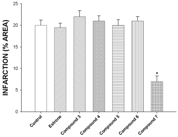
Comparison of cardioprotective effect of the compound 7 at a dose of 1 nM with the compounds 3, 4, 5, 6, estrone and the control conditions on the functional recovery of rat hearts subjected to ischemia and reperfusion.
In order to evaluate this hypothesis, the activity induced by the compound 7 on blood vessel capacity and coronary resistance; translated as changes in perfusion pressure was evaluated in an isolated rat heart model. The results showed that compound 7 significantly increase the perfusion pressure over time (3-18 min) compared with the control conditions (Figure 4). Analyzing this data and evaluating the possibility that functional groups involved in the chemical structure of 7 could be by itself the responsible of their activity on perfusion pressure; in this study, the compounds 3, 4, 5, 6 and estrone were used such pharmacological tool. The results showed that perfusion pressure was not affected in presence of these compounds at a dose of 0.001 nM; these data indicate that functional groups different involved in the compound 7 are the responsible of the activity exerted on perfusion pressure, which could result in changes in coronary resistance. To evaluate this hypothesis, the effects induced by the compounds 3, 4, 5, 6 and estrone on coronary resistance were evaluated (Figure 5). The results showed that coronary resistance in presence of compound 7 was higher (P = 0.05) in comparison the compounds 3, 4, 5, 6, estrone and control conditions.
Figure 4.
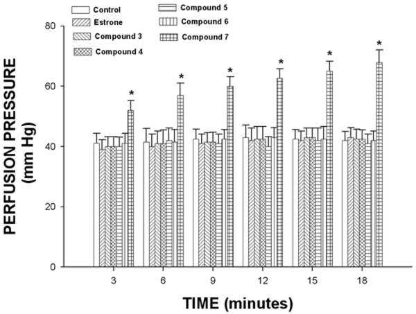
Effect induced by estrone and its derivatives on perfusion pressure. The results show that the compound 7 significantly increase perfusion pressure (P = 0.05) through time in comparison with the compounds 3, 4, 5, 6, estrone and the control conditions. Each bar represents the mean ± S.E. of 9 experiments.
Figure 5.

Activity exerted by estrone and its derivatives on coronary resistance. The results show that coronary resistance was higher (P = 0.05) in the presence of 7 in comparison with the compound 3, 4, 5, 6, estrone and the control conditions. Each bar represents the mean ± S.E. of 9 experiments.
Analyzing these results and other data which indicate that some steroids derivatives can exert effects on left ventricular pressure [44]; in this study, the activity of 7 on left ventricular pressure was evaluated. The results showed that compound 7 higher the left ventricular pressure in a manner dose dependent (Figure 6).
Figure 6.

Effect exerted by the compound 7 on left ventricular pressure (LVP) at dose of 0.001 to 100 nM using an isolated rat heart model.
In the search of molecular mechanism involved in the effect exerted by the compound 7 on left ventricular pressure; in this study, also were analyzed some reports, which indicates that some estrogen derivatives can induce changes on left ventricular pressure via estrogen receptor [45,46]. For this reason, we used tamoxifen, an estrogen receptor blocker to determine if the effects of the compound 7 on left ventricular pressure were via the estrogen receptor. It is noteworthy that interaction of 7 with the estrogen-receptor, may be a key requirement for the biological activity as in the case of other estradiol derivatives [47,48]. Our results (Figure 7) showed that the effect of 7 was not inhibited by tamoxifen, suggesting that the molecular mechanism is not via the estrogen-receptor.
Figure 7.

Effect induced by the compound 7 on LVP through of estrogen receptors. The compound 7 [0.001 to 100 nM] was administered (intracoronary boluses, 50 μl) and the corresponding effect on the LVP was evaluated in the absence and presence of tamoxifen. The results showed that activity induced by 7 on LVP was not inhibited in the presence of tamoxifen. Each bar represents the mean ± S.E. of 9 experiments. LVP = left ventricular pressure.
Analyzing these data and other reports which indicate that some estrogen can exert an indirect tonic effect on adrenal catecholamine synthesis [49,50]; in this study, the activity exerted of compound 7 on left ventricular pressure in the absence or presence of prazosin, metoprolol was evaluated (Figure 8). The results showed that 7 increases the left ventricular pressure in a manner dose dependent and this effect was not inhibited by prazosin, propanolol or metoprolol at dose of 1 nM (Figure 8). These results indicate that the molecular mechanism involved in the effects of this compound 7 on left ventricular pressure was not via adrenergic activity.
Figure 8.
Activity exerted by the compound 7 on LVP through of adrenergic receptors. The compound 7 [0.001 to 100 nM] was administered (intracoronary boluses, 50 μl) and the corresponding effect on the LVP was evaluated in the absence and presence of prazosin or metoprolol. The results showed that activity induced by 7 on LVP was not inhibited in the presence of prazosin or metoprolol. Each bar represents the mean ± S.E. of 9 experiments. LVP = left ventricular pressure.
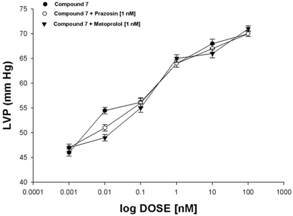
Analyzing other type of reports which indicate that some drugs may induce its effect on left ventricular pressure via prostaglandins synthesis [51]; in this study the activity exerted by the compound 7 on left ventricular pressure in the absence or presence of indomethacin was evaluated to analyze the possibility that the activities exerted by 7 might involve stimulation and secretion of prostaglandins. The results showed that compound 7 increased the left ventricular pressure in a manner dose dependent and this effect was not inhibited with indomethacin (Figure 9); these data suggest that activity exerted by 7 on left ventricular pressure was not through of synthesis and release prostaglandins.
Figure 9.

Effects induced by the compound 7 on LVP through prostaglandins synthesis or calcium channel activation. Intracoronary boluses (50 μl) of the compound 7 [0.001 to 100 nM] were administered and the corresponding effect on the LVP was determined in the absence and presence of indomethacin or nifedipine. The results showed that 7 higher the LVP in a dependent dose manner and this effect was not inhibited in the presence of indomethacin. However, the activity induced by 7 on LVP was blocked by and nifedipine. Each bar represents the mean ± S.E. of 9 experiments. LVP = left ventricular pressure.
In search of a different molecular mechanism induced by 7 on left ventricular pressure; other data which indicate that some estrogen derivatives exert their activity on left ventricular pressure through activation of calcium channel were analyzed [17]. Therefore, in this study the effect exerted by the compound 7 on left ventricular pressure was evaluated using nifedipine as pharmacological tool to characterize whether this mechanism was involved in their pharmacological activity. The results showed that 7 (Figure 9) increases the left ventricular pressure in a dose dependent manner; however this effect was inhibited in presence of nifedipine. All these data indicate that activity exerted by the compound 7 was via calcium channel type L activation.
Conclusions
The estrogen derivative (compound 7) is a particularly interesting drug, because the activity induced for this compound on injury by ischemia/reperfusion involves a molecular mechanism different in comparison with other drugs. This phenomenon may constitute a novel therapy for ischemia-reperfusion injury.
Disclosure of conflict of interest
None.
References
- 1.Yusuf S, Hawken S, Ounpuu S. Obesity and the risk of myocardial infarction in 27,000 participants from 52 countries: a case-control study. Lancet. 2005;366:1640–1649. doi: 10.1016/S0140-6736(05)67663-5. [DOI] [PubMed] [Google Scholar]
- 2.Thygesen K, Alpert J. Universal definition of myocardial infarction. J Am Coll Cardiol. 2007;50:2173–2195. doi: 10.1016/j.jacc.2007.09.011. [DOI] [PubMed] [Google Scholar]
- 3.Pfeffer M. Left ventricular remodeling after acute myocardial infarction. Annu Rev Med. 1995;46:455–466. doi: 10.1146/annurev.med.46.1.455. [DOI] [PubMed] [Google Scholar]
- 4.Klone R, Przyklener K, Whittaker P. Deterious Effects of Oxygen Radicals in Ischemia/Reperfusion. Circulation. 1989;80:1115–1127. doi: 10.1161/01.cir.80.5.1115. [DOI] [PubMed] [Google Scholar]
- 5.Salaris C, Babbs C, Voorhees W. Methylene blue as an inhibitor of superoxide generation by xanthine oxidase: A potential new drug for the attenuation of ischemia/reperfusion injury. Biochem Pharmacol. 1991;42:499–506. doi: 10.1016/0006-2952(91)90311-r. [DOI] [PubMed] [Google Scholar]
- 6.Meng E, Pierce G. Protective effects of 5-(N,N-dimethyl)amiloride on ischemia-reperfusion injury in hearts. Am J Physiol-Heart Circ Physiol. 1999;258:H1615–H1619. doi: 10.1152/ajpheart.1990.258.5.H1615. [DOI] [PubMed] [Google Scholar]
- 7.Chen H, Hung Y, Lee E, Chen T, Chuang I, Wu T. The protective efficacy of magnolol in hind limb ischemia-reperfusion injury. Phytomedicine. 2009;16:976–981. doi: 10.1016/j.phymed.2009.03.001. [DOI] [PubMed] [Google Scholar]
- 8.Cole W, McPherson C, Sontag D. ATP-regulated K+ channels protect the myocardium against ischemia/reperfusion damage. Circ Res. 1991;69:571–581. doi: 10.1161/01.res.69.3.571. [DOI] [PubMed] [Google Scholar]
- 9.Griffiths E, Halestrap A. ATP-regulated K+ channels protect the myocardium against ischemia/ reperfusion damage. J Mol Cel Cardiol. 1993;25:1461–1469. [Google Scholar]
- 10.Rossoni R, Manfredi B, Cavalca V, Razzetti R, Bongrani S, Polvani G. The Aminotetraline Derivative (±)-(R,S)-5,6-Dihydroxy-2-methylamino-1,2,3,4-tetrahydro-naphthalene Hydrochloride (CHF-1024) Displays Cardioprotection in Postischemic Ventricular Dysfunction of the Rat Heart. J Pharmacol Exp Ther. 2003;307:633–639. doi: 10.1124/jpet.103.054700. [DOI] [PubMed] [Google Scholar]
- 11.Hale S, Birnbaum Y, Kloner R. β-Estradiol, but not α-estradiol, reduces myocardial necrosis in rabbits after ischemia and reperfusion. Am Heart J. 1996;132:258–262. doi: 10.1016/s0002-8703(96)90419-6. [DOI] [PubMed] [Google Scholar]
- 12.Kim Y, Chen B, Beauregard J, Kouretas P, Thomas G, Farhat M, Myers A, Lees D. 17ß-Estradiol Prevents Dysfunction of Canine Coronary Endothelium and Myocardium and Reperfusion Arrhythmias After Brief Ischemia/Reperfusion. Circulation. 1996;94:2901–2908. doi: 10.1161/01.cir.94.11.2901. [DOI] [PubMed] [Google Scholar]
- 13.Dhote V, Balaraman R. Gender specific effect of progesterone on myocardial ischemia/reperfusion injury in rats. Life Sci. 2007;81:188–197. doi: 10.1016/j.lfs.2007.05.010. [DOI] [PubMed] [Google Scholar]
- 14.Chang-Her T, Sheng-Fang S, Tsai-Fwu C, Tsung-Ming L. Differential Effects of Sarcolemma and Mitochondrial KATP Channels Activated by 17β-Estradiol on Reperfusion Arrhythmias and Infarct Sizes in Canine Hearts. J Pharmacol Exp Ther. 2002;31:234–240. doi: 10.1124/jpet.301.1.234. [DOI] [PubMed] [Google Scholar]
- 15.Wang M, Crisostomo P, Markel T, Wang Y, Lillemoe K, Meldrum D. Estrogen receptor beta mediates acute myocardial protection following ischemia. Surgery. 2008;144:233–238. doi: 10.1016/j.surg.2008.03.009. [DOI] [PubMed] [Google Scholar]
- 16.Anstead G, Carlson K, Katzenellenbogen J. The estradiol pharmacophore: Ligand structure-estrogen receptor binding affinity relationships and a model for the receptor binding site. Steroids. 1997;62:268–303. doi: 10.1016/s0039-128x(96)00242-5. [DOI] [PubMed] [Google Scholar]
- 17.Figueroa-Valverde L, Diaz-Cedillo F, Lopez-Ramos M, Garcia-Cervera E, Quijano K, Cordoba J. Changes induced by estradiolethylenediamine derivative on perfusion pressure and coronary resistance in isolated rat heart: L-type calcium channel. Biomed Pap. 2011;155:27–32. doi: 10.5507/bp.2011.018. [DOI] [PubMed] [Google Scholar]
- 18.Shiau A, Barstad D, Loria P, Cheng L, Kushner P, Agard D, Greene G. The Structural Basis of Estrogen Receptor/Coactivator Recognition and the Antagonism of This Interaction by Tamoxifen. Cell. 1998;95:927–937. doi: 10.1016/s0092-8674(00)81717-1. [DOI] [PubMed] [Google Scholar]
- 19.Kristek F, Koprdova R. Long-Term Effect of Prazosin Administration on Blood Pressure Heart and Structure of Coronary Artery of Young Spontaneously Hypertensive Rats. J Physiol Pharmacol. 1977;62:295–301. [PubMed] [Google Scholar]
- 20.Bengtsson C, Johnsson G, Regardh CG. Plasma Levels and Effects of Metoprolol on Blood Pressure and Heart Rate in Hypertensive Patients after an Acute Dose and between Two Doses during Long-Term Treatment. Clin Pharmacol Ther. 1975;17:400–408. doi: 10.1002/cpt1975174400. [DOI] [PubMed] [Google Scholar]
- 21.Blouin M, Han Y, Burch J, Farand J, Mellon C, Gaudreault M. The Discovery of 4-{1-[({2,5-Dimethyl-4-[4-(trifluorome-thyl)benzyl] -3-thien yl}carbonyl)amino] cyclopropyl} benzoic Acid (MK-2894), A Potent and Selective Prostaglandin E2 Subtype 4 Receptor Antagonist. J Med Chem. 2010;53:2227–2238. doi: 10.1021/jm901771h. [DOI] [PubMed] [Google Scholar]
- 22.Henry P. Comparative Pharmacology of Calcium Antagonists: Nifedipine, Verapamil and Diltiazem. Am J Cardiol. 1980;46:1047–1058. doi: 10.1016/0002-9149(80)90366-5. [DOI] [PubMed] [Google Scholar]
- 23.Figueroa-Valverde L, Díaz-Cedillo F, García-Cervera E, Pool Gómez E, Rosas-Nexticapa M, Mendoza-López R, López-Ramos M, May-Gil I. Design and Synthesis of Estrogen Derivative. Asian J Chem. 2014;26:1007–1010. [Google Scholar]
- 24.Mannhod R, Waterbeemd H. Substructure and whole approaches for calculating logP. J Comput Aided Mol Des. 2001;15:337–354. doi: 10.1023/a:1011107422318. [DOI] [PubMed] [Google Scholar]
- 25.Bayne K. Revised Guide for the Care and Use of Laboratory Animals Available. Physiologist. 1996;9:208–211. [PubMed] [Google Scholar]
- 26.Figueroa-Valverde L, Díaz-Cedillo F, López-Ramos M, García-Cervera E, Quijano-Ascencio K. Inotropic Activity Induced by Carbamazepine-AlKyne Derivative in an Isolated Heart Model and Perfused to Constant Flow. Biomedica. 2011;31:232–241. doi: 10.1590/S0120-41572011000200011. [DOI] [PubMed] [Google Scholar]
- 27.Booth E, Obeid N, Lucchesi B. Activation of Estrogen Receptor-α Protects the in Vivo Rabbit Heart from Ischemia-Reperfusion Injury. Am J Physiol Heart Circ Physiol. 2005;289:H2039–H2047. doi: 10.1152/ajpheart.00479.2005. [DOI] [PubMed] [Google Scholar]
- 28.Hocht C, Opezzo J, Gorzalczany S. Una Aproxi-mación Cinética y Dinámica de Metildopa en Ratas con Coartación Aórtica Mediante Microdiálisis. Revista Arg Cardiol. 1999;67:769–773. [Google Scholar]
- 29.Hussey S, Muddana S, Peterson B. Synthesis of a β-Estradiol-Biotin Chimera that Potently Heterodimerizes Estrogen Receptor and Streptavidin Proteins in a Yeast Three-Hybrid System. J Am Chem Soc. 2003;125:3692–3693. doi: 10.1021/ja0293305. [DOI] [PubMed] [Google Scholar]
- 30.Djerassi C, Rosenkranz G, Romo J, Pataki J, Kaufmann S. Steroids. VIII.1,2 The Dienone-Phenol Rearrangement in the Steroid Series. Synthesis of a New Class of Estrogens. J Am Chem Soc. 1950;72:4540–4544. [Google Scholar]
- 31.Kopach M, Kelsh L, Stork K, Harman W. Direct 10. beta.-alkylation of .beta.-estradiol: osmium(II) as a dearomatization agent. J Am Chem Soc. 1993;15:5322–5323. [Google Scholar]
- 32.Takekoshi T. Synthesis of high performance aromatic polymers via nucleophilic nitro displacement reaction. Polymer J. 1987;19:191–202. [Google Scholar]
- 33.Leo A, Jow P, Silipo C. Calculation of hydrophobic constant (log P) from pi and f constants. J Med Chem. 1975;18:865–868. doi: 10.1021/jm00243a001. [DOI] [PubMed] [Google Scholar]
- 34.Leo A, Hoekman D. Calculating log P(oct) with no missing fragments; The problem of estimating new interaction parameters. Perspect Drug Discov Des. 2000;18:19–38. [Google Scholar]
- 35.Hansch C, Leo A, Taft R. A survey of Hammett substituent constants and resonance and field parameters. Chem Rev. 1991;91:165–195. [Google Scholar]
- 36.Yoshid K, Shigeoka T, Yamauchi F. Relationship between molar refraction and n-octanol/water partition coefficient. Ecotoxicol Environ Saf. 1983;7:558–565. doi: 10.1016/0147-6513(83)90015-5. [DOI] [PubMed] [Google Scholar]
- 37.Schnackenberg K, Beger D. Whole-molecule calculation of log P based on molar volume, hydrogen bonds, and simulated 13C NMR spectra. J Chem Inf Model. 2005;45:360–365. doi: 10.1021/ci049643e. [DOI] [PubMed] [Google Scholar]
- 38.Takur A. QSAR study on benzenesulfonamide dissociation constant pKa: physicochemical approach using surface tension. Arkivoc. 2005;xiv:49–58. [Google Scholar]
- 39.Dimova V, Perisic-Janjic N. QSAR study by 1,2,4-triazoles using several physicochemical descriptors. Macedonian J Chem Chem Engin. 2009;28:79–89. [Google Scholar]
- 40.Node K, Kitakaze M, Kosaka H. Amelioration of Ischemia- and Reperfusion-Induced Myocardial Injury by 17β-Estradiol. Circulation. 1997;96:1953–1963. doi: 10.1161/01.cir.96.6.1953. [DOI] [PubMed] [Google Scholar]
- 41.Suparto I, Koudy W, Fox J. A comparison of two progestins on myocardial ischemia–reperfusion injury in ovariectomized monkeys receiving estrogen therapy. Coron Artery Dis. 2005;16:301–308. doi: 10.1097/00019501-200508000-00007. [DOI] [PubMed] [Google Scholar]
- 42.Jeanes H, Wanikiat P, Sharif I. A Medroxyprogesterone acetate inhibits the cardioprotective effect of estrogen in experimental ischemia-reperfusion injury. Menopause. 2006;13:80–86. doi: 10.1097/01.gme.0000196593.44335.eb. [DOI] [PubMed] [Google Scholar]
- 43.Bouïs D, Hospers G, Meijer C. Endothelium in vitro: A review of human vascular endothelial cell lines for blood vessel-related research. Angiogenesis. 2000;4:91–102. doi: 10.1023/a:1012259529167. [DOI] [PubMed] [Google Scholar]
- 44.Figueroa-Valverde L, Diaz-Ku E, Diaz-Cedillo F, Baqueiro-Bricaire A, Camacho-Luis A. Effects of danazol and danazol hemisuccinate on perfusión pressure and vascular resistance. Acta Bioquim Clin Latinoam. 2010;44:37–45. [Google Scholar]
- 45.Karas RH, Patterson BL, Mendelsohn ME. Human vascular smooth muscle cells contain functional estrogen receptor. Circulation. 1994;89:1943–1950. doi: 10.1161/01.cir.89.5.1943. [DOI] [PubMed] [Google Scholar]
- 46.Mahmoodzadeh S, Fritschka S, Dworatzek E, Pham T, Becher E, Kuehne A, Davidson M, Regitz-Zagrosek V. Nuclear Factor-κB Regulates Estrogen Receptor-α Transcription in the Human Heart. J Biol Chem. 2009;284:24705–24714. doi: 10.1074/jbc.M109.000463. [DOI] [PMC free article] [PubMed] [Google Scholar]
- 47.Haynes M, Sinha E, Russell K, Collinge M, Fulton D, Morales-Ruiz M, Sessa W, Bender J. Membrane Estrogen Receptor Engagement Activates Endothelial Nitric Oxide Synthase via the PI3-Kinase-Akt Pathway in Human Endothelial Cells. Circ Res. 2000;87:677–682. doi: 10.1161/01.res.87.8.677. [DOI] [PubMed] [Google Scholar]
- 48.Kaspar P, Witzel H. Steroid binding to the cytosolic estrogen receptor from rat uterus. influence of the orientation of substituents in the 17-position of the 8β- and 8α-series. J Steroids Biochem. 1985;23:259–265. doi: 10.1016/0022-4731(85)90403-0. [DOI] [PubMed] [Google Scholar]
- 49.Sudhir K, Esler MD, Garry K, Jennings L, Komesaroff P. Estrogen Supplementation Decreases Norepinephrine-Induced Vasoconstriction and Total Body Norepinephrine Spillover in Perimenopausal Women. Hypertens. 1997;30:1538–1543. doi: 10.1161/01.hyp.30.6.1538. [DOI] [PubMed] [Google Scholar]
- 50.Peng N, Clark JT, Wei C, Wyss JM. Estrogen Depletion Increases Blood Pressure and Hypothalamic Norepinephrine in Middle-Aged Spontaneously Hypertensive Rats. Hypertension. 2003;41:1164–1167. doi: 10.1161/01.HYP.0000065387.09043.2E. [DOI] [PubMed] [Google Scholar]
- 51.Seillan C, Ody C, Russo F, Duval D. Differential aspects of sex steroids on prostaglandin secretion by male and female cultured piglet endothelial cells. Prostaglandins. 1983;26:3–12. doi: 10.1016/0090-6980(83)90069-2. [DOI] [PubMed] [Google Scholar]