


Abstract
Arrhythmogenic cardiomyopathy (AC) is a familial heart muscle disease with mutations of desmosomal gene and its pathogenesis is related with mutations of desmosomal gene and abnormality of connexin43 (Cx43). One of Rho GTPase, RhoA involves in many pathological processes and is regulated by desmosomal gene PKP2. We aim to identify if PKP2 regulate RhoA activity in myocardium of AC patients, the activity change of RhoA in patients’ myocardium and to investigate the effect of active RhoA on the protein expression of Cx43 in myocardial cells. The protein expression level was assessed by western blot and the activity of RhoA was assessed by RhoA protein activation assay. Our results showed that the expression of PKP2 was decreased in myocardium of three patients, one of which carried PKP2 mutations. The activity of RhoA in myocardium was increased in myocardium of AC as compared with healthy control except for the patient with PKP2 mutation, the expression of Cx43 was also increased in those patients with increased activity of RhoA. The results in vitro demonstrated that the increase of active RhoA can cause the change of protein expression of Cx43 in HL-1 cardiomyocytes. In conclusion, regulation of RhoA activity is complex in the myocardium of AC and the activity of RhoA is increased in AC patients without PKP2 mutations. What’s more, the active RhoA affects the protein expression of Cx43 in vivo and in vitro, this may be the possible disease mechanism of AC.
Keywords: Arrhythmogenic cardiomyopathy, RhoA, activity, connexin 43, cytoskeleton
Introduction
Arrhythmogenic cardiomyopathy (AC) is a myocardial disease characterized by fibro-fatty replacement and ventricular arrhythmias. It is a leading cause of sudden cardiac death (SCD) in people aged ≤ 35 years and accounts for up to 10% of deaths from cardiac disease in people aged < 65 years [1]. In recent years, it has been recognized that AC is a genetically determined disorder. The majority of the genes actually known to cause AC are those encoding cardiac desmosomal proteins (plakoglobin, plakophilin-2, desmoplakin, desmoglein, and desmocollin) [2]. One of the desmosomal protein, plakophilin-2 (PKP2), seems to be the most common mutations in these desmosomal genes [3,4]. Cardiomyocytes with dysfunctional desmosomes can be torn apart more easily, which leads to cell death and fibro-fatty replacement as an unspecific repair mechanism. Connexin 43 (Cx43), one of the gap junction in intercalated disc, is structure that underlie the electrical conduction between myocytes [5], and the cellular contact provided by desmosome is necessary for electric conduction and mechanical contraction of the myocardial cells. In addition, parts of the pathogenesis of AC are related with the abnormality of Cx43 [5,6].
RhoA, one isoform of Rho GTPase, involves in many pathological processes [7-9], and it has been reported that one of the desmosomal protein, PKP2, regulated RhoA activity [10]. However, the change of RhoA activity in AC patients and the relationship between RhoA and Cx43 have never been reported.
In this study, we detected the expression level of PKP2 and RhoA activity in myocardium of AC patients who underwent heart transplantation and discussed whether active RhoA was regulated by PKP2, we also demonstrated that active RhoA affects protein expression of Cx43 in vivo and in vitro. Our study may supply a new perspective in research of disease mechanisms of AC.
Material and methods
Ethics statement
The patients enrolled in the present study included definite AC individuals who underwent heart transplantation in Fuwai hospital between 2005 and 2012. These subjects were diagnosed using the Task Force criteria for AC. All patients have signed a written informed consent for this investigation, which was approved by the Institutional Ethical Review Board of Fuwai Hospital (Beijing, China).
Tissue sample preparation and histology
Cardiac tissue samples were collected from these AC patients mentioned above. The control samples were derived from autopsies or donors with no history of heart disease who died in accidents. For histology, 3 µm paraffin sections of cardiac tissue were stained with haematoxylin and eosin and Masson’s trichrome. Frozen sections were stained with oil red O to detect lipids.
Protein extraction and immunoblot analysis
Snap frozen tissues were homogenized in buffer (8 M Urea, 50 mM Tris-HCl, pH 7.5, 100 mM NaCl, 1 mM DTT, 1% Triton-X-100) supplemented with 0.1% PMSF. Then centrifuged for 30 min at 4°C, 13,400 g. Primary antibodies against PKP2 (Abcam, Cambrige, UK), RhoA (cytoskeleton, Denver, USA), Cx43 (Cell Signaling Technology, Inc., Danvers, USA), and the loading control GAPDH (Abmart, Shanghai, China) were used. All blotting experiments were made in triplicates.
Cell culture and test reagents
The HL-1 cells was grown in Dulbeccos modified Eagles medium (DMEM) supplemented with 10% fetal calf serum (FCS) in a humidified atmosphere (95% air/5% CO2) at 37°C. The cultures were used for experiments when grown to confluent monolayers. Calpeptin was bought from Abcam and applied at 100 μg/ml. A specific inhibitor of ROCK, Y-27632 (Calbiochem, CA), was added in 10% FBS/DMEM during the first 1 day to inhibit intracellular RhoA activity, the inhibitor was applied at 10 μM.
RhoA protein activation assay
To test for RhoA protein activation in HL-1 cells upon treatment with calpeptin (100 μg/ml, 10 min, 30 min, and 60 min), the RhoA activation assay kit (Cytoskeleton, Denver, USA) were applied following manufacturer’s instructions. In brief, HL-1 cells were grown to confluence in T75 culture flasks for 3 days, the medium was replaced with DMEM plus 1% FCS and cultured for 24 h, and serum-starved for 16 h-24 h and then incubated in the absence (control) or presence of calpeptin (100 μg/ml, 10 min, 30 min, and 60 min) in DMEM. After rinsing with ice-cold PBS, cells were harvested in lysis buffer containing 50 mM Tris-HCl (pH 7.4), 100 mM NaCl, 1 mM DTT, 1% Triton X-100, 0.1% PMSF. Lysates were centrifuged 15 min at 3000 g at 4°C. Supernatants were incubated with rhotekin binding domain-beads (cytoskeleton) and rotated for 60 min at 4°C, washed one time using washing buffer (cytoskeleton) and then suspended in 2 × Laemmli sample buffer (125 mM Tris PH 6.8, 20% glycerol, 4% SDS, 0.005% Bromophenol blue, 5% beta-mercaptoethanol, cytoskeleton), RhoA was detected by western blot using a monoclonal antibody to RhoA (cytoskeleton).
Statistical analysis
Quantitative data were expressed as mean ± S.E.M. Unpaired Student’s t test was used for comparison of data between groups. P < 0.05 was considered to be statistically significant.
Results
The pathological characteristic of AC patients
Myocardial tissues of right ventricle from independent AC patients who received heart transplantation were obtained in this study. The representative pathological characteristics of them are shown in Figure 1. This results showed that compared with healthy control, cardiocytes in myocardial tissues of AC were arranged more disorderly, the myocardial cells presented cell islets, and surrounded by fibrous tissues (Figure 1E, 1F). There were also lots of infiltrated fat tissues in myocardium (Figure 1H, 1I).
Figure 1.
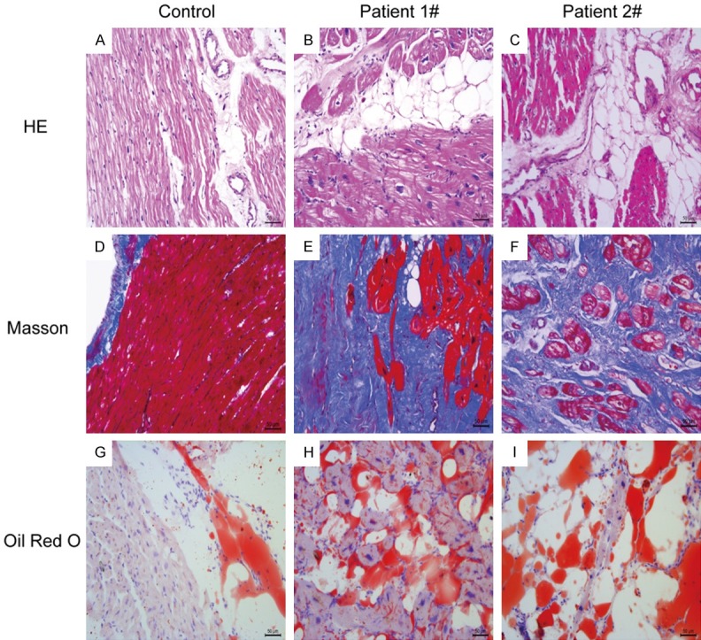
Representative pathological characteristics of right ventricular (RV) myocardium in patients with AC. A, D, G. HE, Masson and Oil Red O staining of myocardium in healthy control. B, C, E, F, H, I. HE, Masson and Oil Red O staining of myocardium of AC patients. Scale bars = 50 μm.
The protein expression of PKP2 and RhoA activity assay in myocardium
Due to that PKP2 is one of the most important desmosomal protein in pathogenesis of AC, we measured the protein expression of PKP2 in myocardium of AC, and found that the expression level of PKP2 in patient 1#, 2#, and 3# was decreased significantly as compared with controls and other AC patients (Figure 2A).
Figure 2.
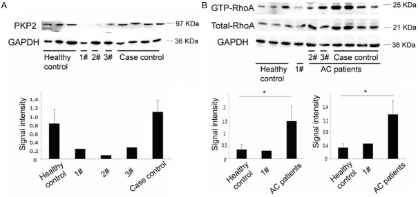
Protein expression analyses of PKP2 and activity analyses of RhoA of RV in AC patients. A. Immunoblot expression analysis of PKP2 in RV myocardium of healthy controls and AC patients and the quantification. Values represent averages ± SE (n = 3, control, n = 4, patients). B. Activity and protein expression analysis of RhoA in RV myocardium of healthy controls and AC patients and the quantification. Values represent averages ± SE (n = 3, control, n = 6, patients). *, P < 0.05. 1#: AC patients with PKP2 mutations; 2#, 3#: AC patients without PKP2 mutations. Case control: AC patients with normal expression of PKP2 and without PKP2 mutations.
Among the three patients in which the expression level of PKP2 decreased, patient 1# carried PKP2 mutations (data not shown), and the result of pull-down assays for GTP-bound RhoA showed that the activity of RhoA was also decreased in myocardium of patient 1#, the other two patients showed increased activity of RhoA in myocardium, what’s more, in myocardium without expression level change of PKP2, RhoA activity was also increased; this result was consistent to the change of the expression level of the total RhoA, which was also increased in AC patients compared with healthy controls (Figure 2B).
The expression level change of Cx43 in myocardium of AC patients
We further detected expression level of Cx43 in myocardium, and found that in myocardium of patient 1#, the expression of Cx43 was decreased, in patient 2#, 3#, and other AC patients’ myocardial tissues, in which the activity of RhoA increased, the protein expression of Cx43 was increased compared with healthy controls (Figure 3).
Figure 3.
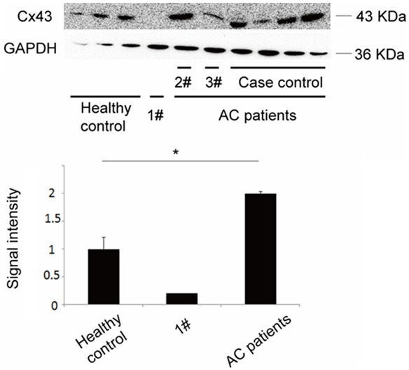
Protein expression analyses of Cx43 in RV myocardium of healthy controls and AC patients and the quantification. Values represent averages ± SE (n = 3, control, n = 6, patients). *, P < 0.05. 1#: AC patients with PKP2 mutations; 2#, 3#: AC patients without PKP2 mutations. Case control: AC patients with normal expression of PKP2 and without PKP2 mutations.
The active RhoA affected the protein expression of Cx43 in HL-1 cells
To simulate the increased activity of RhoA in myocardial tissues, we treated HL-1 cells with calpeptin to activate RhoA (Figure 4A), the result showed that compared with control, the activity of RhoA in HL-1 was increased after stimulated for 30 minutes, after treatment for 60 minutes, the activity of RhoA decreased to normal level.
Figure 4.
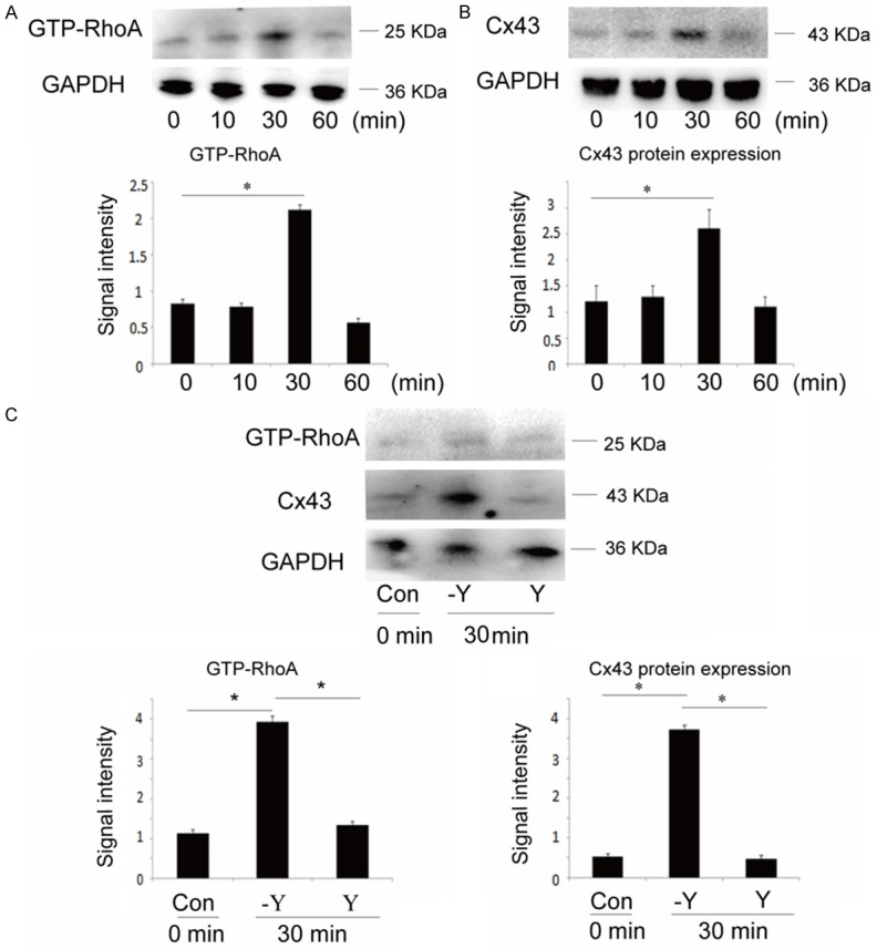
In vitro activity analyses of RhoA and protein expression analyses of Cx43 in HL-1 cells. A. Activity analysis of RhoA in HL-1 cells with treated by calpeptin for 0, 10, 30, and 60 min. B. Cx43 protein expression analysis in HL-1 cells with treated by calpeptin for 0, 10, 30, and 60 min. C. Activity analysis of RhoA and Cx43 protein expression analysis in HL-1 cells which were treated with 10 μm Y-27632 (Y) prior to the stimulation of calpeptin. Values represent averages ± SE (n = 3). *, P < 0.05.
Apart from the activity of RhoA, the protein expression level of Cx43 was increased when stimulated with calpeptin for 30 minutes, in contrast, it had no change with treatment of calpeptin for 10 minutes and 60 minutes compared with control (Figure 4B).
As shown in Figure 4C, the activity of RhoA was inhibited by Rho kinase inhibitor Y-27632, and while the RhoA activity decreased, the Cx43 protein expression markedly reduced when compared with control cells, providing further evidence that the activity of RhoA may regulate protein expression of Cx43 in HL-1.
Discussion
AC is a cardiomyopathy regarded as an important cause of sudden death in young people and particularly in athletes [11], the pathogenesis of AC is progressive replacement of the right ventricular myocardium by fibro-fatty tissue. The fibro-fatty replacement interferes with electrical impulse conduction, it is the key cause of epsilon waves, right bundle branch block, late potentials, and re-entrant ventricular arrhythmias [11]. Though AC is referred to as “a disease of the desmosome”, there are only 50% AC patients carrying pathogenic gene mutations [12], and the pathogenesis of the other 50% patients of AC was unknown.
The pathogenic mechanism of desmosomal gene mutations constitutes the dystrophic theories [13], the most general theory. The discovery of mutations in desmosomal genes proposes that impairment of desmosomal function attributable to genetic defects would result in cardiomyocytes detachment and death, particularly in the setting of mechanical stress [14].
The variants of PKP2, one of the pathogenic genes, are relatively common among AC patients [4], and it plays an essential role in desmosome formation, cell signaling [15] and regulating cardiac sodium channel function [16]. Our result showed that the expression level of PKP2 was decreased in three samples. There are three possibilities about this result: protein degradation, transcriptional defects and translational defects. However, the exact mechanism of the reduction is still unclear, and needs to be verified in further research.
From the study of Godsel et al [10], PKP2 can regulate RhoA activity in desmosomal plaque assembles. To address the possibility that PKP2 could regulate the activity of RhoA in myocardial tissue of AC, we detected RhoA activity in these myocardial tissues. Interestingly, in our report, except the patient who carries PKP2 mutations, the activity of RhoA was increased in myocardium of remaining AC patients whether the PKP2 expression level changed or not. This result was inconsistent with Godsel’s [10], the possible explanation may be that in myocardial tissues, especially in myocardial tissues of AC, the regulation system of RhoA activity is much more complex than we expected, the regulation of PKP2 on RhoA activity is also complex, many factors involved in the regulation process, such as PKP2 mutation; we also speculate that the increase of RhoA activity may involve in the disease mechanism of AC considering the activity of RhoA increased in myocardium of AC patients, this result was consistant with the changes of expression level of total RhoA, it implied that the increase of RhoA activity may partly due to the increase of expression level of total RhoA.
Cx43, a gap junction in intercalated disc, is an important structure in myocardiocytes and it involved in the pathogenesis of AC. It is reported that in AC patients, PKP2 mutation can lead to decreased expression level of Cx43 [5]. In myocardiocytes, knockdown of DSP can cause the change of location and expression level of Cx43 [6]. Apart from desmosomal genes, the pathogenesis of non-desmosomal genes is related with Cx43. One mutation of TMEM43, p S358L, leads to the change of location of Cx43 in intercalated disc, and this effect may involve in the process of AC [17]. From these results, we can speculate that the abnormality of Cx43 is related with the pathogenesis of AC. In our study, the expression level of Cx43 was increased in AC patients in which myocardium the RhoA activity increased, and in HL-1 cells with active RhoA, the expression of Cx43 was also increased. These results suggest that RhoA may involve in the process of AC by influencing protein expression of Cx43.
In heart, changes of the activity of RhoA are significant in myocardial function, it involves in assembling of desmosome, myocardial cell apoptosis [7], and heart failure [18]. In view of pathological characteristics of AC, RhoA regulate a switch between adipogenesis and myogenesis [19]. Apart from affecting protein expression of Cx43, we speculate that RhoA involves in AC in other two ways: (1) affecting NaV1.5 to induce arrhythmia genesis, (2) regulating differentiaion between adipogenesis and myogenesis result in the appearance of adipose tissue. Further research need to explore this conjecture.
AC is characterized genetically as a disease of the desmosome, through several decades of genetics research and discovery, many pathogenic gene mutations have been found. Besides mutational desmosomal genes, there are other potential mechanisms in AC, such as ion channel abnormity [20], inhibiting Wnt signaling pathway [21] and myocyte transdifferentiation [22]. Chen et al [23] suggested that the hippo pathway is causal mechanism in AC, Li et al [24] found that transforming growth factor β-mediated signaling is elevated in Jup mutant cardiomyocytes, and suggested that it is an important pathogenic pathway for Jup-related AC. Studying in new disease mechanism have provided further insights for the pathogenesis of AC and potential therapeutic interventions.
There is a growing study about the pathophysiological role of RhoA in cardiovascular diseases, such as adriamycin-induced cardiomyopathy [25], cardiomyocyte hypertrophy [26]. However, what is the role of RhoA in inherited cardiomyopathy, such as AC remains unknown. As the expression and activity of RhoA increased in myocardium of AC, our report points out a new clue about the role of RhoA in the process of AC, and suggests another new potential pathogenesis of AC.
Acknowledgements
We are grateful to our patients and their families for their support and collaboration. This work was supported by research grants from the National Natural Science Foundation of China (81170206 to Y.J.W.).
Disclosure of conflict of interest
None.
References
- 1.Sen-Chowdhry S, Morgan RD, Chambers JC, McKenna WJ. Arrhythmogenic cardiomyopathy: etiology, diagnosis, and treatment. Annu Rev Med. 2010;61:233–253. doi: 10.1146/annurev.med.052208.130419. [DOI] [PubMed] [Google Scholar]
- 2.Boldt LH, Haverkamp W. Arrhythmogenic right ventricular cardiomyopathy: diagnosis and risk stratification. Herz. 2009;34:290–297. doi: 10.1007/s00059-009-3246-1. [DOI] [PubMed] [Google Scholar]
- 3.Saffitz JE. Desmosome mutations in arrhythmogenic right ventricular cardiomyopathy: important insight but only part of the picture. Circ Cardiovasc Genet. 2009;2:415–417. doi: 10.1161/CIRCGENETICS.109.909366. [DOI] [PubMed] [Google Scholar]
- 4.Gerull B, Heuser A, Wichter T, Paul M, Basson CT, McDermott DA, Lerman BB, Markowitz SM, Ellinor PT, MacRae CA, Peters S, Grossmann KS, Drenckhahn J, Michely B, Sasse-Klaassen S, Birchmeier W, Dietz R, Breithardt G, Schulze-Bahr E, Thierfelder L. Mutations in the desmosomal protein plakophilin-2 are common in arrhythmogenic right ventricular cardiomyopathy. Nat Genet. 2004;36:1162–1164. doi: 10.1038/ng1461. [DOI] [PubMed] [Google Scholar]
- 5.Wang PN, Wu SL, Zhang B, Lin QX, Shan ZX. Function of a novel plakophilin-2 mutation in the abnormal expression of connexin43 in a patient with arrhythmogenic right ventricular cardiomyopathy. Exp Ther Med. 2015;9:967–971. doi: 10.3892/etm.2014.2145. [DOI] [PMC free article] [PubMed] [Google Scholar]
- 6.Zhang Q, Deng C, Rao F, Modi RM, Zhu J, Liu X, Mai L, Tan H, Yu X, Lin Q, Xiao D, Kuang S, Wu S. Silencing of desmoplakin decreases connexin 43/Nav1.5 expression and sodium current in HL1 cardiomyocytes. Mol Med Rep. 2013;8:780–786. doi: 10.3892/mmr.2013.1594. [DOI] [PubMed] [Google Scholar]
- 7.Del Re DP, Miyamoto S, Brown JH. RhoA/Rho kinase up-regulate Bax to activate a mitochondrial death pathway and induce cardiomyocyte apoptosis. J Biol Chem. 2007;282:8069–8078. doi: 10.1074/jbc.M604298200. [DOI] [PubMed] [Google Scholar]
- 8.Dulong C, Fang YJ, Gest C, Zhou MH, Patte-Mensah C, Mensah-Nyagan AG, Vannier JP, Lu H, Soria C, Cazin L, Mei YA, Varin R, Li H. The small GTPase RhoA regulates the expression and function of the sodium channel Nav1.5 in breast cancer cells. Int J Oncol. 2014;44:539–547. doi: 10.3892/ijo.2013.2214. [DOI] [PubMed] [Google Scholar]
- 9.Hosoyama T, Ishiguro N, Yamanouchi K, Nishihara M. Degenerative muscle fiber accelerates adipogenesis of intramuscular cells via RhoA signaling pathway. Differentiation. 2009;77:350–359. doi: 10.1016/j.diff.2008.11.001. [DOI] [PubMed] [Google Scholar]
- 10.Godsel LM, Dubash AD, Bass-Zubek AE, Amargo EV, Klessner JL, Hobbs RP, Chen X, Green KJ. Plakophilin 2 couples actomyosin remodeling to desmosomal plaque assembly via RhoA. Mol Biol Cell. 2010;21:2844–2859. doi: 10.1091/mbc.E10-02-0131. [DOI] [PMC free article] [PubMed] [Google Scholar]
- 11.Basso C, Corrado D, Marcus FI, Nava A, Thiene G. Arrhythmogenic right ventricular cardiomyopathy. Lancet. 2009;373:1289–1300. doi: 10.1016/S0140-6736(09)60256-7. [DOI] [PubMed] [Google Scholar]
- 12.Saffitz JE. Arrhythmogenic cardiomyopathy and abnormalities of cell-to-cell coupling. Heart Rhythm. 2009;6:S62–65. doi: 10.1016/j.hrthm.2009.03.003. [DOI] [PubMed] [Google Scholar]
- 13.Basso C, Thiene G, Corrado D, Angelini A, Nava A, Valente M. Arrhythmogenic right ventricular cardiomyopathy. Dysplasia, dystrophy, or myocarditis? Circulation. 1996;94:983–991. doi: 10.1161/01.cir.94.5.983. [DOI] [PubMed] [Google Scholar]
- 14.Protonotarios N, Tsatsopoulou A. Naxos disease and Carvajal syndrome: cardiocutaneous disorders that highlight the pathogenesis and broaden the spectrum of arrhythmogenic right ventricular cardiomyopathy. Cardiovasc Pathol. 2004;13:185–194. doi: 10.1016/j.carpath.2004.03.609. [DOI] [PubMed] [Google Scholar]
- 15.Chen X, Bonne S, Hatzfeld M, van Roy F, Green KJ. Protein binding and functional characterization of plakophilin 2. Evidence for its diverse roles in desmosomes and beta -catenin signaling. J Biol Chem. 2002;277:10512–10522. doi: 10.1074/jbc.M108765200. [DOI] [PubMed] [Google Scholar]
- 16.Cerrone M, Delmar M. Desmosomes and the sodium channel complex: implications for arrhythmogenic cardiomyopathy and Brugada syndrome. Trends Cardiovasc Med. 2014;24:184–190. doi: 10.1016/j.tcm.2014.02.001. [DOI] [PMC free article] [PubMed] [Google Scholar]
- 17.Siragam V, Cui X, Masse S, Ackerley C, Aafaqi S, Strandberg L, Tropak M, Fridman MD, Nanthakumar K, Liu J, Sun Y, Su B, Wang C, Liu X, Yan Y, Mendlowitz A, Hamilton RM. TMEM43 mutation p. S358L alters intercalated disc protein expression and reduces conduction velocity in arrhythmogenic right ventricular cardiomyopathy. PLoS One. 2014;9:e109128. doi: 10.1371/journal.pone.0109128. [DOI] [PMC free article] [PubMed] [Google Scholar]
- 18.Sah VP, Minamisawa S, Tam SP, Wu TH, Dorn GW 2nd, Ross J Jr, Chien KR, Brown JH. Cardiac-specific overexpression of RhoA results in sinus and atrioventricular nodal dysfunction and contractile failure. J Clin Invest. 1999;103:1627–1634. doi: 10.1172/JCI6842. [DOI] [PMC free article] [PubMed] [Google Scholar]
- 19.Sordella R, Jiang W, Chen GC, Curto M, Settleman J. Modulation of Rho GTPase signaling regulates a switch between adipogenesis and myogenesis. Cell. 2003;113:147–158. doi: 10.1016/s0092-8674(03)00271-x. [DOI] [PubMed] [Google Scholar]
- 20.Sato PY, Musa H, Coombs W, Guerrero-Serna G, Patino GA, Taffet SM, Isom LL, Delmar M. Loss of plakophilin-2 expression leads to decreased sodium current and slower conduction velocity in cultured cardiac myocytes. Circ Res. 2009;105:523–526. doi: 10.1161/CIRCRESAHA.109.201418. [DOI] [PMC free article] [PubMed] [Google Scholar]
- 21.Garcia-Gras E, Lombardi R, Giocondo MJ, Willerson JT, Schneider MD, Khoury DS, Marian AJ. Suppression of canonical Wnt/beta-catenin signaling by nuclear plakoglobin recapitulates phenotype of arrhythmogenic right ventricular cardiomyopathy. J Clin Invest. 2006;116:2012–2021. doi: 10.1172/JCI27751. [DOI] [PMC free article] [PubMed] [Google Scholar]
- 22.D’Amati G, di Gioia CR, Giordano C, Gallo P. Myocyte transdifferentiation: a possible pathogenetic mechanism for arrhythmogenic right ventricular cardiomyopathy. Arch Pathol Lab Med. 2000;124:287–290. doi: 10.5858/2000-124-0287-MT. [DOI] [PubMed] [Google Scholar]
- 23.Chen SN, Gurha P, Lombardi R, Ruggiero A, Willerson JT, Marian AJ. The hippo pathway is activated and is a causal mechanism for adipogenesis in arrhythmogenic cardiomyopathy. Circ Res. 2014;114:454–468. doi: 10.1161/CIRCRESAHA.114.302810. [DOI] [PMC free article] [PubMed] [Google Scholar]
- 24.Li D, Liu Y, Maruyama M, Zhu W, Chen H, Zhang W, Reuter S, Lin SF, Haneline LS, Field LJ, Chen PS, Shou W. Restrictive loss of plakoglobin in cardiomyocytes leads to arrhythmogenic cardiomyopathy. Hum Mol Genet. 2011;20:4582–4596. doi: 10.1093/hmg/ddr392. [DOI] [PMC free article] [PubMed] [Google Scholar]
- 25.Wang N, Guan P, Zhang JP, Chang YZ, Gu LJ, Hao FK, Shi ZH, Wang FY, Chu L. Preventive effects of fasudil on adriamycin-induced cardiomyopathy: possible involvement of inhibition of RhoA/ROCK pathway. Food Chem Toxicol. 2011;49:2975–2982. doi: 10.1016/j.fct.2011.06.080. [DOI] [PubMed] [Google Scholar]
- 26.Rangrez AY, Bernt A, Poyanmehr R, Harazin V, Boomgaarden I, Kuhn C, Rohrbeck A, Frank D, Frey N. Dysbindin is a potent inducer of RhoA-SRF-mediated cardiomyocyte hypertrophy. J Cell Biol. 2013;203:643–656. doi: 10.1083/jcb.201303052. [DOI] [PMC free article] [PubMed] [Google Scholar]