

Abstract
The anaesthetic management of patients with pre-existing pulmonary disease is a challenging task. It is associated with increased morbidity in the form of post-operative pulmonary complications. Pre-operative optimisation of lung function helps in reducing these complications. Patients are advised to stop smoking for a period of 4–6 weeks. This reduces airway reactivity, improves mucociliary function and decreases carboxy-haemoglobin. The widely used incentive spirometry may be useful only when combined with other respiratory muscle exercises. Volume-based inspiratory devices have the best results. Pharmacotherapy of asthma and chronic obstructive pulmonary disease must be optimised before considering the patient for elective surgery. Beta 2 agonists, inhaled corticosteroids and systemic corticosteroids, are the main drugs used for this and several drugs play an adjunctive role in medical therapy. A graded approach has been suggested to manage these patients for elective surgery with an aim to achieve optimal pulmonary function.
Keywords: Asthma, beta 2 agonists, chronic obstructive pulmonary disease, incentive spirometry, inhaled steroids, post-operative pulmonary complications, smoking
INTRODUCTION
Pre-existing pulmonary diseases are increasingly encountered in patients presenting for elective and emergency surgeries. They require meticulous work up and care to reduce the post-operative pulmonary complications (PPCs) that can otherwise occur. PPCs are associated with prolonged hospital stay, increased morbidity, mortality and increased monetary considerations.[1] The challenge of optimally caring for these patients begins in the pre-operative work-up and optimisation of therapy for these patients. Inadequately optimised patients, who develop PPCs, can end up with major problems such as right heart failure and prolonged mechanical ventilator support. Some patients may end up developing pulmonary hypertension and chronic respiratory muscle fatigue leading to increased mortality.
Pre-operative therapy focuses not only on medical management of disease processes such as chronic obstructive pulmonary disease (COPD) and asthma but also on behaviour modification, giving up smoking and incentive spirometry.
SMOKING CESSATION
Most COPD patients share a common risk factor of smoking[2] and stopping this habit 2 months prior to surgery has been proven to be a beneficial intervention before elective surgery. A double-blinded prospective study conducted at Mayo Hospital showed that smoking cessation for a period of 8 weeks or more was very effective in decreasing PPC rate. People who stopped smoking had a reduced PPCs rate of only 12% as opposed to those who continued to smoke having a PPCs rate of 33%.[3] Nicotine in cigarette smoke induces hepatic microsomal enzymes and enhances the activity of cytochrome P-450 mixed oxidase metabolic pathway. This alters greatly the pharmacodynamics and pharmacokinetics of many drugs undergoing hepatic metabolism. An abstinence from smoking for a period of 6 weeks restores the hepatic enzymes and immune system to normal levels thus making drug metabolism more predictable.[4] Carboxy-haemoglobin (Hb) can be as high as 10% in smokers as opposed to only 1% in non-smokers. Smoking cessation even 12 to 24 h pre-operatively can decrease the carboxy-Hb concentrations to normal level.[5] Smoking cessation of at least 2 months is necessary to improve mucus clearance by improved activity of endobronchial ciliary function.[6] Improved ciliary movement produces a significant decrease in sputum. This change requires a minimum 2 weeks of abstinence from smoking. Several published studies in patients undergoing general surgery have shown a 41–21% reduction in PPCs if smoking was stopped 3–4 weeks prior to surgery. Hence, smoking cessation programmes of 4–6 weeks have a high recommendation of class 1B and strong a level of evidence. If the patients are unable or do not have the time to follow a 4–6 weeks smoking cessation, they should still be encouraged to quit smoking for whatever duration is available.[7] The smoking cessation of 4–6 weeks should not be treated as an ‘all or none’ rule.
OPTIMISATION OF OTHER RISK FACTORS
Several other risk factors have been identified in the causation of PPCs, which need to be recognised and corrected. Improvements in general health and nutritional status have been recommended with high protein diet. Low albumin levels in a setting of poor nutrition may have an increased incidence of PPCs.[8,9] Obesity with a body mass index >27 kg/m2 is associated with increased incidence of atelectasis and pneumonia following abdominal surgery.[10] But the evidence is conflicting and requires more studies to quantify the problem. In elective cases, weight reduction may be advised to obese patients.
Patients with COPD develop pulmonary vascular abnormalities and pulmonary hypertension leading to severe right ventricular dysfunction called cor pulmonale. The development of cor pulmonale carries poor prognosis. Pre-optimisation of right ventricular function is therefore imperative and requires diuretics, digoxin, oxygen therapy, vasodilators, anticoagulants and methylxanthines.[11] These drugs improve myocardial contractility, produce pulmonary vasodilatation and prevent thromboembolic episodes during the peri-operative period. Patients may present with super added lung infection and sputum production. These patients need to be started on appropriate antibiotics based on local microbial prevalence and sensitivities. Sputum culture is used in picking the most appropriate antibiotic in problematic cases.
In presence of lung infection with abscess formation and specific areas of atelectasis, postural drainage and conventional percussion chest physiotherapy were previously strongly recommended. In recent times, autogenic drainage that is respiratory self-drainage technique that uses controlled expiratory flow to mobilise secretions is the preferred technique.[12]
PRE-OPERATIVE PHYSIOTHERAPY, INCENTIVE SPIROMETRY
Respiratory muscle dysfunction has been linked to anaesthesia starting from induction and extending into the post-operative period. This dysfunction has been implicated as one of the causative factors of PPCs. Various methods of respiratory physiotherapy including inspiratory muscle training (IMT) and incentive spirometry have been described as effective tools in optimising the respiratory muscle function. Optimising respiratory muscle function can be achieved by respiratory muscle training, neuromuscular electrical stimulation and breathing exercises.[13] Nomori et al.[14] and several other studies have shown that pre-operative IMT decreased incidence of PPCs following coronary artery bypass graft and thoracic surgeries.[15,16]
IMT targets the diaphragm and other accessory muscles of respiration, and it aims to increase the endurance strength and performance of the inspiratory muscles. Usually, practised in ventilated patients in ICU, the IMT technique is undertaken using several mechanisms such as isocapnic hyperpnoea training, resistive flow training and threshold pressure training. The same principles have been applied in pre-operative preparation of patients with an aim of minimising PPCs. Isocapnic hyperpnoea involves patient voluntarily breathing at increased minute ventilation levels for a sustained period of time creating a high flow low-pressure load on the respiratory muscles. Inspiratory resistive flow training involves subjecting the inspiratory muscles to an increased load by making the patient to inhale through an orifice with a reduced diameter. Threshold pressure training involves a spring loaded valve mechanism, which requires the patient to produce a pre-determined negative threshold pressure before the valve opens and allows inspiratory flow to occur.[17]
By encouraging the patient to take long, slow and deep breaths, the incentive spirometry attempts to mimic a yawn or a sigh. Incentive spirometry devices give usually a visual or positive feedback to the patient if they achieve a predetermined flow volume or rate and maintain the inflation for a minimum of 3 s. This manoeuvre increases the inspiratory volumes and transpulmonary pressure, thus improving inspiratory muscle function. The pulmonary hyperinflation that is simulated by these procedures, if repeated regularly and often, maintains airway patency and minimises atelectasis and PPCs.
A systematic review of the effect of incentive spirometry using continuous positive airway pressure and intermittent positive pressure breathing devices on lung function and prevention of PPCs in patients undergoing major abdominal, cardiothoracic and pulmonary surgeries by Carvalho et al. failed to demonstrate any beneficial effect.[18] Several studies have addressed this issue but have failed to demonstrate and beneficial effect of pre-operative incentive spirometry on prevention of PPCs. Despite these findings, the use of incentive spirometry remains a procedure extensively used and recommended by all health professionals. There is no standardisation of the use of these techniques in clinical practice.
In 2011, the American Association of Respiratory Care clinical practice guidelines were issued regarding the use of incentive spirometry. They find that incentive spirometry when used alone either in the pre-operative or post-operative period was inadequate to prevent the development of PPCs. Its routine use added no further benefit in either general surgery, cardiothoracic or pulmonary surgery. Incentive spirometry when combined with other techniques such as deep breathing techniques, directed coughing, early mobilisation and optimal analgesia minimised the incidence of PPCs. If incentive spirometry is planned, a volume oriented device is preferred over other devices.[19]
The most commonly used volume device for administering incentive spirometry in our country is the three ball inspiratory device [Figure 1] which allows inspiratory flow rates between 600 and 1200 cc/min. There is a positive visual feedback to the patient that enables to achieve set endpoints during inspiration and to monitor improvement.
Figure 1.
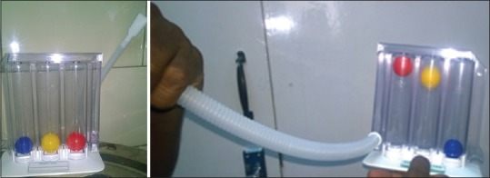
Volume oriented incentive spirometry device
The strength of the expiratory muscles (abdominal and intercostals) is also significantly decreased in patients with COPD making their cough weak and inefficient.[20] Expiratory muscle strength training is achieved using a mouth piece and a one-way spring loaded valve capable of allowing expiratory pressure loads to a maximum of 150 cm of water. By varying the applied force against expiration, the muscle strength is slowly built up. Several commercial devices are available built on this model to train and improve the expiratory muscle strength.
COPD patients due to their ineffective cough are unable to clear mucus effectively. Mechanical insufflation – exsufflation devices also known as ‘cough assist’ devices are very effective in such circumstances. These devices are capable of producing airflow changes that mimic the natural cough and aid sputum clearance.[21] The device uses positive pressure to deliver a maximum lung inhalation followed by abrupt change to negative pressure in the upper airway thus simulating the pressure and flow changes in cough. They produce excellent results when combined with chest physiotherapy.
ASTHMA AND CHRONIC OBSTRUCTIVE PULMONARY DISEASE – PRE-OPERATIVE OPTIMISATION OF THERAPY
Asthma is a chronic pulmonary disease with a wide prevalence across the globe with published prevalence rates ranging from 0.7% to 18.4%.[22] The disease involves airway inflammation and hyper-responsiveness and poses a challenge of a reactive airway to the administration of anaesthesia especially general anaesthesia. These patients typically present with episodes of coughing, wheezing and breathlessness. Investigations such as spirometry and peak expiratory flow rate usually reveal a small airway obstructive disorder with obstruction to airflow. This airflow obstruction is usually reversible at least in the initial stages. Asthmatics carry an increased risk of peri-operative complications during any surgery.[23,24] COPD is also a small airway disease with poor airflow presenting with cough, shortness of breath, infection and sputum production. This repeated inflammation leads to narrowing of small airway and ultimate breakdown of normal lung architecture leading to emphysema. Traditionally, it was considered that asthmatics with reversible airflow obstruction show improvement with bronchodilators while COPD patients show minimal or no improvement. This has been used to differentiate between the two disease processes. Recent evidence has questioned this assumption, and several studies have demonstrated that COPD patients show clinically significant reversibility with bronchodilator therapy.[25] Flow based measurements of pulmonary function like forced expiratory volume in 1 s (FEV1) and inspiratory capacity show significant improvement with bronchodilator therapy in patients with COPD.[26]
Enright suggested a definitive protocol for management of patients with asthma and COPD.[27]
If bronchospasm is present, inhaled beta 2 agonists to be the drugs of choice
A patient at risk for developing complications or having a pre-operative FEV1 <80% to receive oral steroids. The drugs recommended are oral prednisone 40–60 mg/day or IV hydrocortisone 100 mg/8 hourly
Pulmonary infections to be identified and aggressively treated with appropriate antibiotics
Hydration and fluid deficits to be corrected. Hydration helps in easier mobilisation and removal of sputum. Co-existing electrolyte imbalances can occur because beta 2 agonists can predispose to hypokalaemia, hypomagnesaemia and hyperglycaemia. In the presence of these imbalances, patients may fail to respond to beta 2 agonist therapy and also are prone to cardiac arrhythmias
The mast cells are stabilised, and degranulation leading to release of inflammatory mediators was prevented by prophylactic use of drugs as cromolyn
Chest physiotherapy and postural drainage to be used to improve sputum clearance
Smoking should be stopped at least 2 months before elective surgery to decrease carboxy-Hb levels and improve mucociliary clearance of sputum
In long standing patients, right heart dysfunction and cor pulmonale should be suspected. Appropriate evaluation and drug therapy to optimise cardiac function must be started
The global initiative for asthma has recommended a stepwise management protocol for patients coming for surgery under general anaesthesia.[22] Patients are evaluated and placed on a categorised protocol based on symptoms, severity of disease and pulmonary function testing. This approach enables patients to be optimally prepared before surgery and thus reduces the incidence of PPCs.[28]
DRUG THERAPY
Beta 2 agonists are the mainstay of treatment for asthma and COPD. These drugs can be long-acting beta 2 agonists (LABAs) or short-acting beta 2 agonists (SABAs). SABA provides short-term relief and is used for acute exacerbations. LABA is used for the stabilisation of patients with chronic asthma. Beta 2 agonists bind to the beta 2 receptor and activate the adenylcyclase enzyme mechanism leading to an increased production of cyclic adenosine monophosphate (cAMP). At a cellular level, the cAMP produces relaxation of the bronchial smooth muscle and increases the mucociliary clearance of sputum.[29]
Oral and intravenous preparations of beta 2 agonists are available for use. The most preferred route of administration of beta 2 agonists is the inhaled route because small doses of drug are directly administered to the targeted end-organ. It produces the fastest peak bronchodilation with very few systemic effects when compared to other routes of administration.
The currently used SABA includes:
Salbutamol as metered dose inhaler (MDI) in a dose of 100 to 200 μg and can also be nebulised as 5 mg/ml. It is also available in tablet and syrup forms for oral consumption
Terbutaline as MDI in a dose of 400–500 μg and orally in a dose of 2.5–5 mg
Levalbuterol as MDI in a dose of 45–90 μg and can be nebulised in a dose of 0.21–0.42 mg.
In the current clinical practice, the commonly used LABAs are:
Formoterol used as MDI in a dose or 4.5–12 μg and nebulised as 20 μg in 20 ml
Salmeterol as MDI in dose of 25–50 μg
Tulobuterol is available as oral preparation in dose of 2 mg.
Nebulised bronchodilators must be used at least 24–48 h before surgery for them to be effective. Some studies have indicated that bronchodilators are not very effective in COPD patients. While they decrease wheezing, they rarely improve the FEV1 more than 10% of pre-treatment values.[30,31]
Inhaled corticosteroids provide excellent anti-inflammatory effect that makes them one of the first line drugs in management of persistent asthma and COPD. Steroids block inflammatory response to provocative agents and reduce airway oedema and reactivity. Asthmatics who have received pre-operative steroids have shown to have a low complication rate during surgery. There is a fear that use of corticosteroids increases the risk of pneumonia and may delay wound healing. Several studies have addressed this issue and found these fears to be unsubstantiated.[32,33]
The inhaled steroids in common clinical practice include:
Beclomethasone as MDI in dose of 50–400 μg and nebulised in a dose of 0.2–0.4 mg
Budesonide as MDI in dose of 100, 200 and 400 μg and nebulised in a dose of 0.25–0.5 mg
Fluticasone as MDI in a dose of 50–500 μg.
Individuals with severe and uncontrolled bronchospastic symptoms usually are benefitted by systemic corticosteroid therapy.
Systemic corticosteroids used in severe conditions include:
Prednisone in a dose of 5–60 mg PO
Methylprednisolone in a dose of 4–16 mg PO.
Mast cell stabilisers can block the calcium channel and prevent release of inflammatory mediators as histamine that lead to bronchospasm. They are particularly effective in the treatment of exercise-induced bronchospasm. Cromolyn Sodium is used in a nebulised from in a dose of 20 mg 15 min before exposure and 4 times a day. Nedocromil sodium is used by inhalation with a total dose not exceeding 14 mg/day.
Anticholinergics act on the airway muscarinic cholinergic receptors and decrease reflex bronchospasm as well as decreasing mucous hyper-secretion. Asthmatics demonstrate increased response to cholinergics and their stimulation leads to release of acetylcholine that then acts on muscarinic M3 receptors located on bronchial smooth muscles and can increase brochospasm, sputum production and airway oedema. Anticholinergics can block this effect and can be powerful adjuncts in management.
The commonly used anticholinergics include:
Ipratropium (short-acting) as MDI in a dose of 20–40 μg and nebulised as 0.25–0.5 mg/ml
Oxitropium (short-acting) as MDI in a dose of 100 μg and nebulised as 1.5 mg/ml
Tiotropium (long-acting) as MDI in a dose of 18 μg.
Leukotrienes are biologically active fatty acids produced by cell membranes and play an important mediator role in precipitating bronchospasm.
Leukotriene pathway modifiers are useful in specific asthma situations such as exercise induced, viral induced and aspirin-induced asthma. The approved drugs are:
Zafirlukast 10–20 mg BD orally
Montelukast 5–10 mg orally
Zileuton up to 600 mg BD orally.
Methylxanthines are currently not in favour because of their systemic side effects. They do offer mild bronchodilator and anti-inflammatory properties.[34]
The commonly used drugs of this group include:
Theophylline orally in a dose of 100–600 mg. It is also available in injectable form
Aminophylline orally as 200–600 mg. Aminophylline infusions have been junked in favour of newer drugs with safer profiles.
The current treatment protocols for COPD and Asthma focus on combination therapies instead of single drug therapy to obtain the best results. The nebulised route is preferred over systemic administration for drugs to derive the best benefits while minimising the side effects.
The current most commonly used combination therapies include:
Formoterol LABAs + ipratropium (anticholinergic) as MDI 200 + 80 μg and nebulised in a dose of 1.25 + 0.5 mg
Salbutamol SABA + ipratropium (anticholinergic) as MDI 75 + 15 μg and nebulised in a dose of 0.75 + 0.5 mg
Formoterol LABAs + budesonide (corticosteroid) as inhaler 4.5 + 160 μg MDI
Salmeterol LABAs + fluticasone 25–50 μg + 125–250 μg MDI.
A stepwise approach to optimising pulmonary function of patient with bronchial asthma/COPD has been proposed by several authors:
Known asthmatics that are now not on any drugs and are symptom free must be carefully examined and evaluated. If found to be free of any symptoms, they can be taken up for surgery without any empirical therapy being started
Known asthmatics that are symptom free for the last 3 months but are on regular nebulised LABAs and steroids can be proceeded with after careful evaluation. They are advised to continue their inhaled LABAs and steroids, with a nebulised dose being administered before shifting to operation theatre. The same drugs that the patient is taking and an additional SABA may be made available in the operation theatre to be used in case of any bronchospasm
Asthmatic with the recent increase in symptoms require to be put on regular therapy of LABAs and corticosteroids if not already on. SABA may also be considered as short-term bridge to therapy. These patients show good response to a short course of oral corticosteroids for 3 to 5 days prior to surgery. SABA just before shifting to operation theatre may be considered
Asthmatics on nebulised LABAs and corticosteroids but still having persistent daily symptoms need optimisation before surgery. Elective surgery needs to be rescheduled. These patients are given inhaled LABAs and corticosteroids. Additionally, oral corticosteroids and bridging with SABA may be considered to achieve improvement
Asthmatics on regular standard inhaled LABAs and steroids along with daily oral corticosteroids presenting with persistent and severe symptoms pose an extreme challenge to the anaesthetist. While continuing their standard therapy, bridging with frequent nebulised doses of SABA and high dose oral corticosteroids may be considered if surgery is unavoidable.
Liccardi et al. have proposed a pre-anaesthetic assessment and preparation plan [Table 1] to reduce the risk of peri-operative bronchospasm and PPCs in asthmatics undergoing general anaesthesia.[28]
Table 1.
Adopted from Liccardi et al. A proposed protocol for management of asthmatics and chronic obstructive pulmonary disease patients presenting for surgery

SUMMARY
Avoiding PPCs continues to be a challenge when anaesthetising patients with pre-existing lung diseases. A careful evaluation and appropriate investigation is mandatory before proceeding with surgery. Patient education and smoking cessation is shown to be beneficial. Incentive spirometry usually with volume oriented devices is widely practiced, but its stand-alone benefit in reducing PPCs has not been validated. In COPD and asthmatic patients optimisation of therapy with various combinations of drugs offers the best benefit to the patient. Ideally patients headed for elective surgery should show improvement in their clinical condition and measured pulmonary parameters before proceeding for elective surgery.
Financial support and sponsorship
Nil.
Conflicts of interest
There are no conflicts of interest.
REFERENCES
- 1.Warner DO. Preventing postoperative pulmonary complications: The role of the anesthesiologist. Anesthesiology. 2000;92:1467–72. doi: 10.1097/00000542-200005000-00037. [DOI] [PubMed] [Google Scholar]
- 2.Fauci AS, Kasper DL, Longo DL, Braunwald E, Hauser SL, Jameson JL, et al. Harrison's’ Principles of Internal Medicine. 17th ed. New Delhi: McGraw Hill Comp Inc; 2008. [Google Scholar]
- 3.Warner MA, Offord KP, Warner ME, Lennon RL, Conover MA, Jansson-Schumacher U. Role of preoperative cessation of smoking and other factors in postoperative pulmonary complications: A blinded prospective study of coronary artery bypass patients. Mayo Clin Proc. 1989;64:609–16. doi: 10.1016/s0025-6196(12)65337-3. [DOI] [PubMed] [Google Scholar]
- 4.Kurup V. Respiratory diseases. In: Hines RL, Marhal KE, editors. Stoeltings’ Anaesthesia and Co-existing Diseases. 5th ed. New Delhi: Churchill Livingstone/Elsevier; 2009. pp. 168–73. [Google Scholar]
- 5.Ault ML, Stock MC. Respiratory function. In: Barash PG, editor. Clinical Anaesthesia. 6th ed. Philadelphia: Wolters Kluwer/Lippincott, Williams and Wilkins; 2009. p. 252. [Google Scholar]
- 6.Shaikh SI, Nilangekar MT. Perioperative anaesthetic management in asthma. Int J Biomed Res. 2015;6:144–150. [Google Scholar]
- 7.Pearce AC, Jones RM. Smoking and anesthesia: Preoperative abstinence and perioperative morbidity. Anesthesiology. 1984;61:576–84. doi: 10.1097/00000542-198411000-00018. [DOI] [PubMed] [Google Scholar]
- 8.Arozullah AM, Daley J, Henderson WG, Khuri SF. Multifactorial risk index for predicting postoperative respiratory failure in men after major noncardiac surgery. The National Veterans Administration Surgical Quality Improvement Program. Ann Surg. 2000;232:242–53. doi: 10.1097/00000658-200008000-00015. [DOI] [PMC free article] [PubMed] [Google Scholar]
- 9.Arozullah AM, Khuri SF, Henderson WG, Daley J. Participants in the National Veterans Affairs Surgical Quality Improvement Program. Development and validation of a multifactorial risk index for predicting postoperative pneumonia after major noncardiac surgery. Ann Intern Med. 2001;135:847–57. doi: 10.7326/0003-4819-135-10-200111200-00005. [DOI] [PubMed] [Google Scholar]
- 10.Brooks-Brunn JA. Predictors of postoperative pulmonary complications following abdominal surgery. Chest. 1997;111:564–71. doi: 10.1378/chest.111.3.564. [DOI] [PubMed] [Google Scholar]
- 11.Han MK, McLaughlin VV, Criner GJ, Martinez FJ. Pulmonary diseases and the heart. Circulation. 2007;116:2992–3005. doi: 10.1161/CIRCULATIONAHA.106.685206. [DOI] [PubMed] [Google Scholar]
- 12.Davidson AG, Wong LT. Pirie GE Long-term comparative trial of conventional percussion and drainage physiotherapy versus autogenic drainage in CF. Pediatr Pulmonol. 1992;14:298. [Google Scholar]
- 13.Makhabah DN, Martino F, Ambrosino N. Peri-operative physiotherapy. Multidiscip Respir Med. 2013;8:4. doi: 10.1186/2049-6958-8-4. [DOI] [PMC free article] [PubMed] [Google Scholar]
- 14.Nomori H, Kobayashi R, Fuyuno G, Morinaga S, Yashima H. Preoperative respiratory muscle training. Assessment in thoracic surgery patients with special reference to postoperative pulmonary complications. Chest. 1994;105:1782–8. doi: 10.1378/chest.105.6.1782. [DOI] [PubMed] [Google Scholar]
- 15.Hulzebos EH, Helders PJ, Favié NJ, De Bie RA, Brutel de la Riviere A, Van Meeteren NL. Preoperative intensive inspiratory muscle training to prevent postoperative pulmonary complications in high-risk patients undergoing CABG surgery: A randomized clinical trial. JAMA. 2006;296:1851–7. doi: 10.1001/jama.296.15.1851. [DOI] [PubMed] [Google Scholar]
- 16.Savci S, Degirmenci B, Saglam M, Arikan H, Inal-Ince D, Turan HN, et al. Short-term effects of inspiratory muscle training in coronary artery bypass graft surgery: A randomized controlled trial. Scand Cardiovasc J. 2011;45:286–93. doi: 10.3109/14017431.2011.595820. [DOI] [PubMed] [Google Scholar]
- 17.Moodie LH, Reeve JC, Vermeulen N, Elkins MR. Inspiratory muscle training to facilitate weaning from mechanical ventilation: Protocol for a systematic review. BMC Res Notes. 2011;4:283. doi: 10.1186/1756-0500-4-283. [DOI] [PMC free article] [PubMed] [Google Scholar]
- 18.Carvalho CR, Paisani DM, Lunardi AC. Incentive spirometry in major surgeries: A systematic review. Rev Bras Fisioter. 2011;15:343–50. doi: 10.1590/s1413-35552011005000025. [DOI] [PubMed] [Google Scholar]
- 19.Restrepo RD, Wettstein R, Wittnebel L, Tracy M. Incentive spirometry: 2011. Respir Care. 2011;56:1600–4. doi: 10.4187/respcare.01471. [DOI] [PubMed] [Google Scholar]
- 20.Ramírez-Sarmiento A, Orozco-Levi M, Barreiro E, Méndez R, Ferrer A, Broquetas J, et al. Expiratory muscle endurance in chronic obstructive pulmonary disease. Thorax. 2002;57:132–6. doi: 10.1136/thorax.57.2.132. [DOI] [PMC free article] [PubMed] [Google Scholar]
- 21.Vianello A, Corrado A, Arcaro G, Gallan F, Ori C, Minuzzo M, et al. Mechanical insufflation-exsufflation improves outcomes for neuromuscular disease patients with respiratory tract infections. Am J Phys Med Rehabil. 2005;84:83–8. doi: 10.1097/01.phm.0000151941.97266.96. [DOI] [PubMed] [Google Scholar]
- 22.Applegate R, Lauer R, Lenart J, Gatling J, Vadi M. The perioperative management of asthma. J Allergy Ther. 2013;S11:007. [Google Scholar]
- 23.Warner DO, Warner MA, Barnes RD, Offord KP, Schroeder DR, Gray DT, et al. Perioperative respiratory complications in patients with asthma. Anesthesiology. 1996;85:460–7. doi: 10.1097/00000542-199609000-00003. [DOI] [PubMed] [Google Scholar]
- 24.Orestes MI, Lander L, Verghese S, Shah RK. Incidence of laryngospasm and bronchospasm in pediatric adenotonsillectomy. Laryngoscope. 2012;122:425–8. doi: 10.1002/lary.22423. [DOI] [PubMed] [Google Scholar]
- 25.Tashkin DP, Celli B, Decramer M, Liu D, Burkhart D, Cassino C, et al. Bronchodilator responsiveness in patients with COPD. Eur Respir J. 2008;31:742–50. doi: 10.1183/09031936.00129607. [DOI] [PubMed] [Google Scholar]
- 26.Bleecker ER, Emmett A, Crater G, Knobil K, Kalberg C. Lung function and symptom improvement with fluticasone propionate/salmeterol and ipratropium bromide/albuterol in COPD: Response by beta-agonist reversibility. Pulm Pharmacol Ther. 2008;21:682–8. doi: 10.1016/j.pupt.2008.04.003. [DOI] [PubMed] [Google Scholar]
- 27.Enright A. Bronchospastic disease and emergency surgery. Middle East J Anaesthesiol. 2004;17:927–38. [PubMed] [Google Scholar]
- 28.Liccardi G, Salzillo A, Sofia M, D’Amato M, D’Amato G. Bronchial asthma. Curr Opin Anaesthesiol. 2012;25:30–7. doi: 10.1097/ACO.0b013e32834e7b2e. [DOI] [PubMed] [Google Scholar]
- 29.Johnson M. Molecular mechanisms of beta (2)-adrenergic receptor function, response, and regulation. J Allergy Clin Immunol. 2006;117:18–24. doi: 10.1016/j.jaci.2005.11.012. [DOI] [PubMed] [Google Scholar]
- 30.Playford HR. COPD. In: Sladen RN, Coursin DB, Ketzler JT, Playford H, editors. Anaesthesia and Co-existing Disease. Philadelphia: Saunders Elsevier; 2007. pp. 38–41. [Google Scholar]
- 31.Pedersen SE, Hurd SS, Lemanske RF, Jr, Becker A, Zar HJ, Sly PD, et al. Global strategy for the diagnosis and management of asthma in children 5 years and younger. Pediatr Pulmonol. 2011;46:1–17. doi: 10.1002/ppul.21321. [DOI] [PubMed] [Google Scholar]
- 32.National Asthma Education and Prevention Program. Expert panel report 3 (EPR.3): Guidelines for the diagnosis and management of asthma-summary report 2007. J Allergy Clin Immunol. 2007;120(5 Suppl):S94–138. doi: 10.1016/j.jaci.2007.09.043. [DOI] [PubMed] [Google Scholar]
- 33.Smetana GW. Preoperative pulmonary evaluation: Identifying and reducing risks for pulmonary complications. Cleve Clin J Med. 2006;73(Suppl 1):S36–41. doi: 10.3949/ccjm.73.suppl_1.s36. [DOI] [PubMed] [Google Scholar]
- 34.Weinberger M, Hendeles L. Theophylline in asthma. N Engl J Med. 1996;334:1380. doi: 10.1056/NEJM199605233342107. [DOI] [PubMed] [Google Scholar]