Abstract
Introduction:
Incidence of sudden cardiac death (SCD) has been steadily increasing all over the world. While knowing the cause of SCD is one of the favorites of the physicians involved with these cases, it is very difficult and challenging task for the forensic physician. The present report is a prospective study regarding cause of SCDs on autopsy examination in four-year period, Bangalore, India.
Methods:
The present prospective study is based on autopsy observations, carried out for four-year period from 2008 to 2011, and analyzed for cause of SCDs. The cases were chosen as per the definition of sudden death and autopsied. The material was divided into natural and unnatural groups. Finally, on histopathology, gross examination, hospital details, circumstantial, and police reports the cause of death was inferred.
Results:
A total of 2449 autopsy was conducted of which 204 cases were due to SCD. The highest SCDs were reported in 50-60 years age group (62.24%; n-127), followed closely by the age group 60-69 (28.43%; n-58). Male to female ratio was around 10:1. The maximum number of deaths (n=78) was within few hours (6 hours) after the onset of signs and symptoms. In 24 (11.8%) cases major narrowing was noted in both the main coronaries, in 87 (42.6%) cases in the left anterior descending coronary artery (LAD), and in 18 (51.5%) cases in the right coronary artery (RCA). The major cardiac pathology resulting in sudden death was coronary artery disease (n-116; 56.86%) and myocardial infarction (n-104; 50.9%). most of the SCDs occurred in the place of residence (n-80; 39.2%) followed closely by death in hospital (n-49; 24.01%).
Conclusion:
Coronary occlusion was the major contributory cause of sudden death with cardiac origin and the highest number of deaths were reported in the age 50-59 years with male to female ratio of 10:1.
Key Words: Cause of death, sudden, cardiac, coronary artery disease, autopsy
Introduction
Incidence of sudden cardiac death (SCD) has been steadily increasing all over the world particularly in the urban population during last five decades (1, 2). In India incidence of ischemic heart disease has increased, to about 10 percent (3). As revealed in autopsy findings, majority of sudden and unexpected deaths result as a sequel to cardiovascular disease (4). Probably the most common cause of death recorded in autopsy is myocardial infarction (MI) due to coronary artery insufficiency (coronary occlusion due to atheroma and coronary thrombosis) (5). Knowing the cause of sudden death is always one of the favorites of the physicians involved with these cases. On the other hand, a very difficult and challenging task for the forensic physician, conducting the autopsy, is to ascertain cause and nature of death (6, 7). One difficulty which is mostly encountered in sudden unexpected deaths, whether cardiac or
no cardiac, is the lack of precision of collecting antecedent factors involved within the 24-hours, duration prior to death (8). For example, in certain cases of atrial fibrillations and spasms of coronary as a cause of death, the complete relaxation of coronary leaves no trace of such spasm, even though the death was due to coronary ischemia during autopsy. Many time it has been seen that when gross pathology could not help to evaluate the cause of death, the histology came forward to rescue the situation and a conclusive opinion could be given on the involved cardiac pathology (9, 10). Based on the above mentioned, the present report is a prospective study regarding cause of SCD on autopsy examination in four-year period, Bangalore, India.
Methods
Study design and setting
The present study is a prospective cross-sectional study carried during the 4-year period from January 2008 to December 2011 in Kingston, Jamaica and Bangalore, India. A total of 2449 cases were chosen as per definition of sudden death and autopsied. All the cases were handed over for autopsy after completion of police/magistrate inquest. The material was divided into natural and unnatural deaths on the basis of underlying cause after analyzing the autopsy, police reports, and hospital records. Hence, the cause of death was inferred on histopathology, gross examination, hospital details, circumstantial, and police reports.
Participants
The size of the population limited to the jurisdiction for this autopsy study was around 1,200,000-1,500,000. The cases were deemed medico legal and involved individuals of all age group, sex, religion, caste, and nationality. All were referred as sudden onset of death and deemed as suspicious deaths. The study was focused only on SCD and death from causes other than cardiovascular pathology was excluded.
Autopsy Methods
The heart was dissected as per the Davidsons technique (11) and the ventricles were cut transversely to have the view of endocardium, its color, scars and fibrosis. The tissue were preserved in 10% formal saline, thickness of 3-5 millimeters was kept for fast and better fixation and stained with Haematoxylin and Eosin (200 gram aluminium ammonium sulphate, 20 gram haematoxylin, 40 milliliter ethanol, 4 gram sodium iodate, 80 milliliter acetic acid, 1200 milliliter glycerol, 2800 milliliter distilled water). Autopsy analysis was focused on the details of morbid findings of the heart consisting of surface, walls, coronaries, valves, and great vessels, weight of heart, ostias, and papillary mussels. The weight was taken as normal of below 325 grams, in cases of males, and below 275 grams in cases of females (12, 13). The MI was considered recent if hemorrhages, paleness, and softening had been observed in the myocardium, and had the following microscopic changes: cytoplasmic hypereosinophilia, contraction band, granularity, lack of nuclei edema, and hemorrhage. All scars more than 0.5 centimeters (usually triangular in shape directed towards the apex) were considered as an old infarct along with the microscopic findings.
Definitions
Sudden death was defined as one which has taken place within 24 hours from the onset of symptoms and signs of disease, based on world health organization (WHO) definition (3, 4). Also, sudden deaths in a short period was defined as deaths occurred within 30 minutes, either in hospital or when being brought to hospital or died within minutes after development of symptoms. The coronary occlusion was classified on rule of thumb that is slight narrowing, 30%; moderate narrowing 50%; and severe narrowing that is 70% and above (14).
Statistical analysis
Descriptive analyses were performed by statistical software SPSS version 11.0 and graphs drawn by Excel 2013. Data were shown as frequency and percentage.
Results
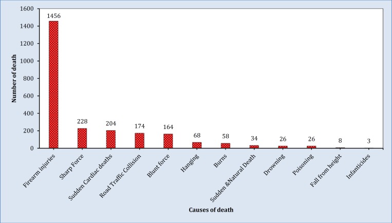
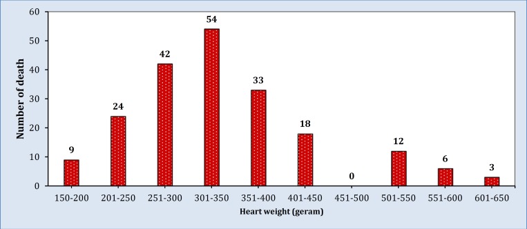
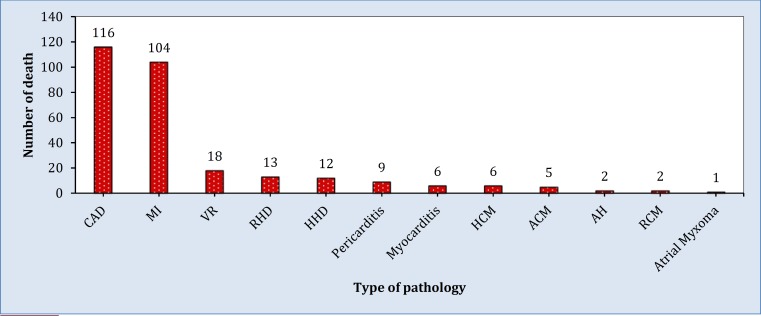
A total of 2449 cases were autopsied during the study period. Two hundred four cases of SCD were enrolled (90.2% male and 76.0% out of hospital) (Figure 1). Male to female ratio was 10:1. Table 1 shows the demographic data of the subjects. Twenty two (10.8%) cases had been found dead without any complaint of illness prior to the admission in casualty. Forty one (20.1%) cases were known as ischemic heart disease but in apparent good health at the time of incidence. Thirty three (16.2%) cases died in short period, 78 (38.2%) within few hours of onset of symptoms, 63 (30.9%) after 24 hours. In 30 (14.7%) cases duration of onset of symptoms and death could not be evaluated, due to non-availability of specific history. The higher income group was more affected [78 (38.2%) cases], followed closely by the lower income group [60 (28.4%) cases] and the least group affected were from the Middle class [27 (13.2%) cases]. The weight of the heart was above normal in 126 (61.8%) cases (minimum 150 grams, maximum 890 grams) (Figure 2). Sweating was noted in 159 (77.9%) cases, dyspnea in 136 (66.7%), precordial pain in 161 (78.9%), vomiting in 157 (77.0%), cyanosis, and exhaust looking face in 110 (53.9%). Coronary artery disease was the most common cardiac pathology in these series [116 (56.86%) cases] (Figure 3). The majority of deaths were occurred out of the hospital [155 (75.99%) cases].
Figure 1.
Distribution of various causes of death both natural and unnatural from Jan 2008 to Dec 2011
Table 1.
Demographic variable of SCD
| Variable | N | % |
|---|---|---|
| Gender | ||
| Male | 184 | 90.2 |
| Female | 20 | 9.8 |
| Age (year) | ||
| 15-19 | 1 | 0.5 |
| 20-29 | 0 | 0.0 |
| 30-39 | 3 | 1.5 |
| 40-49 | 15 | 7.4 |
| 50-59 | 127 | 62.3 |
| 60-69 | 58 | 28.4 |
| 70-above | 0 | 0.0 |
| Socio-economical class | ||
| Low Income Class | 60 | 29.4 |
| Middle Income Class | 27 | 13.2 |
| High Income Class | 78 | 38.2 |
| Unclassified population | 39 | 19.1 |
| Place of death | ||
| Outside the Hospital* | 155 | 76.0 |
| Hospital Deaths | 49 | 24.0 |
| Year of death | ||
| 2008 | 42 | 20.6 |
| 2009 | 50 | 24.5 |
| 2010 | 51 | 25.0 |
| 2011 | 61 | 29.9 |
| Duration of onset of symptoms and death | ||
| Instantaneous(<30min) | 33 | 16.2 |
| Early(<6hrs) | 78 | 38.2 |
| Delayed(<24hrs) | 63 | 30.9 |
| Unknown | 30 | 14.7 |
Death while transportation to Hospital 27 (13.2%) cases; SCD: Sudden cardiac death.
Figure 2.
Weight of Heart in grams
Figure 3.
Type of cardiac pathology. CAD: coronary artery disease; MI: myocardial infarction; VR: ventricular rupture; RHD: rheumatic heart disease; HHD: hypertensive heart disease; HCM: hypertrophic cardiomyopathy; ACM: alcoholic cardiomyopathy; AH: aneurysm of heart; RCM: restrictive cardiomyopathy
Details of Coronary artery pathology
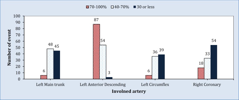
Figure four shows the anatomical location and block percentage of atheromatous and atherosclerotic lesion.
In 24 (11.8%) cases, the major blockage was noted in both main coronaries, in 87 (42.6%) cases in the Left Anterior Descending coronary artery (LAD) and in 18 (51.5%) cases in the Right Coronary Artery (RCA). There were two cases which showed equally severe involvement of the cerebral vessel. The most frequent affected site was proximal 3-5 centimeters of anterior descending branch of LAD from its origin. Types of occlusion observed from concentric central pinhole type to crescentic with lumen pushed to one side of vessel. The highest incidence in all three vessels (circumflex, LAD, and RCA) was encountered in the age group of 50-59 years. The younger cases showed a higher incidence in the LAD. The incidence of severe occlusion in LAD was 48.53% and in RCA 8.82% (Figure 4). The coronary artery least affected by blockage was left circumflex and left main trunk. MI cases were 49 (24.0%) recent and 55 (27.0%) old. In four (2.0%) cases the myocardium showed no evidence of healed or fresh infarcts.
Figure 4.
Percentage of blockage of coronary artery
Discussion:
Based on the results of this study, coronary occlusion was the major contributory cause of SCD, with incidence of 56.9% of all SCD. Male to female ratio has been found to be 10:1. In all cases the atherosclerotic process involved in varying extent, in both coronaries and their major branches. The highest number of SCDs was reported in the age of 50-59 years. The socio-economic status affected equally both low and high income groups.
Kuller et al. showed that nearly two third of sudden non-traumatic deaths are due to arteriosclerotic diseases (15). In addition, Harmon et al. demonstrated that SCD is the leading cause of death in athletes (2). The age-related trend of SCD increases with age regardless of sex or race (16). In this context, the highest prevalence is in the geriatric age group (75-85 years) (17). For example, One study demonstrated that sudden death from coronary artery disease is six times more common at age between 60-69 while for those over 80 it is only two fold (18). The average age in the present series was between ages 50-59 years. Similar age incidence has been observed in the study series of Pentilla to be given 55-66 years (19). It is well-established that men have a higher incidence of SCD than women (16, 17, 20, 21). The present study shows higher male to female ratio (10:1). Framingham studied that SCD was 10 times more among male than females (22). The male to female ratio at the incidence of coronary heart disease was reported from 3:2 to 8:1 (20, 23-25). In addition to age and sex, socio-economic status has substantially impacted on incidence of SCD (26-28).
Our result revealed that the socio-economic status affected low and high income groups. This can be attributed to indiscriminate habit of food and grains as well as other factors like smoking, junk foods, alcohol, and stress in both class of population.
In involvement frequency, LAD observed to have a higher severe narrowing than RCA. Gradwhol quoted the ranges from various published analysis as LAD to be 45-64%, while RCA come next in frequency, 24-46% followed by left circumflex 3-10%, and least affected in left main coronary (29). The observed variations among different series may be due to the regional variations on different cross section of the studied group to different periods.
Limitations
During the comparison of unexpected SCD results of this study with observations of similar field, the certain factors have been considered. Firstly, the population under the study belonged to a cosmopolitan urban population; it is clear that some of them came from different regions of the country. Secondly, though most autopsy investigations are meticulously followed by police authorities, there are skipped cases by omissions which are probably unavoidable in this vast country. While, there were many sophisticated methods of diagnosing myocardial infarction in postmortem examination like three Tesla Magnetic Resonance Imaging (3T-MRI), Immune-Histochemical detection of S100 calcium-binding protein A1 (S100A1), and quantitative myoglobin assay (30-32); however, those investigations were not carried out to find the cause of death in the present study. Lastly, the material available during the last four years was good enough to match with the works done by other researchers in similar field elsewhere.
Conclusion:
Coronary occlusion was the major contributory cause of SCD and the most numbers of deaths were reported in the age 50-59 year with male to female ratio 10:1.
Acknowledgments:
Authors acknowledge department of Pathology Jawaharlal Nehru Institute of Postgraduate Medical Research Institute, Pravara Medical Trust of Loni, India, Kingston Public Hospital of Jamaica, and Sapthagiri Institute of Medical Science and Research Center.
Conflict of interest:
None
Funding support:
None
Authors’ contributions:
Rao D conducted the autopsy, authored the report, grossing of the organs and examined the slides under the microscope. Dongre P extended their histopathology lab and support group. Sood D participated in slide reading and histopathology in few cases. All authors wrote the first draft of the manuscript and approved the final version.
References
- 1.Goldberger JJ, Basu A, Boineau R, et al. Risk stratification for sudden cardiac death a plan for the future. Circulation. 2014;129(4):516–26. doi: 10.1161/CIRCULATIONAHA.113.007149. [DOI] [PubMed] [Google Scholar]
- 2.Harmon KG, Asif IM, Klossner D, Drezner JA. Incidence of sudden cardiac death in national collegiate athletic assoc-iation athletes. Circulation. 2011;123(15):1594–600. doi: 10.1161/CIRCULATIONAHA.110.004622. [DOI] [PubMed] [Google Scholar]
- 3.Reddy KS. Cardiovascular disease in non-Western countr-ies. N Engl J Med. 2004;350:2438–510. doi: 10.1056/NEJMp048024. [DOI] [PubMed] [Google Scholar]
- 4.Pouleur A-C, Barkoudah E, Uno H, et al. Pathogenesis of sudden unexpected death in a clinical trial of patients with myocardial infarction and left ventricular dysfunction, heart failure, or both clinical perspective. Circulation. 2010;122(6):597–602. doi: 10.1161/CIRCULATIONAHA.110.940619. [DOI] [PubMed] [Google Scholar]
- 5.Hurt RD, Weston SA, Ebbert JO, et al. Myocardial infarction and sudden cardiac death in olmsted county, minnesota, before and after smoke-free workplace lawsmi and cardiac death with smoke-free workplace law. Arch Intern Med. 2012;172(21):1635–41. doi: 10.1001/2013.jamainternmed.46. [DOI] [PMC free article] [PubMed] [Google Scholar]
- 6.Liu L, Callinan LS, Holman RC, Blau DM. Determinants for autopsy after unexplained deaths possibly resulting from infectious causes, United States. Emerg Infect Dis. 2012;18(4):549–55. doi: 10.3201/eid1804.111311. [DOI] [PMC free article] [PubMed] [Google Scholar]
- 7.Loff B, Cordner S. Forensic Epidemiology, Forensic Pathology, Ethics and Human Rights. Forensic Epidemiology in the Global Context. Springer; 2013. pp. 121–50. [Google Scholar]
- 8.Rao DS. Sudden and unexpected natural deaths-a four-year autopsy review. J Punjab Acad Forensic Med Toxicol. 2008;8(2):20–4. [Google Scholar]
- 9.Tabib A, Loire R, Chalabreysse L, et al. Circumstances of death and gross and microscopic observations in a series of 200 cases of sudden death associated with arrhythmogenic right ventricular cardiomyopathy and/or dysplasia. Circula-tion. 2003;108(24):3000–5. doi: 10.1161/01.CIR.0000108396.65446.21. [DOI] [PubMed] [Google Scholar]
- 10.Fontaine G, Fornes P. Histology of sudden death in arrhythmogenic right ventricular cardiomyopathy/dysplasia. Circulation. 2004;110(3):e20–e5. doi: 10.1161/01.CIR.0000135590.37950.30. [DOI] [PubMed] [Google Scholar]
- 11.Abrahams C. Current Methods of Autopsy Practice. JAMA. 1979;242(16):1791–2. [Google Scholar]
- 12.McKenna W, Stewart J, Nihoyannopoulos P, McGinty F, Davies M. Hypertrophic cardiomyopathy without hypertro-phy: two families with myocardial disarray in the absence of increased myocardial mass. Br Heart J. 1990;63(5):287–90. doi: 10.1136/hrt.63.5.287. [DOI] [PMC free article] [PubMed] [Google Scholar]
- 13.Michelson N. Bilateral ventricular hypertrophy due to chronic pulmonary disease. CHEST. 1960;38(4):435–46. doi: 10.1378/chest.38.4.435. [DOI] [PubMed] [Google Scholar]
- 14.Topol EJ. Textbook of interventional cardiology. Elsevier Health Sciences; 2003. p. 313. [Google Scholar]
- 15.Kuller L, Cooper M, Perper J. Epidemiology of sudden death. Arch Intern Med. 1972;129(5):714–9. [PubMed] [Google Scholar]
- 16.Deo R, Albert CM. Epidemiology and genetics of sudden cardiac death. Circulation. 2012;125(4):620–37. doi: 10.1161/CIRCULATIONAHA.111.023838. [DOI] [PMC free article] [PubMed] [Google Scholar]
- 17.Chugh SS, Reinier K, Teodorescu C, et al. Epidemiology of sudden cardiac death: clinical and research implications. Prog Cardiovasc Dis. 2008;51(3):213–28. doi: 10.1016/j.pcad.2008.06.003. [DOI] [PMC free article] [PubMed] [Google Scholar]
- 18.Romo M, Ruosteenoja R. Sudden coronary death: incidence and common risk factors. Adv Cardiol. 1978;25:1–8. doi: 10.1159/000401999. [DOI] [PubMed] [Google Scholar]
- 19.Penttilä A. Sudden and unexpected natural deaths of adult males. An analysis of 799 forensic autopsies in 1976. Forensic Sci Int. 1980;16(3):249–59. doi: 10.1016/0379-0738(80)90210-8. [DOI] [PubMed] [Google Scholar]
- 20.Chugh SS, Jui J, Gunson K, et al. Current burden of sudden cardiac death: multiple source surveillance versus retrospec-tive death certificate-based review in a large US community. J Am Coll Cardiol. 2004;44(6):1268–75. doi: 10.1016/j.jacc.2004.06.029. [DOI] [PubMed] [Google Scholar]
- 21.Gerber Y, Jacobsen SJ, Frye RL, Weston SA, Killian JM, roger vl. Secular trends in deaths from cardiovascular diseases a 25-year community study. Circulation. 2006;113(19):2285–92. doi: 10.1161/CIRCULATIONAHA.105.590463. [DOI] [PubMed] [Google Scholar]
- 22.Fox CS, Evans JC, Larson MG, Kannel WB, Levy D. Temporal trends in coronary heart disease mortality and sudden cardiac death from 1950 to 1999 the Framingham Heart Study. Circulation. 2004;110(5):522–7. doi: 10.1161/01.CIR.0000136993.34344.41. [DOI] [PubMed] [Google Scholar]
- 23.Doolan A, Langlois N, Semsarian C. Causes of sudden cardiac death in young Australians. Med J Aust. 2004;180(3):110–2. doi: 10.5694/j.1326-5377.2004.tb05830.x. [DOI] [PubMed] [Google Scholar]
- 24.Kim C, Fahrenbruch CE, Cobb LA, Eisenberg MS. Out-of-hospital cardiac arrest in men and women. Circulation. 2001;104(22):2699–703. doi: 10.1161/hc4701.099784. [DOI] [PubMed] [Google Scholar]
- 25.Kannel WB, Schatzkin A. Sudden death: lessons from subsets in population studies. J Am Coll Cardiol. 1985;5(6s1):141B–9B. doi: 10.1016/s0735-1097(85)80545-3. [DOI] [PubMed] [Google Scholar]
- 26.Hemingway H, Malik M, Marmot M. Social and psychos-ocial influences on sudden cardiac death, ventricular arrhyth-mia and cardiac autonomic function. Eur Heart J. 2001;22(13):1082–101. doi: 10.1053/euhj.2000.2534. [DOI] [PubMed] [Google Scholar]
- 27.Mensah GA, Mokdad AH, Ford ES, Greenlund KJ, Croft JB. State of disparities in cardiovascular health in the United States. Circulation. 2005;111(10):1233–41. doi: 10.1161/01.CIR.0000158136.76824.04. [DOI] [PubMed] [Google Scholar]
- 28.Reinier K, Stecker EC, Vickers C, Gunson K, Jui J, Chugh SS. Incidence of sudden cardiac arrest is higher in areas of low socioeconomic status: a prospective two year study in a large United States community. Resuscitation. 2006;70(2):186–92. doi: 10.1016/j.resuscitation.2005.11.018. [DOI] [PubMed] [Google Scholar]
- 29.Bohrod MG. THe essentials of forensic medicine. JAMA. 1986;255(11):1506–7. [Google Scholar]
- 30.Persson M. Postmortem cardiac 3T magnetic resonance imaging: Visualizing the sudden cardiac death? J Am Coll Cardiol. 2013;62(7):617–29. doi: 10.1016/j.jacc.2013.01.089. [DOI] [PubMed] [Google Scholar]
- 31.Bi H, Yang Y, Huang J, Li Y, Ma C, Cong B. Immunohistoche-mical detection of S100A1 in the postmortem diagnosis of ac-ute myocardial infarction. Diagn Pathol. 2013;8(1):84. doi: 10.1186/1746-1596-8-84. [DOI] [PMC free article] [PubMed] [Google Scholar]
- 32.Solokhin EV, Potemkin AM. The quantitative myoglobin assay for forensic medical expertise. Sud Med Ekspert. 2013 ;56(1):27–30. [Russian] [PubMed] [Google Scholar]


