

Key Clinical Message
Pulmonary artery pseudoaneurysm is an uncommon yet fatal clinical entity. Its presentation can mimic a number of common diseases and can be easily missed. As pseudoaneurysm is associated with a number of fatal complications, clinicians should be aware of imaging features which distinguishes pseudoaneurysms from its close differentials. Early recognition and treatment of pseudoaneurysm can prevent fatal outcomes including hemothorax, rupture, or death.
Keywords: Chemotherapy, coil embolization, pseudoaneurysm
Introduction
Acquired pseudoaneurysm is uncommon finding and is extremely rare after systemic chemotherapy 1,2. Focal dilatation of pulmonary artery can be seen in tuberculosis, pulmonary vascular infections, trauma, and vasculitis. It can also be observed after intra-arterial chemotherapy and Swan–Ganz catheter placement 7. There is some evidence that erosion of vessel wall by tumor can result in a pseudoaneurysm of branches of the pulmonary artery. Rarely pulmonary artery pseudoaneurysms are seen as complication of systemic chemotherapy.
Case Report
About 47-year-old female was recently diagnosed synovial sarcoma of thigh with metastasis to lung with tumor invading the left pulmonary artery (Fig.1). Patient was on systemic chemotherapy with adriamycin and ifosfamide and had completed four cycles of chemotherapy so far. She presented with sudden onset of pleuritic chest pain with subjective fevers. No other positive indicators were found on review of systems. On examination, her vitals were stable, afebrile, and spO2 was 94% on room air. Rest physical examination revealed no focal findings with a benign chest examination. Her laboratory workup revealed anemia (hemoglobin 10 g/dL) and leukopenia. As patient had neutropenia with subjective fevers and recently received a course of chemotherapy, a computerized tomography of chest was done to rule out infection, consolidation, infarct/embolism, or progression of cancer. Computed tomography with contrast revealed 10-mm round isodense enhancement at the base of left main pulmonary artery consistent with pseudoaneurysm and an 8-mm big similar round isodense enhancement was noticed at the base of the subsegmental branches of right pulmonary artery (Fig.2). On further comparison with previous images (imaging done at the time of diagnosis of the metastatic synovial sarcoma), we found that pseudoaneurysm to be a new finding and the site of pseudoaneurysm was same as the site of tumor invasion into pulmonary vasculature. As one of the major vessel had the pseudoaneurysm with a higher chances of rupture resulting in unfavorable outcomes like hemothorax/intraparenchymal hemorrhage or intrabronchiolar hemorrhage, patient underwent angiography and stenting of the pseudoaneurysm. Postprocedure imaging revealed stabilization of the size of pseudoaneurysm and no bleed/leak of the pseudoaneurysm. On follow-up, patient was stable with no progression or new pulmonary artery pseudoaneurysm (Fig.3).
Figure 1.

CT of chest which shows synovial sarcoma invading pulmonary artery.
Figure 2.
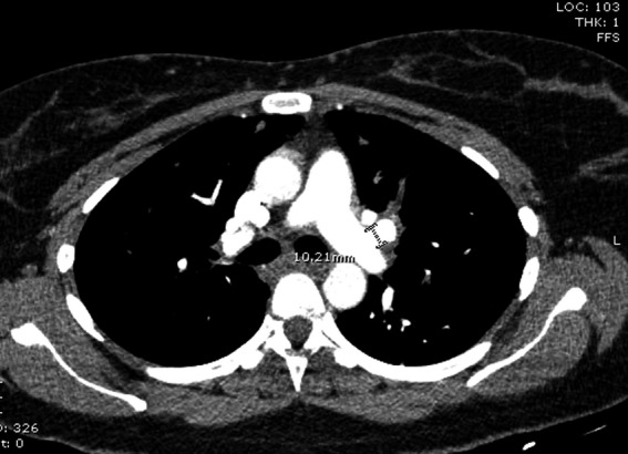
CT showing resolution od cancer invasion after chemotherapy but the same site of invasion now shows a pulmonary artery pseudoaneurysm.
Figure 3.

CT chest showing resolution of pulmonary artery pseudoaneurysm post procedure.
Discussion
Pulmonary artery aneurysms and pseudoaneurysms are uncommon but important to recognize because of the associated morbidity. By definition, an aneurysm is focal dilatation of a blood vessel that involves all three layers of vessel wall. A pseudoaneurysm does not involve all layers of the arterial wall and is therefore at higher risk of rupture. Pseudoaneurysm is commonly described in hepatic artery accounting for more than 65% of all pseudoaneurysms reported in the literature 1,2. Pseudoaneurysm of pulmonary vasculature is infrequently described in the literature and usually remains clinically silent or present with subtle symptoms 1. Because of higher tendency of getting ruptured, on rare occasions these present with hemorrhages and other catastrophic events like intrapulmonary hemorrhage, hemothorax, or intrabronchiolar hemorrhage. Idiopathic pulmonary artery pseudoaneurysm is more commonly found in elderly females, besides that no other demographic event has been attributed to pseudoaneurysm formation in pulmonary vasculature 7. Pulmonary artery pseudoaneurysm are seen either after traumatic event, tuberculosis, pneumonia, vasculitis (few common ones: Behcet's syndrome and Hughes–Stovin syndrome which is characterized by recurrent thrombophlebitis and pulmonary artery aneurysm) or congestive heart failure. There are a handful of cases with invasive cancer presenting with pseudoaneurysm of pulmonary vasculature which resolve after the treatment of cancer with systemic chemotherapy 1,2,4,5. Few case reports mention the occurrence of pseudoaneurysm of pulmonary vasculature after local invasive procedures like placement of Swan–Ganz catheter or after sessions of local chemotherapy 3,7,8. Few cases mention adriamycin as the most commonly associated chemotherapy agent with pseudoaneurysm especially in patients undergoing transarterial chemoemobilzation (TACE) for hepatocellular carcinoma 6–8,10. As pseudoaneurysm is usually a consequence of local procedures involving mechanical trauma to vessel wall or local effect to chemotherapy, it is not clearly mentioned whether adriamycin is the sole culprit 9,10. But development of pseudoaneurysm of pulmonary artery after systemic chemotherapy has not been documented so far in medical literature.
Pseudoaneurysm can present as chest pain, hemoptysis, and shortness of breath. As a number of diseases like pulmonary thromboembolic disease, pulmonary infections, bronchogenic cancer, or pulmonary metastasis can mimic above so it is important for clinicians to know the diagnostic features of pseudoaneurysm on imaging. On a plain radiography image pulmonary artery aneurysm can present as hilar enlargement or lung nodule 12. Computerized tomography with contrast enhancement has high sensitivity of diagnosing pseudoaneurysm and also it gives a precise location of aneurysm for deciding the modality of treatment required. The common presentations on contrast enhanced computerized tomography (CECT) are round enhancement isodense with central pulmonary artery, with central enhancement and no precontrast enhancement are diagnostic of pulmonary artery pseudoaneurysm 1,7. The upper limit of normal diameter of the main pulmonary artery on CT is 29 mm and of the right interlobar artery is 17 mm 11. Aneurysm is defined as focal dilatation of a pulmonary artery beyond its maximal normal caliber, whereas outpocketing from the vessel wall depicts a pseudoaneurysm. MRI also can show arterial wall thickening in connective tissue disease and provide information regarding blood flow direction in cases of poststenotic dilatation due to disease of the pulmonary valve. Besides confirming the diagnosis of pulmonary vasculature pseudoaneurysm, it is also very important to find out the underlying etiology (e.g., tuberculosis-related pseudoaneurysm, mycotic pseudoaneurysm, autoimmune pathology vs. malignant process) of this clinical entity as treatment might vary according to the underlying etiology.
Treatment of pseudoaneurysm is debatable and depends on size of pseudoaneurysm and location of pseudoaneurysm. Because of unpredictable course of pseudoaneurysm, early and rapid treatment is recommended with angiography and coiling/stenting of aneurysm. Other therapeutic options are chemoembolization or surgical procedures like lobectomy which have higher side effects like infections, adhesions, or infarction 9,10. Our case is unique as our patient presented with pseudoaneurysm as a complication of systemic chemotherapy. Early recognition and treatment of pseudoaneurysm prevented mortality and fatal outcomes requiring surgical intervention.
Conclusion
Pulmonary vascular aneurysms and pseudoaneurysms are uncommon entities. It is a potentially life-threatening complication that physicians should be aware of when they evaluate a patient for hemoptysis or chest pain after chemotherapy. Appropriate diagnostic imaging followed by treatment can prevent massive hemorrhage into lungs or mediastinum. Stenting of the involved segment of the pulmonary artery is an option in the management of pseudoaneurysms.
Acknowledgments
This case was presented as a challenging case in scientific community of American Thoracic Society International Conference Abstracts, 2014.
Conflict of Interest
None declared.
References
- Kim S-Y, Kim H-R, Song J-S, Hwang K-E, et al. Pseudoaneurysm due to squamous cell carcinoma of the lung: two cases of spontaneous resolution after chemotherapy. Cancer Res. Treat. 2009;41:237–241. doi: 10.4143/crt.2009.41.4.237. [DOI] [PMC free article] [PubMed] [Google Scholar]
- Markowitz DM, Hughes SH, Shaw C, Denny DF, Wilkinson LA. White RI. Transcatheter detachable balloon embolotherapy for catheter-induced pulmonary artery pseudoaneurysm. J. Thorac. Imaging. 1991;6:75–78. doi: 10.1097/00005382-199104000-00017. [DOI] [PubMed] [Google Scholar]
- Camargo JdeJ, Camargo SM, Machuca TN. Bello RM. Large pulmonary artery pseudoaneurysm due to lung carcinoma: pulmonary artery pseudoaneurysm. J. Thorac. Imaging. 2010;25:W4–W5. doi: 10.1097/RTI.0b013e3181981b40. [DOI] [PubMed] [Google Scholar]
- Oz K, Demirhan R, Onan B. Sancakli I. Pulmonary artery pseudoaneurysm after a vascular access port catheter implantation. Ann. Thorac. Surg. 2009;87:295–297. doi: 10.1016/j.athoracsur.2008.05.061. [DOI] [PubMed] [Google Scholar]
- Bartter T, Irwin RS, Phillips DA, Benotti JR. Worthington-Kirsch RL. Pulmonary artery pseudoaneurysm. A potential complication of pulmonary artery catheterization. Arch. Intern. Med. 1988;148:471–473. [PubMed] [Google Scholar]
- Sakurai J, Mimura H, Gobara H, Hiraki T. Kanazawa S. Pulmonary artery pseudoaneurysm related to radiofrequency ablation of lung tumor. Cardiovasc. Intervent. Radiol. 2010;33:413–416. doi: 10.1007/s00270-009-9565-z. [DOI] [PubMed] [Google Scholar]
- Bao M, Zhou Y, Jiang G. Chen C. Pulmonary artery pseudoaneurysm after a left upper sleeve lobectomy. World J. Surg. Oncol. 2013;11:272. doi: 10.1186/1477-7819-11-272. [DOI] [PMC free article] [PubMed] [Google Scholar]
- Shin TB, Yoon SK, Lee KN, Choi JS, Kim YH, Sung CG, et al. The role of pulmonary ct angiography and selective pulmonary angiography in endovascular management of pulmonary artery pseudoaneurysms associated with infectious lung diseases. J. Vasc. Interv. Radiol. 2007;18:882–887. doi: 10.1016/j.jvir.2007.04.023. [DOI] [PubMed] [Google Scholar]
- Khan AA, Bauer TL, Garcia MJ, Panasuk DB. Davies AL. Angiographic embolization of a traumatic pulmonary pseudoaneurysm. Ann. Thorac. Surg. 2005;79:2136–2138. doi: 10.1016/j.athoracsur.2003.12.067. [DOI] [PubMed] [Google Scholar]
- Dimarakis I, Thorpe JAC. Papagiannopoulos K. Successful treatment of a posttraumatic pulmonary artery pseudoaneurysm with coil embolization. Ann. Thorac. Surg. 2005;79:2134–2136. doi: 10.1016/j.athoracsur.2003.12.023. [DOI] [PubMed] [Google Scholar]
- Nguyen ET, Silva CIS, Seely JM, Chong S, Lee KS. Müller NL. Pulmonary artery aneurysms and pseudoaneurysms in adults: findings at CT and radiography. Am. J. Roentgenol. 2007;188:W126–W134. doi: 10.2214/AJR.05.1652. [DOI] [PubMed] [Google Scholar]
- Pulmonary arterial aneurysm. Dr Ahmed Abd Rabou and Dr Yuranga Weerakkody. Radiopedia.org.