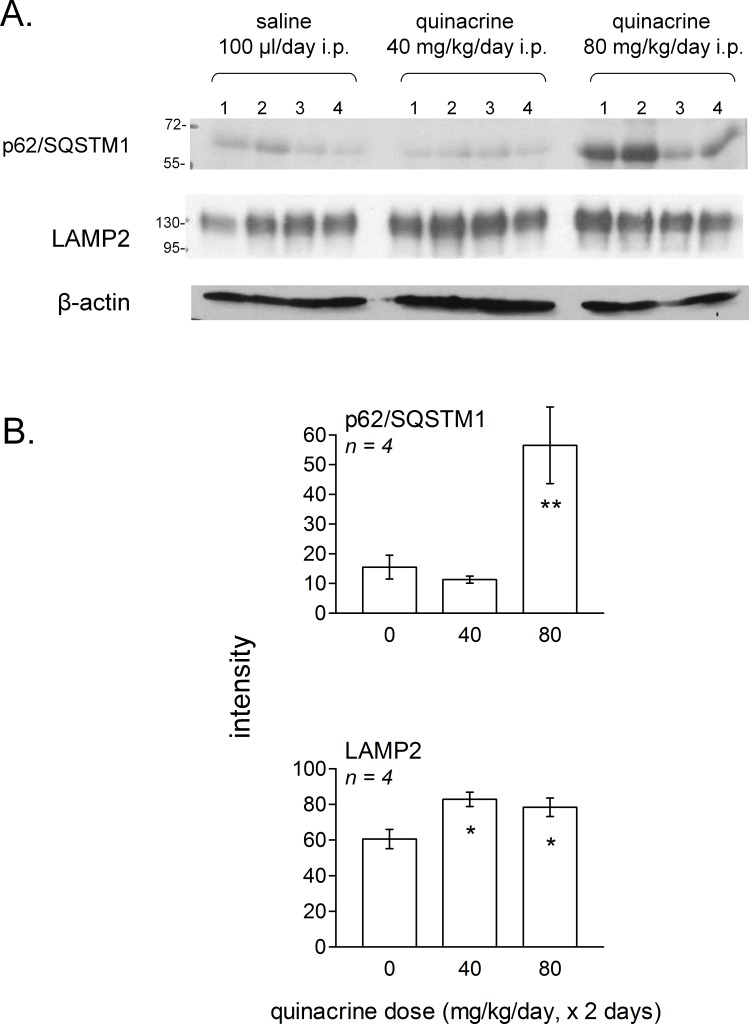
Figure 11. Effect of in vivo treatment with quinacrine on autophagic and lysosomogenesis markers as assessed by immunoblotting of lung tissue extracts.
(A) Groups of 4 mice were treated twice at 24-hr interval, as indicated, and euthanized 24 h after the last dose. Numbers 1–4 in each experimental group represent extracts derived from each experimental animal. (B) averaged quantities of p62/SQSTM1 and LAMP2 were heterogeneous between groups (ANOVA: P < 0.01 and 0.05, respectively). Comparison of values with the common control group for each protein: ∗ P < 0.05; ∗∗ P < 0.01 (Dunnett’s test).

