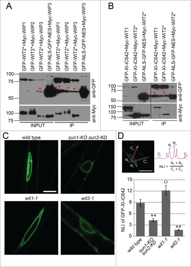
Figure 3.

WIT2 connects myosin XI-i to the SUN-WIP NE bridges.(A) WIT2* interacts with WIP1, WIP2, and WIP3. GFP-tagged proteins were immunoprecipitated and detected with anti-GFP antibody. Myc-tagged proteins were detected with an anti-Myc antibody. Input/IP ratio was 1:9. Numbers on the left indicate molecular mass in kilodaltons. Red arrow heads indicate possible truncated GFP-fusion proteins. (B) WIT1 and WIT2* interacts with XI-iC642. GFP-tagged proteins were immunoprecipitated and detected with anti-GFP antibody. Myc-tagged proteins were detected with an anti-Myc antibody. Input/IP ratio was 1:18. Numbers on the left indicate molecular mass in kilodaltons. Red arrow heads indicate possible truncated GFP-fusion proteins. Vertical back lines represent intervening lanes removed for display purposes. (C) Localization of GFP-XI-iC642 in wild type, sun1-KO sun2-KD, wit1-1, and wit2-1. Scale bar equals 10 μm. All images are single optical sections at the same magnification. (D) NLI of GFP-XI-iC642 in wild type, sun1-KO sun2-KD, wit1-1, and wit2-1. The sum of the 2 maximum NE intensities divided by the sum of the 2 maximum cytoplasmic intensities was used as an NLI (illustrated in the top panel). Double asterisks, P < 0.01, and “O,” P > 0.05, when compared to wild type. Two-tailed t-test was used. For each genotype, 3 transgenic lines and 10 nuclei from each line were analyzed (total n = 30). Error bars represent SE.
