Abstract
AIM: To assess the feasibility and efficacy of laparoscopic total mesorectal excision (LTME) of low rectal cancer with preservation of anal sphincter.
METHODS: From June 2001 to June 2003, 82 patients with low rectal cancer underwent laparoscopic total mesorectal excision with preservation of anal sphincter. The lowest edge of tumors was below peritoneal reflection and 1.5-7 cm from the dentate line (1.5-5 cm in 48 cases, 5-7 cm in 34 cases).
RESULTS: LTME with anal sphincter preservation was performed on 82 randomized patients with low rectal cancer, and 100% sphincter preservation rate was achieved. There were 30 patients with laparoscopic low anterior resection (LLAR) at the level of the anastomosis below peritoneal reflection and 2 cm above from the dentate line; 27 patients with laparoscopic ultralow anterior resection (LULAR) at the level of anastomoses 2 cm below from the dentate line; and 25 patients with laparoscopic coloanal anastomoses (LCAA) at the level of the anastomoses at or below the dentate line. No defunctioning ileostomy was created in any case. The mean operating time was 120 min (ranged from 110-220 min), and the mean operative blood loss was 20 mL (ranged from 5-120 mL). Bowel function was restored and diet was resumed on day 1 or 2 after operation. The mean hospital stay was 8 d (ranged from 5-14). Postoperative analgesics were used in 45 patients. After surgery, 2 patients had urinary retention, one had anastomotic leakage, and another 2 patients had local recurrence one year later. No interoperative complication was observed.
CONCLUSION: LTME with preservation of anal sphincter is a feasible, safe and minimally invasive technique with less postoperative pain and rapid recovery, and importantly, it has preserved the function of the sphincter.
INTRODUCTION
The optimal operation for rectal cancer still remains controversial. Surgical management of rectal cancer has undergone a significant change during the past two decades, a new concept of total mesorectal excision (TME) was introduced[1], and its feasibility and efficacy had been confirmed by a series of clinical trials[2-5]. Compared with conventional procedure, TME markedly improved both oncological and functional outcomes of rectal cancer[6-9], therefore, this procedure has been used as a golden standard for rectal cancer.
Laparoscopic approach has been employed in colorectal surgery for ten years, and its feasibility has been shown in a variety of colorectal operations[10-14]. However, for inadequate operative vision and limitation of the narrow pelvis, total laparoscopic TME with construction of colo-anal anastomosis for low rectal cancer has been regarded as being difficult and time-consuming, and mainly used for upper rectal cancer for a long time[15,16]. Few cases about laparoscopic TME with anal sphincter preservation (SP) for low rectal cancer were reported[17]. The current study was performed to assess the feasibility and efficacy of laparoscopic total mesorectal excision (LTME) for low rectal cancer with preservation of anal sphincter.
MATERIALS AND METHODS
Patients
Between June 2001 and June 2003, randomized 82 patients with low rectal cancer underwent laparoscopic total mesorectal excision with anal sphincter preservation at Department of General Surgery of West China Hospital. The lowest edges of tumors were below peritoneal reflection and 1.5-7 cm from the dentate line (1.5-5 cm in 46 cases, and 5-7 cm in 36 cases). Patients with previous abdominal surgery, obese body, and other surgical benign diseases were not excluded from the laparoscopic procedure. Clinical and demographic data including age, sex, tumor diameter, distances of tumor from the dentate line, concomitant diseases are shown in Table 1.
Table 1.
Clinical and demographic data
| Parameters | Data |
| Age, mean (years) | 26-85, 44 |
| Sex (No. of patients) | |
| Male | 46 |
| Female | 36 |
| Tumor diameter, mean (cm) | 1.5-13, 5.6 |
| Distance of the tumor from the dentate line (cm) | 1.5-7 |
| 1.5-5 cm from lowest edge of tumors to the dentate line (No. of patients) | 48 |
| 5-7 cm from lowest edge of tumors to the dentate line (No. of patients) | 34 |
| Concomitant diseases (No. of patients) | |
| Chronic cholecystitis, cholecystolithiasis, torsion of ovarian cyst and diabetes | 2 |
| Chronic cholecystitis and cholecystolithiasis | 6 |
| Previous lower abdominal operation | 7 |
Preoperative examinations including flexible endoscopes, biopsy, ultrasonography, CT scan, radiography of the chest were performed routinely. All patients underwent preoperative bowel preparation (1L 10% mannite electrolyte solution). Prophylactic antibiotics (ciprofloxacin and metronidazole) were given three days before operation routinely. A urinary catheter and a nasogastric tube were used.
Operative techniques
Under general anesthesia, 82 patients were operated in lithotomic position with 15° head-down tilt by the same surgeon (ZZ ZHOU) with two assistants (YY. YU and Y. SHU). Pneumoperitoneum was introduced through subumbilical incision to maintain pressure at 12-14 mmHg (1 mmHg = 0.133 Kpa). A camera port was created in subumbilical zone with trocar, then an operative port in the right midclavicular line at the level of umbilicus, and an other two operative ports in the left and right McBurney point were created.
Laparoscope was inserted at 25° or 30° into abdominal cavity via the camera port. Routine intra-abdominal exploration was performed. All sharp dissections and divisions on peritoneum, fascia, and connective tissue in retroperitoneal space were performed with a harmonic scalpel. Left lateral peritoneal was divided first, and then sigmoid and descending colon were mobilized completely to ensure the subsequent colo-anal anastomosis free of tension. Then the bowel and its mesentery by a cotton tape at the level 8-10 cm above the upper margin of the tumor were tied, lymph nodes around inferior mesenteric vessels were dissected, and inferior mesenteric vessels were ligated at the high level.
With the operation proceeding of total mesorectal excision, division was moved downward into the pelvis along the anatomic space between visceral and parietal endopelvic fascia. Lateral ligaments of the rectum containing the middle rectal artery or its branches (Figure 1) were gradually divided with the harmonic scalpel from the inner limit of the inferior hypogastric nerve fibers (Figure 2). The pelvic splanchnic nerves were preserved intact as far as possible. Anteriorly, Denonvilliers fascia was dissected and the seminal vesicles and prostate or the posterior wall of the vagina were exposed (Figure 3). At posterior, the rectosacral ligament, anococcygeal ligament, and pubococcygeus muscle were divided, and S2, S3, and S4 sacral splanchnic nerves were identified and protected carefully. The mesorectum, especially the distal mesorectum (DMR), was excised completely till levator ani. Thus, longitudinal muscle layer of the distal precutting rectum and levator ani could be clearly visualized under laparoscopic view, so-called 'denudation and ' uscularization. For low bulky tumor, the'denudation' should be performed under intra-anal finger-guidance to avoid inadvertent damage of adjacent structures. The rectal cross clamping was performed 1.5-3.5 cm below the tumor with endo-cutter (Figure 4).
Figure 1.
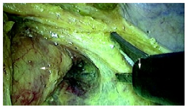
The "lateral" ligaments (→) of rectum containing middle rectal artery or its branches, and mesorectum (←) were dissected completely with a harmonic scalpel (↓).
Figure 2.
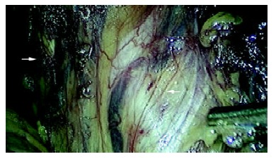
The left pelvic splanchnic nerves were preserved intact as far as possible. Inset shows the inferior hypogastric nerve nerve fibers (←) and the ureter (→).
Figure 3.
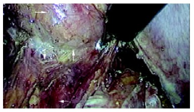
Denonvilliers fascia (↓)was dissected along the space (↑) between the posterior wall of vagina (→) and the rectum (←).
Figure 4.
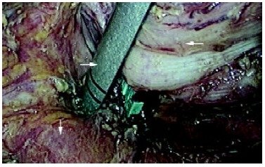
The cross clamping of the rectum (←) was performed 1.5~3.5cm below the tumor with endo-cutter (→). Pelvic floor 'muscularization' was shown (↓).
To extract the bowel loop of the tumor, the port incision was extended at the left McBurney's point to about 3.5 cm long, and isolated the tumor routinely by inserting in a sheath-shaped plastic bag through the incision. The tumor and the proximal colon was extracted through the bag, and then transected the bowel at the level of 10-15 cm above upper margin of the tumor. After inserting the anvil of 29 or 30 mm-sized circular stapler into the end of proximal bowel and securing with 2/0 prolene purse-string suture, the proximal bowel was internalized and the extended incision was closed. Pneumoperitoneum was induced again. Laparoscopic colo-anal or colo-rectal anastomosis was done with CDH 29 circular stapler. After the circular stapler was inserted into the anus, its puncturing cone was pricked through the midpoint of the distal occluding line of the rectum (Figure 5), and fitted into the anvil of the stapler in the pelvic cavity. The stapler was then closed slowly with extreme cautions to avoid inadvertent stapling of adjacent important structures. In this way, the low/ultralow/colo-anal anastomoses were accomplished smoothly. A 10 mm-sized latex tube was routinely put into pelvic cavity through the port at the right McBurney point. No defunctioning ileostomy was created in any case. Distal clearance measurements were taken in an unfixed and unpinned status of surgical specimen in the operating room. The specimen was routinely checked if the visceral endopelvic fascia was dissected completely, and then sent for pathologic examination (Figure 6).
Figure 5.
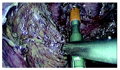
The puncturing cone (→) of the circular stapler pricked through the midpoint of occluding line of the distal rectum(←). Levator ani muscles were exposed (↓).
Figure 6.
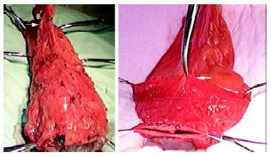
The dorsal mesorectum (→) and distal mesorectum (↓) of the rectal specimen were shown (6a); The anterior side of the specimen and distal margin (←) were shown (6b).
Laparoscopic cholecystectomy and ovariectomy could be performed simultaneously for patients with cholecystolithiasis, chronic cholecystitis, ovarian cyst, and torsion of the ovary.
RESULTS
TME with anal sphincter preservation was accomplished with laparoscope in 82 randomized patients with low rectal cancer, and 100% sphincter preservation rate was achieved. There were 30 patients by laparoscopic low anterior resection (LLAR) at the level of the anastomosis below peritoneal reflection and 2 cm above the dentate line; 27 patients by laparoscopic ultralow anterior resection (LULAR) at the level of the anastomosis 2 cm below the dentate line; and 25 patients by laparoscopic coloanal anastomoses (LCAA) at the level of the anastomosis at/below the dentate line (Figure 7). No defunctioning ileostomy was created in any case. The mean operating time was 120 min (ranged from 110-220 min), and the mean operative blood loss was 20 mL (ranged from 5-120 mL). Both bowel function recovery and diet resumption occurred within 1-2 d after surgery, and the mean hospital stay were 8 d (ranged from 5-14). Postoperative analgesics were used in 45 patients. One patient was converted to open surgery due to dysfunction of coagulation. After operation, 2 patients had urinary retention, one had anastomotic leakage, 2 patients had local recurrence one year later, and no interoperative complication was observed. Clinical and surgical details in this study including tumor and anastomotic levels from anal verge, stage of disease, duration of surgery, length of specimen removed, duration of parenteral analgesia, time to passage of flatus, time to resumption of liquids and solids, and length of post-operative stay are shown in Table 2, Table 3 and Table 4.
Figure 7.
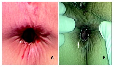
The anastomotic ring (→) could be shown easily in the patient receiving colo-anal anastomosis (a); Satisfactory contractive function of the saved anus (↑) was achieved in the patients receiving laparoscopic TME with anal sphincter preservation at the first day after operation (b).
Table 2.
Clinical parameters for patients with LTME and SP
| Parameters | Data |
| Dukes stage (No. of patients) | |
| A | 5 |
| B | 10 |
| C1 | 33 |
| C2 | 30 |
| D | 4 |
| Pathologic types (No. of patients) | |
| High differentiated adenocarcinoma | 24 |
| Moderately differentiated adenocarcinoma | 37 |
| Low differentiated adenocarcinoma | 21 |
| Multiple primary carcinomas on the bowel wall | 3 |
| Distance of the tumor from the section edge (cm) | 1.5-4 |
| Cancer cell found in the cut margins (No. of patients) | 1 |
| Colo-rectal/anal anastomotic height (cm) (No. of patients) | |
| LLAR, below peritoneal reflection and 2 cm above the dentate line | 30 |
| LULAR, 2 cm below the dentate line | 27 |
| LCAA, at/below the dentate line | 25 |
Table 3.
Early results for laparoscopic TME and SP
| Parameters | Data |
| Operation time (min) | 120(110-220) |
| Operative bleeding (ml) | 20(5-120) |
| Time for bowel function recovery (d) | 1-2 |
| Time to resume normal diet (d) | 1-2 |
| Post-operative analgesic requirement (No. Of patients) | 45 |
| Total hospital stay (d) | 8 (5-14) |
| Sphincter preservation rate (%) | 100% |
| Mortality (%) | 0 |
Table 4.
Complications of laparoscopic TME and SP
| Complications | No. of patients |
| Total number of patients | 82 |
| Perforation of rectum | 0 |
| Urethra damage | 0 |
| Intra-abdominal bleeding | 0 |
| Pelvic abscess | 0 |
| Urinary retention | 2 |
| Anastomotic Leakage | 1 |
| Local recurrence | 2 |
The patients were mobilized two days after operation. Oral intake was gradually increased with the recovery of intestinal function. Most of the patients with low anastomosis as well as about 1/2 of the patients with ultralow or colo-anal anastomosis experienced a quick recovery in anal sphincter's function and controlling of defecation, while 50% of patients with ultralow anastomosis or colo-anal anastomosis suffered from urgent defecation about 5-10 times per day, and their defecation was controlled gradually by proper medication and functional exercise of anal sphincter and levator ani around half a year.
There were no portal site recurrence and mortality observed during follow-up, which ranged from 1 to 24 mo.
DISCUSSION
Study on the management of rectal cancer has progressed greatly in both clinical practice[18-23] and basic research[24-27] in recent years. Multiple clinical studies have demonstrated the correlation of high pelvic recurrence with the degree of mesorectal excision[28]. Residual mesorectum, especially inadequate excision of distal mesorectum (DMR), contributed to poor oncologic outcomes. Regarding DMR, histopathological evidence revealed a high metastasis rate in this region, and it was also found that patients with metastasis in this region would experience a poor prognosis. This is why the principle of TME should be followed in the treatment of rectal cancer[29-31]. Since clinical application of TME, the local recurrence rate of the cancer has decreased dramatically to 5%-7.1%[7,32,33], while that of conventional operative procedure remains 18.5%.
Up to now, there are only a few reports on laparoscopic procedure for low rectal cancer in the literatures, which was mainly due to inadequate surgical vision and limitation of the narrow pelvis. The area 5-8 cm from anal verge and below peritoneal reflection has ever been considered as a blind zone and that within 5 cm from anal verge as a forbidden zone. The current study revealed that the so-called blind and forbidden zones could be broken through, and minimally invasive TME with anal sphincter preservation could be performed safely for patients with rectal cancer based on our success of the large number of open TME and low/ultralow/colo-anal anastomosis and proficient laparoscopic skill. This study is concerned with our experience in colorectal surgery and deals with special laparoscopic colorectal techniques including LLAR, LULAR and LCAA.
Based on our clinical experiences, laparoscopic TME and SP have the following advantages: (1) it helps surgeons identify accurately the interspace of loose connective tissue between visceral and parietal pelvic fascia, and select operative access by amplifying inner image on the monitors. (2) 25° or 30° laparoscope, the third eye of the surgeon, can reach the narrow lesser pelvis and magnify the local vision; (3) it is more definite to identify and protect the pelvic autonomic nerve fiber and plexus due to its magnifying function. (4) hemostatic benefit was owed to minimally invasive sharp dissection with minimal blood loss under laparoscopic view.
However, the laparoscopic technique has its disadvantages, such as long operative time, short of direct hand feeling, technical constraints of the narrow pelvis, difficulty in assessing adequate surgical margins and in ultralow rectal cross clamping, and a long learning course for surgeons.
Good experience with laparoscopic surgery leads to shorter operating time and encouraging results. A bulky tumor or a thickening mesentery usually occupies the most space of narrow pelvic cavity and often influence the operation. To avoid this impact, the camera operator should try his best to adjust laparoscope constantly by 25-30°, which keeps the operator at a correct position. Crack sight and smog are other troublesome problems. Crack sight often occurs after the operation moving into lesser pelvis and it can be solved by adjusting the angle of laparoscope properly. Smog results from the operation of using the harmonic scalpel or cautery, which often distracts operator's vision, even breaks the operative process. The camera operator should withdraw the laparoscope and disperse the smog in time when smog is too heavy or obscures the lens of laparoscope. Therefore, the role of the camera operator is so important that his skill can directly influence the operative processes and results.
The anastomosis is a critical step for the success of this minimally invasive technique with anal sphincter preservation[28]. Double stapling technique (DST) is the remarkable progress in the anastomosis for the operation of low rectal cancer in recent years. Research showed that local recurrence rate was much lower in patients treated with DST than those treated with conventional anastomosis[34]. Low/ultralow/colo-anal anastomoses in all cases of laparoscopic procedure were achieved with DST at this hospital. Based on our experience, there are two special points regarding the anastomosis with DST: (1) denudation of the distal rectal tube, and (2) selection of the pricking point on the occluding line. By denuding the pre-cutting part of the rectal tube, the fat and lymphatic tissue within the mesorectum of distal rectum could be thoroughly excised, the denuded rectal longitudinal muscle could be visualized, and the distal rectum could easily be divided and occluded with endo-stapler. The pricking point of the cone on the stapler should locate at midpoint of the occluding line, because too much displacement of the pricking point may result in ischemia and leakage of the anastomosis, or stapling of adjacent important structures. These two steps can effectively prevent rectal wall from damage and dehiscence of the anastomosis, reduce the use of endo-staplers, and decrease local recurrence.
Special training on TME technique is necessary for surgeons to have enough experience of TME and SP[35]. Proficient skills of laparoscopic operation in pelvis and plentiful experience of open TME are important factors for the success of LTME and SP.
Footnotes
Supported by the Key Project of National Outstanding Youth Foundation of China, No. 39925032
Edited by Ren SY
References
- 1.Heald RJ, Husband EM, Ryall RD. The mesorectum in rectal cancer surgery--the clue to pelvic recurrence. Br J Surg. 1982;69:613–616. doi: 10.1002/bjs.1800691019. [DOI] [PubMed] [Google Scholar]
- 2.Goldberg S, Klas JV. Total mesorectal excision in the treatment of rectal cancer: a view from the USA. Semin Surg Oncol. 1998;15:87–90. doi: 10.1002/(sici)1098-2388(199809)15:2<87::aid-ssu5>3.0.co;2-1. [DOI] [PubMed] [Google Scholar]
- 3.MacFarlane JK, Ryall RD, Heald RJ. Mesorectal excision for rectal cancer. Lancet. 1993;341:457–460. doi: 10.1016/0140-6736(93)90207-w. [DOI] [PubMed] [Google Scholar]
- 4.Ceelen W, Pattyn P. Total mesorectal excision in the treatment of rectal cancer: a review. Acta Chir Belg. 2000;100:94–99. [PubMed] [Google Scholar]
- 5.Kapiteijn E, van De Velde CJ. European trials with total mesorectal excision. Semin Surg Oncol. 2000;19:350–357. doi: 10.1002/ssu.5. [DOI] [PubMed] [Google Scholar]
- 6.Lazuskas T, Lelcuk S, Michowitz M, Rabau M. [Anterior resection with colo-anal anastomosis for low rectal cancer] Harefuah. 1994;126:505–506, 563. [PubMed] [Google Scholar]
- 7.Killingback M, Barron P, Dent OF. Local recurrence after curative resection of cancer of the rectum without total mesorectal excision. Dis Colon Rectum. 2001;44:473–483; discussion 483-6. doi: 10.1007/BF02234317. [DOI] [PubMed] [Google Scholar]
- 8.Bolognese A, Cardi M, Muttillo IA, Barbarosos A, Bocchetti T, Valabrega S. Total mesorectal excision for surgical treatment of rectal cancer. J Surg Oncol. 2000;74:21–23. doi: 10.1002/1096-9098(200005)74:1<21::aid-jso5>3.0.co;2-6. [DOI] [PubMed] [Google Scholar]
- 9.Kapiteijn E, Putter H, van de Velde CJ. Impact of the introduction and training of total mesorectal excision on recurrence and survival in rectal cancer in The Netherlands. Br J Surg. 2002;89:1142–1149. doi: 10.1046/j.1365-2168.2002.02196.x. [DOI] [PubMed] [Google Scholar]
- 10.Monson JR, Darzi A, Carey PD, Guillou PJ. Prospective evaluation of laparoscopic-assisted colectomy in an unselected group of patients. Lancet. 1992;340:831–833. doi: 10.1016/0140-6736(92)92694-b. [DOI] [PubMed] [Google Scholar]
- 11.Huscher C, Silecchia G, Croce E, Farello GA, Lezoche E, Morino M, Azzola M, Feliciotti F, Rosato P, Tarantini M, et al. Laparoscopic colorectal resection. A multicenter Italian study. Surg Endosc. 1996;10:875–879. doi: 10.1007/BF00188473. [DOI] [PubMed] [Google Scholar]
- 12.Kwok SP, Lau WY, Carey PD, Kelly SB, Leung KL, Li AK. Prospective evaluation of laparoscopic-assisted large bowel excision for cancer. Ann Surg. 1996;223:170–176. doi: 10.1097/00000658-199602000-00009. [DOI] [PMC free article] [PubMed] [Google Scholar]
- 13.Bärlehner E, Decker T, Anders S, Heukrodt B. [Laparoscopic surgery of rectal carcinoma. Radical oncology and late results] Zentralbl Chir. 2001;126:302–306. doi: 10.1055/s-2001-14745. [DOI] [PubMed] [Google Scholar]
- 14.Watanabe M, Teramoto T, Hasegawa H, Kitajima M. Laparoscopic ultralow anterior resection combined with per anum intersphincteric rectal dissection for lower rectal cancer. Dis Colon Rectum. 2000;43:S94–S97. doi: 10.1007/BF02237232. [DOI] [PubMed] [Google Scholar]
- 15.Hartley JE, Mehigan BJ, Qureshi AE, Duthie GS, Lee PW, Monson JR. Total mesorectal excision: assessment of the laparoscopic approach. Dis Colon Rectum. 2001;44:315–321. doi: 10.1007/BF02234726. [DOI] [PubMed] [Google Scholar]
- 16.Weiser MR, Milsom JW. Laparoscopic total mesorectal excision with autonomic nerve preservation. Semin Surg Oncol. 2000;19:396–403. doi: 10.1002/ssu.10. [DOI] [PubMed] [Google Scholar]
- 17.Chung CC, Ha JP, Tsang WW, Li MK. Laparoscopic-assisted total mesorectal excision and colonic J pouch reconstruction in the treatment of rectal cancer. Surg Endosc. 2001;15:1098–1101. doi: 10.1007/s00464-001-9000-2. [DOI] [PubMed] [Google Scholar]
- 18.Cao GW, Qi ZT, Pan X, Zhang XQ, Miao XH, Feng Y, Lu XH, Kuriyama S, Du P. Gene therapy for human colorectal carcinoma using human CEA promoter contro led bacterial ADP-ribosylating toxin genes human CEA: PEA & amp; DTA gene transfer. World J Gastroenterol. 1998;4:388–391. doi: 10.3748/wjg.v4.i5.388. [DOI] [PMC free article] [PubMed] [Google Scholar]
- 19.Mao AW, Gao ZD, Xu JY, Yang RJ, Xiao XS, Jiang TH, Jiang WJ. Treatment of malignant digestive tract obstruction by combined intraluminal stent installation and intra-arterial drug infusion. World J Gastroenterol. 2001;7:587–592. doi: 10.3748/wjg.v7.i4.587. [DOI] [PMC free article] [PubMed] [Google Scholar]
- 20.Makin GB, Breen DJ, Monson JR. The impact of new technology on surgery for colorectal cancer. World J Gastroenterol. 2001;7:612–621. doi: 10.3748/wjg.v7.i5.612. [DOI] [PMC free article] [PubMed] [Google Scholar]
- 21.Deng YC, Zhen YS, Zheng S, Xue YC. Activity of boanmycin against colorectal cancer. World J Gastroenterol. 2001;7:93–97. doi: 10.3748/wjg.v7.i1.93. [DOI] [PMC free article] [PubMed] [Google Scholar]
- 22.Wang SH, Zheng YQ. A clinicopathologic analysis of 354 cases of large intestinal cancer in western. Hunan Xin Xiaohuabingxue Zazhi. 1996;4:325–326. [Google Scholar]
- 23.Kapiteijn E, Marijnen CA, Nagtegaal ID, Putter H, Steup WH, Wiggers T, Rutten HJ, Pahlman L, Glimelius B, van Krieken JH, et al. Preoperative radiotherapy combined with total mesorectal excision for resectable rectal cancer. N Engl J Med. 2001;345:638–646. doi: 10.1056/NEJMoa010580. [DOI] [PubMed] [Google Scholar]
- 24.Yuan HY, Li Y, Yang GL, Bei DJ, Wang K. Study on the causes of local recurrence of rectal cancer after curative resection: analysis of 213 cases. World J Gastroenterol. 1998;4:527–529. doi: 10.3748/wjg.v4.i6.527. [DOI] [PMC free article] [PubMed] [Google Scholar]
- 25.Qing SH, Jiang HY, Qi DL, Zhou ZD, Huang XC, Zhang FM, Sheng QG. Relationship between related factors with lymph node metastasis of colorectal cancer. Shijie Huaren Xiaohua Zazhi. 2000;8:654–657. [Google Scholar]
- 26.Gao ZS, Yin CH, Song DY, Liu FZ, Gu YZ, Liu YP. Clinical significance of pedicled greater omentum transplantation in radical resection of rectal cancer. Huaren Xiaohua Zazhi. 1998;6:875–876. [Google Scholar]
- 27.Yang JH, Rao BQ, Wang Y, Tu XH, Zhang LY, Chen SH, Ou Yang XN, Dai XH. Clinical significance of detecting the circulating cancer cells in peripheral blood from colorectal cancer. Shijie Huaren Xiaohua Zazhi. 2000;8:187–189. [Google Scholar]
- 28.Wexner SD, Rotholtz NA. Surgeon influenced variables in resectional rectal cancer surgery. Dis Colon Rectum. 2000;43:1606–1627. doi: 10.1007/BF02236751. [DOI] [PubMed] [Google Scholar]
- 29.Choi JS, Kim SJ, Kim YI, Min JS. Nodal metastasis in the distal mesorectum: need for total mesorectal excision of rectal cancer. Yonsei Med J. 1996;37:243–250. doi: 10.3349/ymj.1996.37.4.243. [DOI] [PubMed] [Google Scholar]
- 30.Tocchi A, Mazzoni G, Lepre L, Liotta G, Costa G, Agostini N, Miccini M, Scucchi L, Frati G, Tagliacozzo S. Total mesorectal excision and low rectal anastomosis for the treatment of rectal cancer and prevention of pelvic recurrences. Arch Surg. 2001;136:216–220. doi: 10.1001/archsurg.136.2.216. [DOI] [PubMed] [Google Scholar]
- 31.Scott N, Jackson P, al-Jaberi T, Dixon MF, Quirke P, Finan PJ. Total mesorectal excision and local recurrence: a study of tumour spread in the mesorectum distal to rectal cancer. Br J Surg. 1995;82:1031–1033. doi: 10.1002/bjs.1800820808. [DOI] [PubMed] [Google Scholar]
- 32.Heald RJ. Total mesorectal exsicion (TME) Acta Chir Iugosl. 2000;47:17–18. [PubMed] [Google Scholar]
- 33.McCall JL, Cox MR, Wattchow DA. Analysis of local recurrence rates after surgery alone for rectal cancer. Int J Colorectal Dis. 1995;10:126–132. doi: 10.1007/BF00298532. [DOI] [PubMed] [Google Scholar]
- 34.Law WL, Chu KW. Impact of total mesorectal excision on the results of surgery of distal rectal cancer. Br J Surg. 2001;88:1607–1612. doi: 10.1046/j.0007-1323.2001.01929.x. [DOI] [PubMed] [Google Scholar]
- 35.Heald RJ. Total mesorectal excision is optimal surgery for rectal cancer: a Scandinavian consensus. Br J Surg. 1995;82:1297–1299. doi: 10.1002/bjs.1800821002. [DOI] [PubMed] [Google Scholar]


