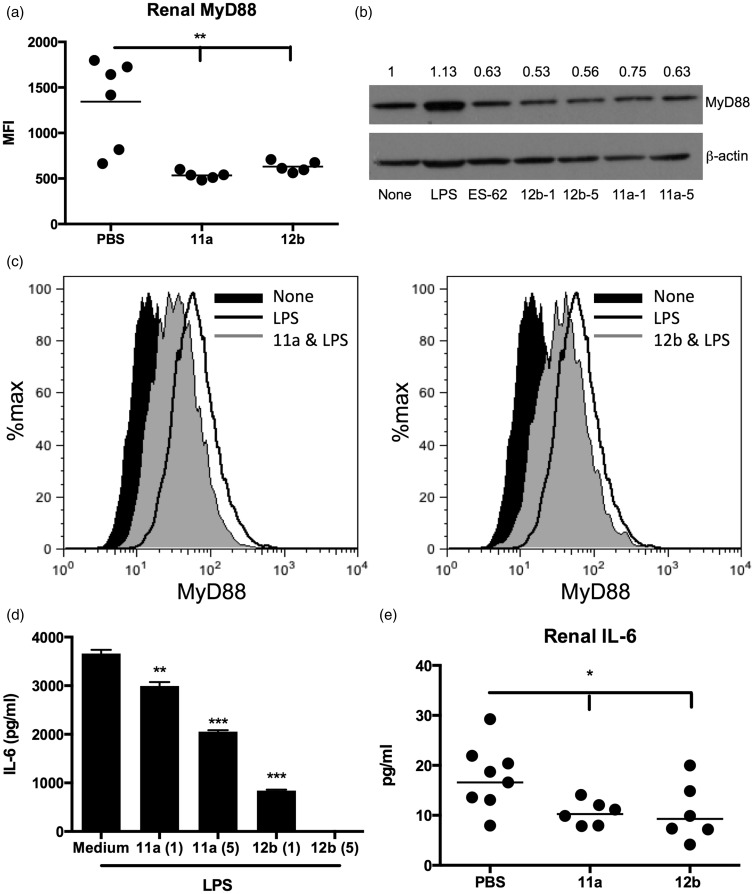
Figure 3.
SMAs 11a and 12b suppress MyD88 signalling and downstream effector IL-6 responses in the kidney. (a) Flow cytometric analysis (MFI) of kidney MyD88 expression in individual mice (week 22). (b) MyD88 expression by bmM5 stimulated with LPS (1 µg/ml), ES-62 (2 µg/ml), 11a or 12b (both at 1 and 5 µg/ml) for 18 hours: exemplar Western blot and quantitation of MyD88/β-actin expression normalized to the ‘None’ (medium) control. (c) Flow cytometric analysis of MyD88 expression by bmM treated with 11a or 12b (5 µg/ml, two hours) prior to stimulation with LPS for 18 hours. IL-6 release by these cells (d), and in the kidney supernatants of PBS-, 11a- or 12b-treated (1 µg/dose) MRL/Lpr mice (e). SMAs: small molecule analogues; MFI: mean fluorescence intensity; LPS: lipopolysaccharide; IL-6: interleukin-6; PBS: phosphate-buffered saline; bmM: bone marrow-derived macrophages; MyD88: myeloid differentiation factor 88.

