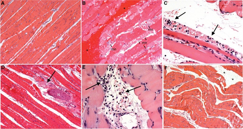
Fig. 2.
Pathology in H&E stained muscle tissue sections.

A, Muscle tissue from non-blasted (control) animals displayed normal morphology with no evidence of background pathology. B, Muscle tissues from many of the blast exposed animals displayed varying degrees of hemorrhage (∗) and muscle fiber necrosis (mn). C, Necrosis was frequently associated with concentrations of polymorphonuclear cells (heterophil) infiltrating the muscle tissues (↑). D, Examples of inflammatory cell infiltration (↑) and hemorrhage (∗) were also observed in adipose and connective tissues surrounding some nerve fiber bundles (N) within the area of injured muscle tissue. E, Some areas of connective and adipose tissue within the muscle were also seen to contain mononuclear lymphocytes (↑). F, Oedema was observed between muscle fiber bundles in some animals exposed to blast injury (∗). Images A, B, D and F scale = 400 μm, C scale = 50 μm, E scale = 100 μm.
