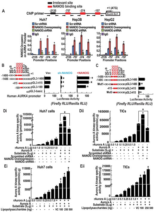
Figure 3. NANOG acts as a transcriptional activator of AURKA and modulates kinase activity.
(A) ChIP-qPCR demonstrates direct association of NANOG at the AURKA promoter. Huh7, HepG2 and Hep3B cells stably expressing NANOG or harboring a non-targeting, scrambled shRNA, or NANOG-targeting shRNA were subjected to Chromatin immunoprecipitation using ChIP grade NANOG antisera or isotype-matched control IgG. Immunoprecipitated chromatin was analyzed by qPCR using primer sets designed to amplify the indicated regions. (B) AURKA promoter reporter assay. Huh7 cells were transfected with the indicated promoter-reporter constructs. (C) Mutation of NANOG-binding sites reduces levels of transactivation. Promoter activity is expressed as relative light units (RLU) normalized to the activity of cotransfected Renilla luciferase. Error bars represent the SD from at least three independent biological replicates (*P < 0.05). (Di–ii) Figures represent the effect of NANOG-silenced and overexpression of the kinase activity of AURKA in Huh7 cells (Di) and TICs (Dii). (Ei–ii) The histogram represents the effect of Lipopolysaccharides (LPS) on AURKA activity Huh7 cells (Ei) and TICs (Eli). Each bar in the histogram represents mean ± SD of three independent experiments, * represents P < 0.05.

