

Abstract
Background
Due to their genetic proximity, chimpanzees share with human several diseases including bacterial, fungal and viral infections, such as candidiasis, acquired immune deficiency syndrome (AIDS), Ebola virus disease. However, in its natural environment, chimpanzees are tolerant to several pathogens including simian immunodeficiency virus (SIV), virus related to human immunodeficiency virus (HIV) that contribute to the emergence of opportunistic diseases such as microbial infections.
Methods
Twenty seven species of plants consumed by chimpanzees were evaluated for their antimicrobial potential against Escherichia coli, Pseudomonas aeruginosa, Staphylococcus aureus, Candida albicans, Candida tropicalis and Candida glabrata using the agar diffusion technique and micro-dilution in 96-well plates. In total 132 extracts (33 dichloromethane, 33 methanol, 33 ethyl acetate and 33 aqueous) were tested.
Results
The results showed that 24 extracts (18 %) showed activity against bacteria and 6 extracts (5 %) were active against yeasts. The minimal inhibitory concentrations (MICs) values of active extracts ranged between 23 and 750 μg/ml for bacteria and between 188 and 1500 μg/ml for yeasts.
Conclusion
Tristemma coronatum was the most promising on the studied microorganisms followed by Beilschmiedia mannii. The extracts of the two plants indicated by chimpanzees have potential for antimicrobial use in human.
Keywords: Chimpanzee’s diet, Antimicrobial, Bacteria, Yeast, Beilschmiedia mannii, Tristemma coronatum, Ivory Coast
Background
The use of plants in herbal medicine is very old and is experiencing a resurgence interest with the public [1]. According to World Health Organization (WHO), approximately, 75–80 % of the world’s population use plant medicine either in part or entirely for their health and care needs [2]. Chimpanzees are mainly frugivorous. This practice seems to have a beneficial effect on their health and well-being. Thus, it is worth exploring their behavioral trend to self-medicate in order to identify plants with therapeutic potential to fight certain human diseases including bacteria and fungi. These diseases are getting more common in humans with the development of drug resistance.
Infectious diseases caused by bacteria and fungi affect millions of people worldwide [3]. According to Soro et al. [4], bacterial and fungal infections, especially candidiasis, are among the most dangerous and deadly opportunistic diseases for vulnerable people such as children, the elderly and immunocompromised persons.
Their control becomes complex due to the emergence of multiresistant bacteria and fungi to many conventional antibiotics and antifungals [5–12]. Combined with the scarcity of new drugs on the market in recent years, this increase in bacterial and fungal resistance worldwide is a major threat to public health. According to the 2014 WHO report, in Africa, there is strong resistance of Escherichia coli to cephalosporins and third generation fluoroquinolones; two types of essential and widely used antibacterial drugs. It is also noted that 80 % of Staphylococcus aureus are resistant to methicillin (MRSA) in some parts of this region [13]. Also, there is an increased resistance to azoles such as fluconazole, the antifungal drug of choice in many countries, and the recently introduced antifungal agent, echinocandins [14]. In Ivory Coast, many cases of multidrug-resistant bacteria are reported [6–10]. According to Guessennd et al. [15], the frequency of bacterial strains resistant to imipenem increased from 1.9 % in 2005 to 5.9 % in 2009 and that of bacteria producing beta-lactamase with extended spectrum, from 5.3 % in 2005 to 16.8 % in 2009.
With the growing concern generated by the use of drugs, both in therapy and in the food industry, there has been in recent years an increase in interest in medicinal plants [16]. Many medicinal plants have shown their effectiveness against microbial infections across Africa, particularly in Ivory Coast [17–20]. However, the consumption pattern of chimpanzees, essentially composed of plant organs, remains poorly explored in this field. Chimpanzee remains morphologically and genetically the closest animal to humans, with 98 % of common DNA [21]. This genetic proximity expressed at many levels, including sharing with humans many zoonotic infections such as candidiasis and other bacterial infections. However, it is known that chimpanzees in their natural habitat are tolerant or resilient to SIV (Simian immunodeficiency virus) while humans are vulnerable to HIV, virus related to the simian virus [22, 23]. This tolerance or resilience of chimpanzee to SIV could be of genetic origin but also due to the environment. Their consumption pattern and feeding habit could indeed greatly contribute to their health, because in captivity, these animals develop many diseases [24]. It is recognized in pathophysiology that the presence of HIV in the body leads to a weakening of the immune system, promoting the emergence of opportunistic infections such as bacterial diseases and candidiasis [25].
A preliminary study carried out by Ahoua et al. [26] on the therapeutic potential, including the antioxidant capacity of eight plant species from Taï’s chimpanzee’s diet and harvested in the same area, but different from those investigated in this work, showed that these plants have strong antioxidant activity.
Based on the “One Health” concept [26–29], that promotes interdisciplinary and intersectorial collaborations at the human-animal-environment interface, the current study explores chimpanzees eating habits and behaviors towards self-medication to identify plants with strong antimicrobial potential able to play a role in the control of infections in humans. These plants could bring added value to the herbal medicine to solve health problems in humans. The main objective is to assess the antimicrobial potential of plants falling within the diet of chimpanzees in Tai National Park (Ivory Coast).
Methods
Selection of plant species
The plant species used for antimicrobial investigations came from the diet of chimpanzees (Pan troglodytes verus) (Fig. 1) from Taï National Park (TNP), located in the Southwestern region of Ivory Coast (Fig. 2). The research authorization was provided by the Ministry of Higher Education and Scientific Research of Ivory Coast while access and work in the park was issued by the Ministry of the Environment and Sustainable Development through the Ivorian Parks and Reserves Office.
Fig. 1.
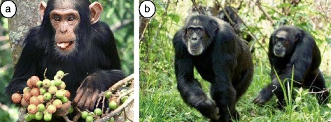
Chimpanzees (Pan troglodytes verus) in Taï National Park. a Young chimpanzee eating Ficus fruits (Source: http://pin.primate.wisc.edu/factsheets/french/chimpanzee); b-Group of chimpanzees (Source: http://www.sunservices.org/sun/index.php/tourisme/nos-parcs-et-reserves)
Fig. 2.
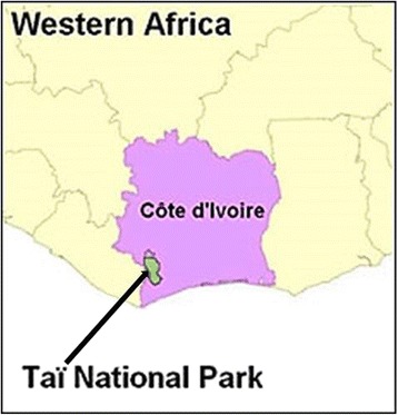
Localization of Taï National Park on Ivory Coast map (Source: wildchimps.org)
These plants were identified through direct observations in response to a previous study carried by N’Guessan [30] on Taï’s National Park chimpanzee’s diet. Many criteria including the consumption frequency of plant species, the consumed quantity and the duration of consumption allowed us to select 27 plant species among 131 reported. These plants were harvested in TNP in May and November 2012. The different parts harvested were leaves, fruits (pericarp, mesocarp and endocarp) and marrows (Fig. 3). Herbarium specimens were deposited in the herbarium of Swiss Centre of Scientific Research in Ivory Coast. Table 1 shows the different harvested plants.
Fig. 3.
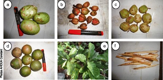
Example of plant organs consumed by chimpanzees. a Magnistipula butayei fruits; b Pycnanthus angolensis fruits; c Duboscia viridiflora fruits; d Panda oleosa fruits; e Nauclea diderrichii leaves; f Ancistrophyllum secondiflorum marrows
Table 1.
List of tested plant species
| N° | Plant species | Family | Plant part used |
|---|---|---|---|
| 1 | Afzelia bella Harms | Leguminosae | Leaves |
| 2 | Ancistrophyllum secundiflorum (P.Beauv.) G.Mann & H.Wendl. | Arecaceae | Marrow |
| 3 | Beilschmiedia mannii (Meisn.) Benth. & Hook. f. | Lauraceae | Whole fruits; Pericarp; Mesocarp; Endocarp |
| 4 | Calpocalyx aubrevillei Pellegr. | Leguminosae | Leaves |
| 5 | Chrysophyllum taiense Aubrév. & Pellegr. | Sapotaceae | Leaves |
| 6 | Coula edulis Baill. | Olacaceae | Fruits (Endocarp) |
| 7 | Dacryodes klaineana (Pierre) H.J.Lam. | Burseraceae | Fruits |
| 8 | Dialium aubrevillei Pellegr. | Leguminosae | Fruits |
| 9 | Dichapetalum pallidum (Oliv.) Engl. | Dichapetalaceae | Leaves |
| 10 | Dioscorea multiflora Mart. ex Griseb. | Dioscoreaceae | Leaves |
| 11 | Duboscia viridiflora (K.Schum.) Mildbr. | Malvaceae | Fruits |
| 12 | Glyphaea brevis (Spreng.) Monach. | Tiliaceae | Leaves |
| 13 | Halopegia azurea (K.Schum.) K.Schum. | Marantaceae | Marrow |
| 14 | Irvingia grandifolia (Engl.) Engl. | Irvingiaceae | Fruits |
| 15 | Keayodendron bridelioides Gilg & Mildbr. ex Hutch. & Dalziel | Euphorbiaceae | Leaves |
| 16 | Klainedoxa gabonensis Pierre ex Engl. | Irvingiaceae | Fruits (Pericarp) |
| 17 | Landolphia hirsuta (Hua) Pichon | Apocynaceae | Leaves |
| 18 | Magnistipula butayei De Wild. | Chrysobalanaceae | Fruits (Pericarp and endocarp) |
| 19 | Manniophyton fulvum Müll. Arg. | Euphorbiaceae | Leaves |
| 20 | Musanga cecropiodes R. Br. ex Tedlie | Moraceae | Leaves |
| 21 | Nauclea diderrichii (De Wild.) Merr. | Rubiaceae | Fruits; Leaves |
| 22 | Panda oleosa Pierre | Pandaceae | Fruits (Pericarp) |
| 23 | Parinari excelsa Sabine | Chrysobalanaceae | Fruits (Pericarp and endocarp) |
| 24 | Platysepalum hirsutum (Dunn) Hepper | Leguminosae | Leaves |
| 25 | Sarcophrynium brachystachys (Benth.) K. Schum. | Marantaceae | Marrow |
| 26 | Sterculia oblonga Mast. | Sterculiaceae | Leaves |
| 27 | Tristemma coronatum Benth. | Melastomataceae | Leaves |
Extracts preparation
The samples were cleaned immediately after collection by washing them with water, dried under air at 18 °C for 3 weeks for the leaves and marrows. Fruits were lyophilized and then pulverized. Afterwards, 20 g of plant powder were macerated successively in 200 ml of organic solvent (dichloromethane, ethyl acetate and methanol) twice for 24 hours. Otherwise, 20 g of plant powder were macerated in 200 ml of water in the same conditions. The macerates obtained were then filtered and evaporated with a rotavapor at 40 °C. The methanol and aqueous extracts were then lyophilized. Dichloromethane and ethyl acetate extracts were evaporated to dryness.
Assessment of the antimicrobial activity
Tested microorganisms
The antimicrobial activity was assessed against seven strains of bacteria including Escherichia coli ATCC 25922, Escherichia coli CIP 54127AF, Pseudomonas aeruginosa CIP 103467, Pseudomonas aeruginosa ATCC 27853, Staphylococcus aureus sensitive to penicillin, Staphylococcus aureus ATCC 25923, Staphylococcus aureus CIP 4.83 and four strains of yeast (Candida albicans (1), Candida albicans (2), Candida tropicalis and Candida glabrata). These reference and clinical strains were provided by the National Laboratory of Public Health of Ivory Coast and the Microbiology Laboratory of Swiss Centre of Scientific Research in Ivory Coast.
The antimicrobial activity was evaluated according to the protocol described by Koné et al. [18] using the agar diffusion technique. Stock solutions of plant extracts were prepared at 30 mg/ml in dimethyl sulfoxide (DMSO) and at 1 mg/ml (in distilled water) for antibiotics (tetracycline and gentamicin) and antifungals (nystatin and amphotericin B).
Antibacterial activity
Sensitivity test
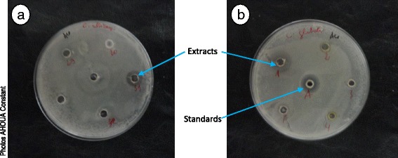
Mueller-Hinton agar in Petri dishes (thickness = 4 mm) were soaked with an inoculum equivalent to 0.5 of McFarland. After drying, wells (diameter = 6 mm) were made in the agar using sterile Pasteur pipette. Fifty microliters (50 μl) of extract (1.5 mg/ml) or antibiotic (0.025 mg/ml) was poured in the wells. Plates were left at ambient laboratory temperature for 15 to 30 min for a pre-diffusion of the solutions, and then incubated at 37 °C for 18 h. After incubation, the diameters (mm) of inhibition zones were measured (Fig. 4). The tests were carried out twice.
Fig. 4.
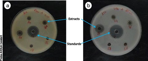
Inhibitory diameters of some ethyl acetate extracts on two bacteria Pseudomonas aeruginosa CIP 103467 (a) and Staphylococcus aureus ATCC 25923 (b) 24 = Musanga cecropioides; 25 = Landolphia hirsuta; 26 = Endocarp of Beilschmiedia mannii fruits; 28 = Duboscia viridiflora; 31 = Klainedoxa gabonensis; G = gentamicin
Determination of Minimum Inhibitory Concentration (MIC)
The extracts showing an inhibitory diameter of at least 10 mm were selected to determine the minimum inhibitory concentrations (MICs) using broth microdilution method in 96-wells microplates [18]. The MIC is the lowest concentration at which the visible growth of a strain was completely inhibited (no visible turbidity in wells). The plant extracts were solubilized in DMSO (30 mg/ml) and serially diluted in Mueller-Hinton medium, from 1500 to 1.5 μg/ml. The final concentrations were 25 to 0.05 μg/ml for antibiotics. All the tested bacteria were used with an initial inoculum of 3 × 10⁶ bacteria/ml. The microplates were incubated at 37 °C for 18 h.
Determination of Minimum Bactericidal Concentration (MBC)
MBC is the lowest concentration of antibiotic or crude extract in which less than 0.01 % of the initial inoculum survived after 18–24 h. Medium from wells with no visible growth and from the initial inoculum (dilution 10−1, 10−2, 10−3; 10−4 and 10−5) were plated on agar, and colonies counted. The value MBC/MIC allowed to determine whether an extract was bacteriostatic (MBC/MIC > 4) or bactericidal (MBC/MIC < 4) [31].
Antifungal activity
Sensitivity test
Tryptone Soya Agar in Petri dishes was soaked with a yeast suspension. After drying, wells (diameter = 6 mm) were made in the agar using a sterile Pasteur pipette. A fraction (50 μl) of plant extract at 1.5 mg/ml or standard antifungal at 0.025 mg/ml was poured in the wells. Plates were left at ambient laboratory temperature for 15 to 30 min for a pre-diffusion of the solutions and then incubated at 30 °C for 48 to 72 h. After incubation, the diameters (mm) of inhibition zones were measured (Fig. 5). The tests were carried out twice for result validation.
Fig. 5.
Inhibitory diameters of some ethyl acetate extracts on two yeasts Candida albicans ATCC 10231 (a) and Candida glabrata (b). 1 = Dacryodes klaineana; 2 = Chrysophyllum taiense; 3 = Pericarp of Panda oleosa fruits; 4 = Dichapetalum pallidum; 5 = Mesocarp of Beilschmiedia mannii fruits; 29 = Pericarp of Beilschmiedia mannii fruits; 30 = Endocarp of Parinari excelsa fruits; 31 = Klainedoxa gabonensis
Determination of Minimum Inhibition Concentration (MIC)
Extracts with inhibitory diameters greater than or equal to 10 mm, were selected to determine the minimum inhibitory concentrations (MICs) by using a microdilution method in liquid medium [16]. Plants extracts (1500 to 1.5 μg/ml) or nystatin and amphotericin B, positive controls (25 to 0.05 μg/ml) were serially diluted in Sabouraud broth in microplates (96 wells). This was followed by the addition of 50 μl of the inoculum to each well. The microplates were incubated at 30 °C for 48 to 72 h. After incubation, 40 μl of chloride methylthiazoyltetrazolium (MTT) at 0.2 mg/ml (in water) were added to each well, and further incubated again for 30 min at room temperature. The wells with no growth of yeast were colored in yellow and the lowest concentration (MIC) was determined [32]. The experiment was performed in duplicate in the same plate and tests were repeated twice.
Statistical analysis
Microsoft Access 2007 was used for data entry. Data were then exported to Excel for analysis. The software STATISTICA 7.1 was used for data analysis [33]. Results were presented as mean ± SD of duplicate experiments. One-way analysis of variance (ANOVA) was used to compare extract’s inhibitory diameters. Significant differences between extracts were determined at P < 0.05. The least significant difference (LSD) between extracts was performed by the HDS test of Tukey.
Results and discussion
The results of this study showed that out of the 132 tested extracts, 24 (18 %) were active against bacteria (Tables 2 and 3) and 6 (5 %) against fungi (Table 4). The inhibition zones diameters were between 10 and 20 mm. The MICs values ranged between 23 and 750 μg/ml for extracts and between 0.10 and 3 μg/ml for standards (0.10 to 3 μg/ml for gentamicin; 0.20 to 1.6 μg/ml for tetracycline). The active extracts were bactericidals and bacteriostatics (Table 3). A MIC value of 188 μg/ml or lower was considered a reasonable cut-off point for crude extracts.
Table 2.
Inhibitory diameters (mm) of extracts on tested bacteria*
| Bacteria | |||||||||
|---|---|---|---|---|---|---|---|---|---|
| Plant species | Plant parts used | Extracts | S. a. CIP 4.83 | S. a. Sen | S. a. ATCC 25923 | E.coli CIP 54127AF | E. coli ATCC 25925 | P. a. CIP 103467 | P. a. ATCC 27853 |
| Beilschmiedia mannii | Pericarp | DCM | 15 ± 0.5cd | 7 ± 0.5lm | 0 | 0 | 0 | 9 ± 0.0gj | 0 |
| Mesocarp | DCM | 15 ± 1.0cd | 14 ± 1.0c | 13 ± 2.0fg | 0 | 0 | 12 ± 1.5cf | 0 | |
| EtOAc | 12 ± 0.5e | 12 ± 0.5de | 10 ± 0.0hj | 0 | 0 | 13 ± 0.5ce | 0 | ||
| MeOH | 10 ± 0.5fg | 11 ± 0.5eg | 9 ± 0.5jk | 0 | 0 | 10 ± 0.0fi | 0 | ||
| Aqueous | 0 | 10 ± 0.0eh | 0 | 0 | 0 | 0 | 0 | ||
| Endocarp | DCM | 0 | 9 ± 0.5hk | 0 | 0 | 0 | 10 ± 0.0fi | 0 | |
| Whole fruits | EtOAc | 10 ± 0.0f | 11 ± 0.0df | 9 ± 0.5jk | 0 | 0 | 11 ± 0.5eh | 0 | |
| MeOH | 10 ± 1.0f | 11 ± 0.0df | 11 ± 0.5hi | 0 | 0 | 11 ± 0.0eh | 0 | ||
| Chrysophyllum taiense | Leaves | EtOAc | 0 | 11 ± 0.5eg | 0 | 0 | 0 | 11 ± 0.5eh | 0 |
| MeOH | 8 ± 0.5ij | 10 ± 0.0eh | 9 ± 0.0ik | 0 | 0 | 10 ± 0.5fj | 0 | ||
| Nauclea diderrichii | Leaves | DCM | 0 | 13 ± 0.5cd | 0 | 0 | 0 | 14 ± 2.5c | 0 |
| MeOH | 10 ± 0.5fg | 10 ± 0.0eh | 9 ± 0.0ik | 0 | 0 | 9 ± 0.5fj | 0 | ||
| Manniophyton fulvum | Leaves | MeOH | 0 | 9 ± 0.50hk | 15 ± 0.5ef | 0 | 0 | 9 ± 0.5hj | 0 |
| Calpocalyx aubrevillei | Leaves | EtOAc | 13 ± 0.5e | 13 ± 0.5cd | 12 ± 0.5gh | 0 | 0 | 13 ± 0.0cd | 0 |
| MeOH | 9 ± 0.5gi | 11 ± 0.5eg | 9 ± 0.0ik | 0 | 0 | 10 ± 1.0fj | 0 | ||
| Dacryodes klaineana | Fruits | EtOAc | 0 | 0 | 18 ± 0.5cd | 0 | 0 | 9 ± 1.5gj | 0 |
| Landolphia hirsuta | Leaves | MeOH | 9 ± 0.5gi | 10 ± 0.5fi | 7 ± 0.5l | 0 | 0 | 10 ± 0.5fj | 0 |
| Platysepalum hirsutum | Leaves | EtOAc | 0 | 9 ± 0.5fi | 0 | 0 | 0 | 10 ± 0.0fi | 0 |
| MeOH | 9 ± 0.5gi | 10 ± 0.5fi | 8 ± 0.5kl | 0 | 0 | 10 ± 0.5fj | 0 | ||
| Klainedoxa gabonensis | Pericarp | EtOAc | 15 ± 0.5cd | 10 ± 0.0eh | 16 ± 0.0de | 0 | 0 | 11 ± 0.0dg | 0 |
| Sterculia oblonga | Leaves | DCM | 0 | 11 ± 0.0df | 0 | 0 | 0 | 0 | 0 |
| Magnistipula butayei | Pericarp | DCM | 15 ± 0.0cd | 9 ± 0.0gj | 9 ± 0.5ijk | 0 | 0 | 9 ± 1.5gj | 0 |
| Tristemma coronatum | Leaves | EtOAc | 16 ± 1.0c | 9 ± 0.5fi | 19 ± 1.0c | 0 | 0 | 10 ± 0.5fj | 0 |
| MeOH | 0 | 9 ± 0.0fi | 17 ± 1.5d | 0 | 0 | 8 ± 0.0ij | 0 | ||
| Gentamicin | 20 ± 0.5b | 22 ± 1.5a | 22 ± 0.5b | 21 ± 2.5a | 0 | 29 ± 1.0a | 29 ± 1.5a | ||
| Tetracycline | 23 ± 0.5a | 19 ± 1.0b | 30 ± 1.5a | 17 ± 1.0b | 0 | 21 ± 0.5b | 24 ± 1.0b | ||
| F | 432.11 | 268.08 | 290.02 | 97.082 | 176.88 | 423.89 | |||
| P | < 0.001 | ||||||||
*: Mean ± SD of two replicates; DCM: Dichloromethane; EtOAc: Ethyl Acetate; MeOH: Methanol; S. a.: Staphylococcus; E: Escherichia; P. a.: Pseudomonas aeruginosa; ATCC: American Type Culture Collection; CIP: Centre Institut Pasteur; Sen: Sensitive; F: Fisher statistical; P: Probability; Values with the same superscript letter are not significantly different (P < 0.001)
Table 3.
Antibacterial powers of active extracts
| Bacteria | |||||||
|---|---|---|---|---|---|---|---|
| Plant species | Plant part used | Extracts | S. a. CIP 4.83 | S. a. Sen | S. a. ATCC 25923 | P. a. CIP 103467 | |
| Chrysophyllum taiense | Leaves | MeOH | MIC | >1500 | 188 | >1500 | 188 |
| MBC | 1500 | 750 | |||||
| MBC/MIC | 8 | 4 | |||||
| APw | bacstat | bact | |||||
| EtOAc | MIC | >1500 | 188 | >1500 | 188 | ||
| MBC | 750 | 750 | |||||
| MBC/MIC | 4 | 4 | |||||
| APw | bact | bact | |||||
| Manniophyton fulvum | Leaves | MeOH | MIC | >1500 | >1500 | 47 | >1500 |
| MBC | 1500 | ||||||
| MBC/MIC | 32 | ||||||
| APw | bacstat | ||||||
| Calpocalyx aubrevillei | Leaves | MeOH | MIC | >1500 | 94 | >1500 | 94 |
| MBC | 1500 | 750 | |||||
| MBC/MIC | 16 | 8 | |||||
| APw | bacstat | bact | |||||
| EtOAc | MIC | 188 | 23 | 23 | 47 | ||
| MBC | 750 | 375 | 375 | 375 | |||
| MBC/MIC | 4 | 16 | 16 | 8 | |||
| APw | bact | bacstat | bacstat | bacstat | |||
| Beilschmiedia mannii | Mesocarp | DCM | MIC | 47 | 47 | 47 | 94 |
| MBC | 47 | 94 | 1500 | 94 | |||
| MBC/MIC | 1 | 2 | 32 | 1 | |||
| APw | bact | bact | bacstat | bac | |||
| EtOAc | MIC | 188 | 188 | 94 | 94 | ||
| MBC | >1500 | 375 | 375 | 375 | |||
| MBC/MIC | >8 | 2 | 4 | 4 | |||
| APw | bacstat | bact | bact | bact | |||
| MeOH | MIC | 94 | 188 | >1500 | 188 | ||
| MBC | >1500 | >1500 | >1500 | ||||
| MBC/MIC | >16 | >8 | >8 | ||||
| APw | bacstat | bacstat | bacstat | ||||
| Aqueous | MIC | >1500 | 94 | >1500 | >1500 | ||
| MBC | >1500 | ||||||
| MBC/MIC | >16 | ||||||
| APw | bacstat | ||||||
| Platysepalum hirsutum | Leaves | MeOH | MIC | >1500 | 375 | >1500 | 375 |
| MBC | >1500 | 1500 | |||||
| MBC/MIC | >4 | 4 | |||||
| APw | bacstat | bact | |||||
| EtOAc | MIC | >1500 | >1500 | >1500 | 187 | ||
| MBC | 750 | ||||||
| MBC/MIC | 4 | ||||||
| APw | bact | ||||||
| Landolphia hirsuta | Leaves | MeOH | MIC | >1500 | 375 | >1500 | 375 |
| MBC | 1500 | 750 | |||||
| MBC/MIC | 4 | 2 | |||||
| APw | bact | bact | |||||
| Beilschmiedia mannii | Whole fruits | MeOH | MIC | 94 | 375 | 94 | 188 |
| MBC | 375 | 750 | >1500 | 750 | |||
| MBC/MIC | 4 | 2 | >16 | 4 | |||
| APw | bact | bact | bacstat | bact | |||
| EtOAc | MIC | 188 | 47 | >1500 | 188 | ||
| MBC | >1500 | 750 | 1500 | ||||
| MBC/MIC | >8 | 16 | 8 | ||||
| APw | bacstat | bacstat | bacstat | ||||
| Nauclea diderrichii | Leaves | DCM | MIC | >1500 | 375 | >1500 | 375 |
| MBC | 1500 | 750 | |||||
| MBC/MIC | 4 | 2 | |||||
| APw | bact | bact | |||||
| MeOH | MIC | 94 | 188 | >1500 | >1500 | ||
| MBC | 750 | 750 | |||||
| MBC/MIC | 8 | 4 | |||||
| APw | bacstat | bact | |||||
| Tristemma coronatum | Leaves | EtOAc | MIC | 188 | >1500 | 47 | 188 |
| MBC | >1500 | 1500 | 750 | ||||
| MBC/MIC | >8 | 32 | 4 | ||||
| APw | bacstat | bacstat | bact | ||||
| MeOH | MIC | >1500 | >1500 | 23 | >1500 | ||
| MBC | 375 | ||||||
| MBC/MIC | 16 | ||||||
| APw | bacstat | ||||||
| Beilschmedia mannii | Endocarp | DCM | MIC | >1500 | >1500 | >1500 | 750 |
| MBC | >1500 | ||||||
| MBC/MIC | >2 | ||||||
| APw | bacstat | ||||||
| Beilschmedia mannii | Pericarp | DCM | MIC | 94 | >1500 | >1500 | >1500 |
| MBC | 750 | ||||||
| MBC/MIC | 8 | ||||||
| APw | bacstat | ||||||
| Sterculia oblonga | Leaves | DCM | MIC | >1500 | 94 | >1500 | >1500 |
| MBC | 1500 | ||||||
| MBC/MIC | 16 | ||||||
| APw | bacstat | ||||||
| Magnistipula butayei | Pericarp | DCM | MIC | 94 | >1500 | >1500 | >1500 |
| MBC | 750 | ||||||
| MBC/MIC | 8 | ||||||
| APw | bacstat | ||||||
| Dacryodes klaineana | Fruits | EtOAc | MIC | >1500 | >1500 | 23 | >1500 |
| MBC | 750 | ||||||
| MBC/MIC | 33 | ||||||
| APw | bacstat | ||||||
| Klainedoxa gabonensis | Pericarp | EtOAc | MIC | 188 | 94 | 47 | 188 |
| MBC | 1500 | 750 | 750 | 750 | |||
| MBC/MIC | 8 | 8 | 16 | 4 | |||
| APw | bacstat | bacstat | bacstat | bact | |||
| Gentamicin | MIC | 0.10 | 0.8 | 0.10 | 3 | ||
| MBC | 0.4 | 0.8 | 0.10 | 3 | |||
| MBC/MIC | 4 | 1 | 1 | 1 | |||
| APw | bact | bact | bact | bact | |||
| Tetracycline | MIC | 0.8 | 0.2 | 0.4 | 1.6 | ||
| MBC | 6 | 6 | 0.4 | 3 | |||
| MBC/MIC | 8 | 30 | 1 | 2 | |||
| APw | bacstat | bacstat | bact | bact | |||
MIC minimal inhibitory concentration (μg/ml); CMB minimum bactericidal concentration (μg/ml); APw antibacterial power; Bacstat bacteriostatic; Bact bactericidal; S. a. Staphylococcus; P. a. Pseudomonas aeruginosa; ATCC American type culture collection; CIP Centre Institut Pasteur; Sen sensitive
Table 4.
Inhibitory diameters (mm) of active extracts on Candida strains*
| Fungi | ||||||
|---|---|---|---|---|---|---|
| Plant species | Plant part used | Extracts | C. albicans (1) | C. albicans (2) | C. tropicalis | C. glabrata |
| Ancistrophyllum secundiflorum | Marrow | DCM | 0 | 0 | 0 | 0 |
| EtOAc | 0 | 0 | 0 | 0 | ||
| MeOH | 0 | 0 | 0 | 11 ± 0.5c | ||
| Aqueous | 0 | 0 | 0 | 0 | ||
| Manniophyton fulvum | Leaves | DCM | 0 | 0 | 0 | 0 |
| EtOAc | 0 | 0 | 0 | 0 | ||
| MeOH | 12 ± 0.0b | 0 | 0 | 0 | ||
| Aqueous | 0 | 0 | 0 | 0 | ||
| Dacryodes klaineana | Fruits | DCM | 0 | 0 | 0 | 0 |
| EtOAc | 12 ± 0.5bc | 6 ± 0.0c | 0 | 15 ± 0.5a | ||
| MeOH | 0 | 0 | 0 | 0 | ||
| Aqueous | 0 | 0 | 0 | 0 | ||
| Klainedoxa gabonensis | Pericarp | DCM | 0 | 0 | 0 | 0 |
| EtOAc | 12 ± 0.0b | 8 ± 0.5a | 10 ± 0.0c | 10 ± 0.0c | ||
| MeOH | 0 | 0 | 0 | 0 | ||
| Aqueous | 0 | 0 | 0 | 0 | ||
| Tristemma coronatum | Leaves | DCM | 0 | 0 | 0 | 0 |
| EtOAc | 12 ± 0.0b | 0 | 0 | 13 ± 0.5b | ||
| MeOH | 12 ± 0.5bc | 0 | 11 ± 0.0b | 12 ± 0.0b | ||
| Aqueous | 0 | 0 | 0 | 0 | ||
| Amphotericin B | 14 ± 0.0a | 0 | 16 ± 0.5a | 15 ± 0.0a | ||
| Nystatine | 11 ± 1.0c | 0 | 11 ± 1.0b | 13 ± 0.5b | ||
| F | 495.94 | 993.87 | 404.49 | 1091.4 | ||
| P | < 0.001 | |||||
*: Mean ± SD of two replicates; DCM: Dichloromethane; EtOAc: Ethyl Acetate; MeOH: Methanol; C: Candida; F: Fisher statistical; P: Probability; Values with the same superscript letter are not significantly different (P < 0.001)
In short, eight extracts were the most active showing MICs below this limit. These extracts showed activities against more than two strains of bacteria with low MIC values, namely the ethyl acetate extract of the leaves from C. aubrevillei (23–188 μg/ml), the ethyl acetate and dichloromethane extracts of the fruits mesocarp from B. mannii (94–188 μg/ml and 47–94 μg/ml, respectively), the ethyl acetate extract of the fruits pericarp from K. gabonensis (47–188 μg/ml) and the methanol extract of the whole fruits from B. mannii (94–375 μg/ml). These extracts were followed by those with the methanol extract of the fruits mesocarp from B. mannii (94–188 μg/ml) and the ethyl acetate extracts from the whole fruits of B. mannii (47–188 μg/ml) and the leaves of T. coronatum (47–188 μg/ml). For most of these plants, mainly B. mannii and C. aubrevillei, it is the first time that their antimicrobial activity is demonstrated. However, B. mannii was previously studied for other activities. Thus, the fruits of B. mannii were reported to have low antioxidant property [34]. Moreover, antibacterial activity of related species of B. mannii was screened. So, Fankam et al. [35] showed that the methanol extract of fruits from Beilschmiedia obscura exhibited antimicrobial activity on various bacterial strains including P. aeruginosa, a multiresistant bacteria. Compounds isolated from the stem bark of Beilschmiedia anacardioides showed activity against Bacillus subtilis, Micrococcus luteus and Streptococcus faecalis [36]. Phytochemical studies of several species of Beilschmiedia genus revealed the presence of antibacterial compounds such as alkaloids [37], flavonoids [38, 39] and phenolics, and related compounds [40]. These compounds could explain the bactericidal activity of B. mannii where inhibitory diameters (10–15 mm) found with the extracts were the closest to those of gentamicin (20–29 mm) and tetracycline (17–30 mm). The activities observed in various species of Beilschmiedia showed that this genus has the potential to fight against bacteria and could be exploited to search for potential antibacterial molecules.
Concerning C. aubrevillei, no biological and phytochemical studies were reported on this species. However, it was reported that this plant is also consumed by Bossou chimpanzees in Guinea [41]. On the opposite, in the western region of Ivory Coast, it fits in the diet of people [42]. According to the results obtained in this study, this plant has mainly bacteriostatic effects with inhibitory diameters mainly around 13 mm and MICs values between 23 and 188 μg/ml.
Nevertheless, none of the tested extracts showed activity against the two strains of E. coli and that of P. aeruginosa ATCC 27853. Gentamicin and tetracycline showed activities (17–30 mm) on all strains.
The methanol extract of the leaves from T. coronatum and the ethyl acetate extracts from K. gabonensis fruits showed activity against C. tropicalis, C. albicans (1) and C. glabrata (Table 4). The ethyl acetate extract of the leaves from T. coronatum and the fruits from D. klaineana were effective against C. albicans (1) and C. glabrata while the methanolic extracts of the leaves from M. fulvum and the marrow of Ancistrophyllum secundiflorum were only active against C. albicans (1) and C. glabrata, respectively. Amphotericin B and nystatin did not show any activity against C. albicans (2), while the ethyl acetate extracts from D. klaineana fruits and the fruits pericarp of K. gabonensis showed low activity in this strain with respectively 6 and 8 mm inhibitory diameter. The MICs values of the active extracts ranged between 188 and 1500 μg/ml and were greater or equal to 25 μg/ml for the positive controls (>25 μg/ml for amphotericin B; ≥ 25 μg/ml for nystatin) (Table 5). The methanol and ethyl acetate extracts of the leaves from T. coronatum as well as the ethyl acetate extract from the pericarp of K. gabonensis were the most active.
Table 5.
Minimal Inhibitory Concentration (MIC) of active extracts on yeasts
| MIC (μg/ml) | |||||
|---|---|---|---|---|---|
| Plant species | Plant part used | Extracts | C. albicans (1) | C. tropicalis | C. glabrata |
| Manniophyton fulvum | Leaves | MeOH | 1500 | nd | nd |
| Tristemma coronatum | Leaves | EtOAc | 750 | nd | 375 |
| MeOH | >1500 | 375 | 375 | ||
| Ancistrophyllum secundiflorum | Marrow | MeOH | nd | nd | >1500 |
| Dacryodes klaineana | Fruits | EtOAc | 188 | nd | nd |
| Klainedoxa gabonensis | Pericarp | EtOAc | 750 | 188 | 750 |
| Amphotericin B | >25 | >25 | >25 | ||
| Nystatine | 25 | >25 | >25 | ||
EtOAc ethyl acetate, MeOH methanol, C Candida, nd non determined
The ethyl acetate extract from K. gabonensis fruits and from the leaves of T. coronatum showed an activity against fungi as well as, bacteria and were mainly bacteriostatics. These two plant extracts showed the greatest inhibitory diameters on bacteria. For T. coronatum it is also the first time, to our knowledge, that the antimicrobial activity is reported. This plant showed the greatest inhibitory diameters specifically on S. aureus CIP 4.83 (16 mm) and S. aureus ATCC 25923 (17–19 mm) and also yeasts (11–13 mm) with MICs values between 375 and 1500 μg/ml. K. gabonensis was already studied for its antimicrobial properties. Wansi et al. [43] showed the effect of some compounds isolated from the stem bark of this plant, in particular ellagic acid, 3,3′-dimethyl ether ellagic acid, gallic acid and the methyl gallate on various sources of microbes including E. coli, S. aureus and C. albicans. These compounds could be the basis of the bacteriostatic effects or the antifungal activities (MIC between 188 and 750 μg/ml) of the studied fruits in this work. In Cameroon, K. gabonensis is used to treat various diseases including bacterial diseases [44]. On the opposite, in Congo, it is used by traditional healers for the treatment of dermatitis incurable by modern medicine [45]. In Ivory Coast, any indication of the therapeutic use of this plant species has not yet been reported. Therefore, the results obtained in this study with the fruits of this plant confirm the traditional uses already described above.
Conclusion
The results of this study showed that the plants consumed by chimpanzees of TNP possess strong antimicrobial activity. The use of plants by chimpanzees confirms the self-medication in the natural setting. These plants are poorly exploited in various areas, including their antimicrobial potential. Faced with the resistance of microorganisms to various conventional drugs, it would be interesting to explore the feeding behavior of primates in order to make profits. These plants could help to fight efficiently against several pathologies especially opportunistic diseases. Interestingly, promising plants such as T. coronatum and B. mannii were identified. The extracts of those plants could be used for further investigation in human medicine. We plan to investigate other biological activities of these species and their phytochemicals.
Acknowledgements
We thank PASRES (Programme d’Appui Stratégique à la Recherche Scientifique) for the financial support and Mr TERE Henri for botanical assistance. We are also grateful to the Swiss Confederation for training fellowship in Geneva and Professor CUENDET Muriel for her critical editing of the paper.
Abbreviations
- DCM
Dichloromethane
- EtOAc
Ethyl acetate
- MeOH
Methanol
- S. a.
Staphylococcus
- E
Escherichia
- P. a.
Pseudomonas aeruginosa
- C
Candida
- ATCC
American type culture collection
- CIP
Centre Institut Pasteur
- Sen
Sensitive
- F
Fisher statistical
- P
Probability
- MIC
Minimal inhibitory concentration (μg/ml)
- CMB
Minimum bactericidal concentration (μg/ml)
- APw
Antibacterial power
- Bacstat
Bacteriostatic
- Bact
Bactericidal
Footnotes
Competing interests
No competing interests exists for this work.
Authors’ contributions
K-MW and B-B conceived the study and designed the experiments. A-ARC performed the experiments. K-MW and A-ARC analyzed the data and discussed the results. K-MW supervised the project and all authors including K-AG wrote the manuscript. All authors read and approved the final manuscript.
Contributor Information
Angora Rémi Constant Ahoua, Email: constant.ahoua@csrs.ci, Email: ahouaconstant@yahoo.fr.
Amoin Georgette Konan, Email: georgette.konan@csrs.ci, Email: georgetteka@yahoo.fr.
Bassirou Bonfoh, Email: bassirou.bonfoh@csrs.ci, Email: bassirou.bonfoh@gmx.ch.
Mamidou Witabouna Koné, Email: mamidou.kone@csrs.ci, Email: witabouna@yahoo.fr.
References
- 1.Diallo A. Etude de la phytochimie et des activités biologiques de Syzygium guineense Willd. (Myrtaceae) Université de Bamako, Mali: Faculté de Médecine, de Pharmacie et d’Odoto-Stomatologie; 2005. [Google Scholar]
- 2.Blythe S. An introduction to medicines from plants. 1999. [Google Scholar]
- 3.Chanda S, Rakholiya K. Combination therapy: Synergism between natural plant extracts and antibiotics against infectious diseases. In: Méndez-Vials A, editor. Science against microbiology pathogens: communicating current research and technological advances. 2011. pp. 520–9. [Google Scholar]
- 4.Soro D, Kone MW, Kamanzi K. Evaluation des activités antimicrobiennes et anti-radicaux libres de quelques taxons bioactifs de Côte d’Ivoire. Eur J Sci Res. 2010;40:307–17. [Google Scholar]
- 5.Akinyemi KO, Oladapo O, Okwara CE, Ibe CC, Fasure KA. Screening of crude extracts of six medicinal plants used in South-West Nigerian unorthodox medicine for anti-methicillin resistant Staphylococcus aureus activity. BMC Complement Altern Med. 2005;5:6. doi: 10.1186/1472-6882-5-6. [DOI] [PMC free article] [PubMed] [Google Scholar]
- 6.Akoua-Koffi C, Guessennd N, Gbonon V, Faye-Ketté H, Dosso M. Methicillin-resistance of Staphylococcus aureus in Abidjan (1998–2001): a new hospital problem. Med Mal Infect. 2004;34:132–6. doi: 10.1016/j.medmal.2003.12.001. [DOI] [PubMed] [Google Scholar]
- 7.Balkis MM, Leidich SD, Mukherjee PK, Ghannoum MA. Mechanisms of fungal resistance: an overview. Drugs. 2002;62:1025–40. doi: 10.2165/00003495-200262070-00004. [DOI] [PubMed] [Google Scholar]
- 8.Benbachir M, Benredjeb S, Boye CS, Dosso M, Belabbes H, Kamoun A, et al. Two-year surveillance of antibiotic resistance in Streptococcus pneumoniae in four African cities. Antimicrob Agents Chemother. 2001;45:627–9. doi: 10.1128/AAC.45.2.627-629.2001. [DOI] [PMC free article] [PubMed] [Google Scholar]
- 9.Ju J, Polhamus C, Marr K, Holland S, Bennett JE. Efficacies of fluconazole, caspofungin, and amphotericin B in Candida glabrata-infected p47phox−/−knockout mice. Antimicrob Agents Chemother. 2002;46:1240–5. doi: 10.1128/AAC.46.5.1240-1245.2002. [DOI] [PMC free article] [PubMed] [Google Scholar]
- 10.Kacou-N’Douba A, Bouzid SA, Guessennd KN, Kouassi-M’Bengue AA, Faye-Kette AY, Dosso M. Antimicrobial resistance of nasopharyngeal isolates of Streptococcus pneumoniae in healthy carriers: report of a study in 5-year-olds in Marcory, Abidjan, Côte d’Ivoire. Ann Trop Paediatr. 2001;21:149–54. doi: 10.1080/02724930120058223. [DOI] [PubMed] [Google Scholar]
- 11.Ramage G, Bachmann S, Patterson T, Wickes B, López-Ribot J. Investigation of multidrug efflux pumps in relation to fluconazole resistance in Candida albicans biofilms. J Antimicrob Chemother. 2002;49:973–80. doi: 10.1093/jac/dkf049. [DOI] [PubMed] [Google Scholar]
- 12.Rodero L, Cuenca-Estrella M, Cordoba S, Cahn P, Davel G, Kaufman S, et al. Transient fungemia caused by an amphotericin B-resistant isolate of Candida haemulonii. J Clin Microbiol. 2002;40:2266–9. doi: 10.1128/JCM.40.6.2266-2269.2002. [DOI] [PMC free article] [PubMed] [Google Scholar]
- 13.OMS . Premier rapport de l’OMS sur la résistance aux antibiotiques: une menace grave d’ampleur mondiale. 2014. [Google Scholar]
- 14.WHO. Antimicrobial resistance: Global Report on surveillance 2014. WHO Press. 2014. http://www.who.int/mediacentre/news/releases/2014/amr-report/fr/. Accessed 30 Oct 2014.
- 15.Guessennd N, Gbonon V, Tiékoura K, Kakou-N’douba A, Ouattara D, Boni-Cissé C, et al. Evolution de la résistance bactérienne à l’imipénème en Côte d’Ivoire de 2005 à 2009. 2009. [Google Scholar]
- 16.Senhaji O, Faid M, Elyachioui M, Dehhaoui M. Étude de l’activité antifongique de divers extraits de cannelle. J Mycol Med. 2005;15:220–9. doi: 10.1016/j.mycmed.2005.07.002. [DOI] [Google Scholar]
- 17.Atindehou K, Koné M, Terreaux C, Traoré D, Hostettmann K, Dosso M. Evaluation of the antimicrobial potential of medicinal plants from the Ivory Coast. Phytother Res. 2002;16:497–502. doi: 10.1002/ptr.970. [DOI] [PubMed] [Google Scholar]
- 18.Koné WM, Atindéhou KK, Terreaux C, Hostettmann K, Traore D, Dosso M. Traditional medicine in North Cote-d’Ivoire: screening of 50 medicinal plants for antibacterial activity. J Ethnopharmacol. 2004;93:43–9. doi: 10.1016/j.jep.2004.03.006. [DOI] [PubMed] [Google Scholar]
- 19.Kouadio F, Guessennd N, Karamoko O, Bahi C, Adama C, Dosso M. Action antibactérienne de l’extrait éthanolique 70 % de Clerodendrum splendens (G. Don) (Verbenacae) sur des souches bactériennes isolées de selles chez des enfants diarrhéiques. Inter J Biol Chem Sci. 2013;7:1332–7. doi: 10.4314/ijbcs.v7i3.38. [DOI] [Google Scholar]
- 20.Tra Bi FH, Koné MW, Kouamé NF. Antifungal activity of Erigeron floribundus (Asteraceae) from Côte d’Ivoire, West Africa. Trop J Pharm Res. 2008;7:975–9. doi: 10.4314/tjpr.v7i2.14681. [DOI] [Google Scholar]
- 21.Sibley CG, Ahlquist JE. The phylogeny of the hominoid primates, as indicated by DNA-DNA hybridization. J Mol Evol. 1984;20:2–15. doi: 10.1007/BF02101980. [DOI] [PubMed] [Google Scholar]
- 22.Kurth R, Norley S. Why don’t the natural hosts of SIV develop simian AIDS? J NIH Res. 1996;8:33–7. [Google Scholar]
- 23.Weiss RA. Gulliver’s travels in HIVland. Nature. 2001;410:963–7. doi: 10.1038/35073632. [DOI] [PubMed] [Google Scholar]
- 24.Gas V. SIDA: pourquoi les singes résistent au VIH? 2004. [Google Scholar]
- 25.ONUSIDA . Maladies opportunistes liées au VIH : Actualisation ONUSIDA. Collection Meilleures Pratiques de l’ONUSIDA. 1999. [Google Scholar]
- 26.Ahoua ARC, Koné MW, Konan AG, Tra Bi FH, Bonfoh B. Antioxidant activity of eight plants consumed by great apes in Côte d’Ivoire. Afr J Biotechnol. 2012;11:11732–40. [Google Scholar]
- 27.Winding W. One Health, pulling animal health and public health together. 2007. [Google Scholar]
- 28.Zinsstag J, Schelling E, Roth F, Bonfoh B, de Savigny D, Tanner M. Human benefits of animal interventions for zoonosis control. Emerg Infect Dis. 2007;13:527–31. doi: 10.3201/eid1304.060381. [DOI] [PMC free article] [PubMed] [Google Scholar]
- 29.Zinsstag J, Schelling E, Wyss K, Mahamat MB. Potential of cooperation between human and animal health to strengthen health systems. Lancet. 2005;366:2142–5. doi: 10.1016/S0140-6736(05)67731-8. [DOI] [PubMed] [Google Scholar]
- 30.N’Guessan KA. Aspects quantitatifs et qualitatifs du régime alimentaire des chimpanzés (Pan Troglodytes Verus, Blumenbach 1779) au Parc National De Taï, Côte D’Ivoire. Université Félix Houphouët-Boigny–Cocody, Abidjan Côte d’Ivoire: UFR Biosciences; 2012. [Google Scholar]
- 31.Kamanzi A. Plantes médicinales de Côte d’Ivoire: investigations phytochimiques guidées par des essais biologiques. Université de Cocody-Abidjan (Côte d’Ivoire): UFR Biosciences; 2002. [Google Scholar]
- 32.Rakotoniriana EF, Rajaonarison JF, Raoelison EG, Rajaonarivelo JP, Manga N, Solofoniaina M, et al. Antimicrobial activity of 23 endemic plants in Madagascar. Trop J Pharm Res. 2010;9:165–71. doi: 10.4314/tjpr.v9i2.53704. [DOI] [Google Scholar]
- 33.Statistica . Statistica pour Windows, release 5.5. France: Statoft Inc; 1999. [Google Scholar]
- 34.Yao K, Koné M, Bonfoh B, Kamanzi K. Antioxidant activity and total phenolic content of nine plants from Côte d’Ivoire (West Africa) J Appl Pharm Sci. 2014;4:36–41. [Google Scholar]
- 35.Fankam AG, Kuiate JR, Kuete V. Antibacterial activities of Beilschmiedia obscura and six other Cameroonian medicinal plants against multi-drug resistant Gram-negative phenotypes. BMC Complement Altern Med. 2014;14:241. doi: 10.1186/1472-6882-14-241. [DOI] [PMC free article] [PubMed] [Google Scholar]
- 36.Chouna JR, Nkeng-Efouet PA, Lenta BN, Devkota KP, Neumann B, Stammler H-G, et al. Antibacterial endiandric acid derivatives from Beilschmiedia anacardioides. Phytochemistry. 2009;70:684–8. doi: 10.1016/j.phytochem.2009.02.012. [DOI] [PubMed] [Google Scholar]
- 37.Paulo Mde Q, Barbosa-Filho J, Lima E, Maia R, Barbosa Rde C, Kaplan M. Antimicrobial activity of benzylisoquinoline alkaloids from Annona salzmanii D.C. J Ethnopharmacol. 1992;36:39–41. doi: 10.1016/0378-8741(92)90058-Y. [DOI] [PubMed] [Google Scholar]
- 38.Fitzgerald DJ, Stratford M, Gasson MJ, Narbad A. Structure-function analysis of the vanillin molecule and its antifungal properties. J Agric Food Chem. 2005;53:1769–75. doi: 10.1021/jf048575t. [DOI] [PubMed] [Google Scholar]
- 39.Lenta BN, Tantangmo F, Devkota KP, Wansi JD, Chouna JR, Soh RC, et al. Bioactive constituents of the stem bark of Beilschmiedia zenkeri. J Nat Prod. 2009;72:2130–4. doi: 10.1021/np900341f. [DOI] [PubMed] [Google Scholar]
- 40.Park J, Lee J, Jung E, Park Y, Kim K, Park B, et al. In vitro antibacterial and anti-inflammatory effects of honokiol and magnolol against Propionibacterium sp. Eur J Pharmacol. 2004;496:189–95. doi: 10.1016/j.ejphar.2004.05.047. [DOI] [PubMed] [Google Scholar]
- 41.Sugiyama Y, Koman J. A preliminary list of chimpanzees’ alimentation at Bossou, Guinea. Primates. 1987;28:133–47. doi: 10.1007/BF02382192. [DOI] [Google Scholar]
- 42.Kouamé N, Gnahoua G, Kouassi KE, Traore D. Plantes alimentaires spontanées de la région du Fromager (Centre-Ouest de la Côte d’Ivoire): flore, habitats et organes consommés. Sci Nat. 2008;5:61–70. [Google Scholar]
- 43.Wansi JD, Chiozem DD, Tcho AT, Toze FAA, Devkota KP, Ndjakou BL, et al. Antimicrobial and antioxidant effects of phenolic constituents from Klainedoxa gabonensis. Pharm Biol. 2010;48:1124–9. doi: 10.3109/13880200903486644. [DOI] [PubMed] [Google Scholar]
- 44.Bouquet A. Traditional doctors and traditional medicines of Congo Brazzaville. Paris: Orstom; 1969. [Google Scholar]
- 45.Onanga M, Ekouya A, Ouabonzi A, Itoua GB. Ethnobotanical, pharmacological and chemical studies of plants used in the treatment of ‘Mwandza’dermatites. Fitoterapia. 1999;70:579–85. doi: 10.1016/S0367-326X(99)00103-3. [DOI] [Google Scholar]