Abstract
Purpose
The anti-programmed death-1 (PD-1) antibody nivolumab (BMS-936558) has clinical activity in patients with metastatic melanoma. Nivolumab plus vaccine was investigated as adjuvant therapy in resected stage IIIC and IV melanoma patients.
Experimental Design
HLA-A*0201 positive patients with HMB-45, NY-ESO-1, and/or MART-1 positive resected tumors received nivolumab (1 mg/kg, 3 mg/kg, or 10 mg/kg i.v.) with a multi-peptide vaccine (gp100, MART-1, and NY-ESO-1 with Montanide ISA 51 VG) every 2 weeks for 12 doses followed by nivolumab maintenance every 12 weeks for 8 doses. Primary objective was safety and determination of a maximum tolerated dose (MTD). Secondary objectives included relapse-free survival (RFS), overall survival (OS), and immunologic correlative studies.
Results
Thirty-three patients were enrolled. Median age was 47 years; 55% were male. Two patients had stage IIIC disease; 31 patients had stage IV disease. Median follow-up was 32.1 months. MTD was not reached. Most common related adverse events (>40%) were vaccine injection site reaction, fatigue, rash, pruritus, nausea, and arthralgias. Five related grade 3 adverse events [hypokalemia (1), rash (1), enteritis (1), and colitis (2)] were observed. Ten of 33 patients relapsed. Estimated median RFS was 47.1 months; median OS was not reached. Increases in CTLA-4+/CD4+, CD25+Treg/CD4+, and tetramer specific CD8+ T-cell populations were observed with treatment (P < 0.05). Trends for lower baseline myeloid-derived suppressor cell and CD25+Treg/CD4+ populations were seen in nonrelapsing patients; PD-L1 tumor status was not significantly associated with RFS.
Conclusions
Nivolumab with vaccine is well tolerated as adjuvant therapy and demonstrates immunologic activity with promising survival in high-risk resected melanoma, justifying further study.
Introduction
Within the tumor microenvironment, the function of T-cells is thought to be impaired due in part to engagement of the programmed death 1 (PD-1) receptor found on T-cells with its ligand, programmed death receptor ligand (PD-L1), which is expressed by antigen-presenting cells such as dendritic cells and macrophages, as well as tumor and other cells (1–3). Tumor cells can “hijack” this pathway by ectopically expressing PD-L1 on their surface, which often is associated with a poor outcome (4–7). This interaction within the tumor microenvironment inhibits immune cell function leading to T-cell “exhaustion,” thereby inhibiting T-cell function and promoting tumor growth. A promising immunotherapy strategy being evaluated in multiple cancers is inhibition of this interaction between PD-1 and PD-L1 by the use of blocking antibodies, thereby overcoming a critical immune checkpoint to facilitate tumor cell destruction (8, 9).
Recent results from clinical trials of PD-1 and PD-L1 abrogating antibodies suggest that they can induce significant rates of tumor regression in melanoma, as well as renal cell, non–small cell lung, and bladder cancer (10–15). Objective response rates in ipilimumab-naive and ipilimumab-refractory metastatic melanoma patients treated with anti–PD-1 agents (nivolumab and pembrolizumab) range from 25% to 43%. The toxicity profile of these drugs has shown that they can induce immune-related adverse events, including hypophysitis, colitis, rash, hepatitis, and pneumonitis, with a rate of related severe (grade 3–4) adverse events that is less than 15%. Overall, anti–PD-1 and anti–PD-L1 therapies are well tolerated and toxicities are generally easily managed with supportive care and/or high-dose steroids.
Adjuvant therapy for resected high-risk melanoma continues to be an area in need of more effective strategies. Patients with resected stage IV melanoma have no FDA-approved adjuvant therapy option. Median relapse-free survival (RFS) has been reported to be as short as 5 months, with median overall survival (OS) ranging from 12 to 36 months (16–19). Similarly, subset analysis of resected stage IV patients on the ECOG4697 study comparing GM-CSF versus placebo demonstrated a median disease-free survival of 12 months and 6 months, respectively (20). Stage IIIC melanoma patients also have a poor prognosis, although in the United States, high-dose and pegylated interferon alpha2b are approved as adjuvant therapies for that subgroup (21–25). Because of the high relapse rate (>80%), long-term survival of less than 30% and the need for evaluation of new adjuvant treatments for these resected melanoma populations, we tested the monoclonal anti–PD-1 antibody nivolumab given with a vaccine every 2 weeks for 24 weeks, followed by maintenance therapy with nivolumab alone administered every 12 weeks for a total treatment duration of 2.3 years. Nivolumab was used in escalating doses with the multi-peptide vaccine to evaluate safety of this treatment, define its maximum tolerated dose (MTD), and its ability to augment specific T-cell responses directed against melanoma antigens.
Materials and Methods
Patients
Patients ages 18 years or older with stage IIIC or IV melanoma (staged by the American Joint Commission of Cancer Staging 7th edition) were required to be completely, surgically resected. Patients were required to be HLA-A*0201 positive and have tumor staining by immunohistochemistry for HMB-45, NY-ESO-1, and/or MART-1 in at least 10% of tumor cells, as previously performed (26). Patients were required to be free of disease by computed tomography (CT) of the neck, chest, abdomen, and pelvis, and magnetic resonance imaging (MRI) of the brain (or CT scan of the brain if unable to have an MRI) within 4 weeks before study entry. Prior systemic therapy (if given before surgery) and radiation were allowed except for anti–PD-1, anti–PD-L1/PD-L2, or anti–CTLA-4 agents. Further eligibility criteria included adequate renal, hepatic, and hematologic function. Patients were excluded for prior history of autoimmune disease or if steroid dependent (requiring prednisone 10 mg or more per day or equivalent).
Treatment
Treatment included two 12-week induction periods of nivolumab combined with the multi-peptide vaccine administered every 2 weeks, followed by maintenance dosing of nivolumab alone every 12 weeks. Nivolumab (BMS-936558) is a fully human IgG4-blocking monoclonal antibody directed against PD-1 and was provided by Bristol-Myers-Squibb. The gp100209-217 (210M) and MART-126-35 (27L) peptides were provided by the Cancer Therapy Evaluation Program of the National Cancer Institute. The GMP-grade gp100280-288 (288V) and NY-ESO-1157-165 (165V) peptides were produced by Clinalfa. All four peptides were emulsified in Montanide ISA 51 VG (Seppic), and were included to facilitate an assessment of the effects of PD-1 blockade on antigen-specific T-cell reactivity. The protocol was approved by the University of South Florida Institutional Review Board and conducted as an investigator-sponsored trial under investigator IND BB 13704.
Patients in cohort 1 were treated at 1 mg/kg, cohort 2 at 3 mg/kg, and cohort 3 at 10 mg/kg of nivolumab. Each of two induction phases consisted of intravenous nivolumab administered on the same day with subcutaneously injected multi-peptide vaccine in alternating thighs every 2 weeks for 6 doses. Patients remaining disease free by exam and scans continued on maintenance nivolumab at the same dose without vaccine, administered every 12 weeks for a total of 8 doses. Imaging studies (CT scan of the neck, chest, abdomen, and pelvis and MRI or CT of the brain) were performed at baseline and every 12 weeks during the trial (2.3 years), then every 6 months for 3 years.
The primary objective of the protocol was to determine the safety and tolerability of nivolumab in combination with a multi-peptide vaccine in resected stage IIIC and IV melanoma patients. Common Terminology Criteria for Adverse Events (CTCAE) version 4.0 was used for event grading. Dose-limiting toxicity (DLT) was defined as a grade 3 or greater related adverse event within the first 12 weeks. Secondary endpoints included RFS, OS, and evaluation of biomarker studies to determine associations between immune assays and treatment/relapse. Sample size (10 patients per cohort) was selected based on sufficient samples for immune assessments. Patients were replaced if not evaluable for safety and leukapheresis collection after the first induction phase. Patients were followed until relapse and/or death.
Biomarker studies
Correlative studies were performed on leukapheresis blood samples collected at baseline, week 12 (postinduction cycle 1) and week 24 (postinduction cycle 2). Leukapheresis samples were processed to purify peripheral blood mononuclear cells (PBMC) and frozen at – 168° C as previously described (26). After thawing, PBMC samples were batched and run blinded as to treatment time point and clinical outcome. Cells were stained with Live/Dead dye (Invitrogen) at 4° C for 20 to 30 minutes. After two washes, PBMCs were assessed for phenotypic expression by staining with fluoro-chrome-conjugated anti-CD3, CD8, CD4, CD25, CTLA-4, and PD-1 antibodies. T-regulatory cells (Tregs) were characterized as CD3+/CD4+/CD127Low/FoxP3+ by staining with anti-CD3, CD4, CD8, CD127, and FoxP3 antibodies. Myeloid-derived suppressor cells (MDSC) were stained for CD3, CD19, CD56, CD11b, HLA-DR, CD14, CD33, and CD15. CD11b+/HLA-DRLow/CD14+/, Lin−/CD14−/CD11b+/CD15− monocytic MDSCs were assessed. For evaluation of tetramer assays, antigen-specific CD8+ T-cell populations for the gp100, MART-1, and NY-ESO-1 peptides were identified as previously described with a dump gate on CD4, CD14, CD19, and CD56 (26). Data were acquired on an LSR II flow cytometer (BD Biosciences) and analyzed with the FlowJo software (TreeStar).
Immunohistochemical staining for PD-L1 was performed as previously described (15). Briefly, an automated assay developed by Dako North America that incorporated an anti–PD-L1 rabbit monoclonal antibody (clone 28-8) was used. All sections were independently read by two pathologists and final scores were confirmed through an adjudication process. PD-L1 positivity was defined at two thresholds: ≥1% or ≥5% of tumor cells with a minimum of 100 evaluable tumor cells. Associations between PD-L1 tumor expression status and relapse event or RFS were explored.
Statistical analysis
Primary endpoints of the study were toxicity and tolerability. An adverse event was considered to be “significant” if the event required initiation of systemic steroids and/or cessation of therapy. Multiple immune parameters, relapse status, RFS, and OS were secondary endpoints. Differences in the pre- and post-treatment responses to MART-126–35 (27L), NY-ESO-1157-165 (165V), gp100209–217 (210M), and gp100280-288 (288V) peptides in tetramer assays (Beckman Coulter) by flow cytometry were evaluated by the Wilcoxon signed rank test. Association between categorical variables and relapse status was examined using the Fisher exact test. The Kaplan–Meier (KM) product limit method was used to estimate the distribution of a time-to-event endpoint such as RFS and OS; and the log-rank test was used to informally compare the KM curves between two groups of patients. Statistical analyses were performed using GraphPad Prism version 6.0, SPSS version 21.0, and R version 3.1. A P value of less than 0.05 was considered to be statistically significant. No multiple testing adjustments were made due to the exploratory nature of the study.
Results
Patient characteristics
A total of 33 patients were enrolled on protocol (12 patients on cohort 1, 10 patients on cohort 2, and 11 patients on cohort 3; Table 1). Three replacement patients were included (2 in cohort 1, 1 in cohort 3) to obtain leukapheresis specimens through the first induction phase of 12 weeks (toxicity and demographic data on 33 patients was included in the analysis). The median age was 47 years (range, 29–76); 18 of 33 patients (55%) were male.
Table 1.
Baseline characteristics
| Characteristic | Cohort 1 (nivo 1 mg/kg) | Cohort 2 (nivo 3 mg/kg) | Cohort 3 (nivo 10 mg/kg) | All cohorts |
|---|---|---|---|---|
| Subject number | 12 | 10 | 11 | 33 |
| Age median (y) | 55 | 43 | 52 | 47 |
| Range | (37–76) | (29–68) | (31–69) | (29–76) |
| Gender (males) | 5/12 | 6/10 | 7/11 | 18/33 |
| ECOG PS = 0 | 9/12 | 6/10 | 4/11 | 19/33 |
| Disease stage | ||||
| Stage IIIc | 0 | 0 | 2 | 2 |
| Stage IV | 12 | 10 | 9 | 31 |
| M1a | 1/12 | 5/10 | 1/9 | 7/31 |
| M1b | 2/12 | 3/10 | 2/9 | 7/31 |
| M1c | 9/12 | 2/10 | 6/9 | 17/31 |
| Prior treatments | ||||
| Systemic | 8/12 | 4/10 | 4/11 | 16/33 |
| Radiation | 8/12 | 3/10 | 7/11 | 18/33 |
| Received all 20 nivolumab treatments | 7/12 | 5/10 | 0/11 | 12/33 |
| Median nivolumab doses | 20 (1–20 range) | 19.5 (6–20 range) | 17 (5–19 range) | 18 (1–20 range) |
Nineteen patients had ECOG performance status (PS) 0 and 14 patients had PS 1. Baseline lactate dehydrogenase (LDH) was elevated in only 1 of 33 patients. Thirty-one patients had stage IV melanoma, including 17 patients with resected M1c disease; the remaining two patients had resected stage IIIC disease. Two patients had ocular melanoma, and two patients had mucosal primaries. Ten patients had resected brain metastases. The remaining M1C patients had resected metastatic disease from the pharynx, lung, trunk, liver, spleen, and mesentery (2 patients). Radio-therapy was administered to 18 patients before or after surgery, including nine with resected brain metastases. Sixteen patients received one or more systemic therapies before surgery and study enrollment. The most common regimens were chemotherapy/biochemotherapy (6 patients), adjuvant interferon (6 patients), vaccine (3 patients), and high-dose IL2 (2 patients). All patients underwent surgery to be rendered free of disease (no evidence of disease by clinical exam and imaging studies).
Dosing
Twelve of 33 patients (36%) so far have received all 20 treatments of nivolumab with vaccine followed by nivolumab maintenance. Two additional patients completed the full course but skipped 1 to 2 doses due to transient grade 2 toxicities. The median number of nivolumab doses was 20 in cohort 1, 19.5 in cohort 2, and 17 in cohort 3 (group median of 18; range, 1–20). Six of 11 patients enrolled on cohort 3 continue to receive maintenance nivolumab. Of 13 patients who prematurely discontinued therapy, 8 patients discontinued due to relapse and 5 patients discontinued due to toxicity and/or withdrawal. Two patients relapsed after finishing all planned therapy.
Toxicities
Across the three nivolumab dosing cohorts, 481 grade 1–5 adverse events were reported with 286 (59.5%) attributed to treatment (Table 2); each represents the highest CTCAE grade per patient for a particular event. Adverse event distribution was similar across cohorts (cohort 1 = 81, cohort 2 = 104, and cohort 3 = 101 related adverse events). The most common related adverse events included vaccine injection site reaction (discomfort, granuloma, and/or reactive lymphadenopathy; 94% of patients), fatigue (82%), rash (55%), pruritus (42%), nausea (42%), arthralgias (42%), diarrhea (36%), and headache (36%). Nineteen grade 3–5 adverse events were observed in 9 patients. Of these events, only 5 were related to study drug(s) and at the grade 3 level (no related grade 4 or 5) among 4 patients. These included hypokalemia (1 in cohort 2), dermatitis/rash (1 in cohort 1), enteritis (1 in cohort 3), and colitis (2 in cohorts 2 and 3). The only toxicities meeting the DLT criteria were the two colitis events. These serious adverse events responded to courses of high-dose steroids and supportive care. However, only the patient with grade 3 rash was rechallenged with study drug after recovery; there was no recurrence of serious rash. Other immune-related events of interest include grade 2 hypophysitis (n = 2) leading to adrenal insufficiency in both patients, grade 2 thyroiditis (n = 7) leading to primary hypothyroidism, and grade 1 pneumonitis (n = 1) without clinical sequelae. Adrenal insufficiency and hypothyroidism were successfully managed with hormone replacement.
Table 2.
Safety data—related adverse events
| Related adverse events (N = 33) | Grade 1 | Grade 2 | Grade 3 | All grades |
|---|---|---|---|---|
| General | ||||
| Anorexia | 4 | 0 | 0 | 4 (3.0%) |
| Fatigue | 15 | 12 | 0 | 27 (81.8%) |
| Flu-like symptoms | 3 | 2 | 0 | 5 (15.2%) |
| Night sweats | 3 | 0 | 0 | 3 (9.1%) |
| Weight loss | 1 | 2 | 0 | 3 (9.1%) |
| Other (edema limbs, fever, hypertension, insomnia, pain) | 5 | 3 | 0 | 8 (24.2%) |
| Administration | ||||
| Infusion-related reaction (nivolumab) | 4 | 5 | 0 | 9 (27.3%) |
| Injection site reaction (vaccine) | 20 | 11 | 0 | 31 (93.9%) |
| Endocrine | ||||
| Hypophysitis | 0 | 2 | 0 | 2 (6.1%) |
| Adrenal insufficiency | 0 | 2 | 0 | 2 (6.1%) |
| Hypothyroidism | 0 | 7 | 0 | 7 (21.2%) |
| Gastrointestinal | ||||
| Abdominal pain | 4 | 2 | 0 | 6 (18.2%) |
| ALT increased | 0 | 1 | 0 | 1 (3.0%) |
| AST increased | 1 | 0 | 0 | 1 (3.0%) |
| Colitis | 0 | 0 | 2 | 2 (6.1%) |
| Constipation | 5 | 0 | 0 | 5 (15.2%) |
| Diarrhea | 9 | 3 | 0 | 12 (36.4%) |
| Dry mouth | 8 | 0 | 0 | 8 (24.2%) |
| Enteritis | 0 | 0 | 1 | 1 (3.0%) |
| Oral mucositis | 4 | 2 | 0 | 6 (18.2%) |
| Nausea | 12 | 2 | 0 | 14 (42.4%) |
| Vomiting | 4 | 1 | 0 | 5 (15.2%) |
| Other (bloating, dyspepsia, flatulence, gastritis, GERD, proctitis) | 6 | 2 | 0 | 8 (24.2%) |
| Respiratory | ||||
| Pneumonitis | 1 | 0 | 0 | 1 (3.0%) |
| Other (cough, dyspnea, sore throat) | 4 | 0 | 0 | 4 (12.1%) |
| Skin | ||||
| Pruritus | 10 | 4 | 0 | 14 (42.4%) |
| Rash | 12 | 5 | 1 | 18 (54.5%) |
| Vitiligo | 7 | 1 | 0 | 8 (24.2%) |
| Other (Dry skin, erythroderma, hypohydrosis, PPE, photosensitivity) | 6 | 1 | 0 | 7 (21.2%) |
| Musculoskeletal | ||||
| Arthralgia | 9 | 5 | 0 | 14 (42.4%) |
| Myalgia | 4 | 2 | 0 | 6 (18.2%) |
| Muscle weakness (generalized/lower limbs) | 3 | 1 | 0 | 4 (12.1%) |
| Nervous | ||||
| Dizziness/Vertigo | 6 | 1 | 0 | 7 (21.2%) |
| Dysgeusia | 3 | 0 | 0 | 3 (9.1%) |
| Headache | 11 | 1 | 0 | 12 (36.4%) |
| Other (cognitive disturbance, memory impairment) | 2 | 1 | 0 | 3 (9.1%) |
| Ocular | ||||
| Eye disorders (blurred vision, dry eyes, periorbital edema) | 8 | 0 | 0 | 8 (24.2%) |
| Investigations | ||||
| Anemia | 3 | 0 | 0 | 3 (9.1%) |
| Hypokalemia | 0 | 0 | 1 | 1 (3.0%) |
| Other (Alk phos increased, lymph increased, neutro increased) | 3 | 0 | 0 | 3 (9.1%) |
| Total | 200 | 81 | 5 | 286 |
Clinical results
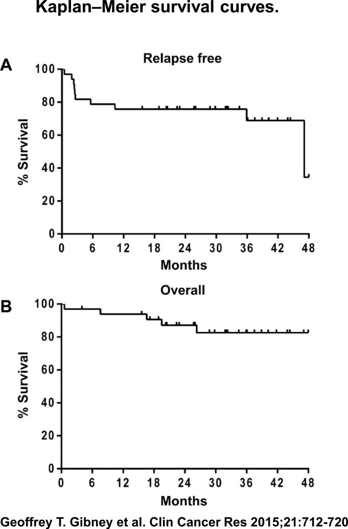
The median follow-up time from study enrollment was 32.1 months. Of 33 patients, 10 (30%) have had a relapse event. Six patients relapsed during induction phase I (weeks 1–12); 1 after induction phase II (weeks 12–24); 1 during the maintenance phase (weeks 24–120); and 2 patients after completing all treatment (>2.3 years). All other patients remain disease free, and 14 (42%) patients have finished all treatment. The estimated median RFS was 47.1 months (Fig. 1). Median OS has not yet been reached. The estimated 12- and 24-month OS rates are 87% and 82%, respectively. Of the 10 relapsed patients, 5 died due to metastatic melanoma; 3 were rendered free of disease surgically and remain disease-free at 2, 27, and 54 weeks after relapse. One patient (described in detail below) had spontaneous regression of disease after a biopsy-proven relapse and has been free of disease for more than 3 years. One additional relapsed patient is alive and on active therapy with dabrafenib plus trametinib.
Figure 1.
Kaplan–Meier survival curves. A, RFS calculated from time of study enrollment. A total of 10 relapse events occurred with fewer events observed in the more recently accrued cohorts. Median RFS was 47.1 months. B, OS calculated from time of study enrollment. A total of 5 death events were observed. Median OS was not reached at a median follow-up time of 32.1 months.
Brain metastases subgroup
Ten patients with resected CNS disease were enrolled. This included 3 patients in cohort 1, 2 patients in cohort 2, and 6 patients in cohort 3. Only 2 of 10 patients have relapsed after a median follow-up time of 22.5 months. Both patients were in cohort 1. The first patient completed all 20 doses of study drugs and developed a new solitary lung metastasis that was resected after 47 months on protocol; she again has no clinical evidence of disease. The second patient was diagnosed with recurrent CNS disease (multiple-subdural metastases and subdural hematoma) after her first treatment with study drugs and expired 3 weeks from initiation on protocol from a CNS hemorrhage.
Postrelapse spontaneous regression
Cases of regression of melanoma after RECIST progression have been well documented for ipilimumab and anti–PD-1 antibodies, and have led to new criteria for antitumor response called immune-related response criteria (irRC; ref. 27). This may complicate the evaluation of patients on an adjuvant trial, as illustrated below. A patient with resected chest wall and pulmonary disease initiated treatment in the 3 mg/kg cohort. At the week 12 evaluation, CT scans showed new chest wall disease and a new splenic nodule of 2.7 cm, biopsy-proven by fine needle aspirate to be melanoma. While waiting to initiate treatment for metastatic disease, a repeat CT scan 4 weeks later showed shrinkage of both lesions. Over the next 24 weeks, repeat CT scans showed further regression and eventual disappearance of both lesions; he remains free of disease 38.8 months since relapse.
Correlative studies
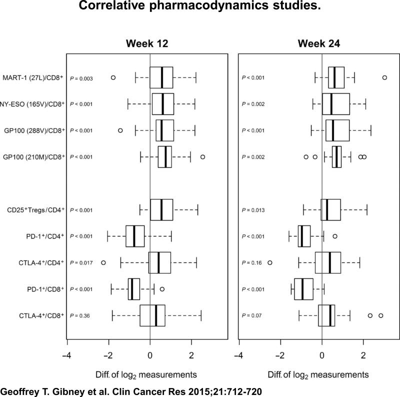
Pharmacodynamic studies were performed to assess changes in T-cell and MDSC populations during therapy (baseline, n = 33; postinduction week 12, n = 32; and postinduction week 24, n = 25). Flow cytometric assays were used to analyze CD4+ and CD8+ T-cell populations. As shown in Fig. 2, tetramer data for antigen-specific T-cell populations demonstrated significant increases in MART-1 (27L), NY-ESO (165V), GP100 (288V), and GP100 (210M) CD8+ T-cells after each 12-week induction phase (P < 0.05 for all groups). Significant increases in CD25+Treg/CD4+ T-cell populations were also observed after 12 and 24 weeks of therapy (P < 0.001 and P = 0.013, respectively). Similar increases in CTLA4+/CD4+ T-cell populations were seen (statistically significant at week 12 only). PD-1+/CD4+ and PD-1+/CD8+ T-cell groups decreased during therapy (P < 0.001 for all groups). No significant differences in tetramer or flow cytometric analyses were observed between cohorts.
Figure 2.
Correlative pharmacodynamics studies. Patient peripheral blood samples were collected by leukapheresis at baseline and after 12 weeks and 24 weeks of therapy with nivolumab plus multi-peptide vaccine. Tetramer assays showed significant increases in MART-1, NY-ESO+, and GP100+/CD8+ T-cell populations after 12 and 24 weeks of treatment with nivolumab and vaccine. Flow cytometry demonstrated significant increases in CD25+Treg/CD4+ and CTLA-4+ T-cell populations with therapy. Decreased levels of PD-1+ T-cell populations were observed with therapy. Open circles, outlier values.
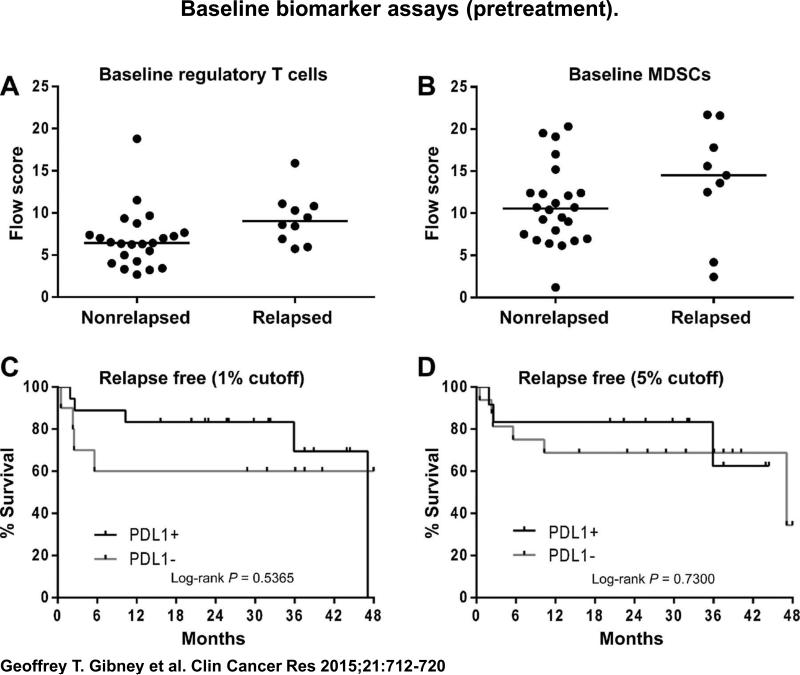
The identification of potential biomarkers to predict relapse was explored (Fig. 3). All cohorts were grouped together. There was a trend toward lower baseline CD25+Treg/CD4+ T-cell (P = 0.0583) and MDSC levels (P = 0.1718) in nonrelapsing patients compared with relapsing patients. No clear association was observed in other baseline T-cell populations or postinduction patient samples and relapse events. Baseline PD-L1 tumor expression was assessed on archived tumor specimens from 28 of the 33 enrolled patients. Two PD-L1 tumor positivity cutoffs were chosen to assess any association between PD-L1 status and relapse. Using a 1% PD-L1 cutoff, 18 samples were positive and 10 samples were negative. Relapse occurred in 5 of 18 (28%) PD-L1–positive patients and 4 of 10 (40%) PD-L1–negative patients (Fisher exact test = 0.677, two-sided). At a threshold of 5% PD-L1 staining, 12 samples were positive for PD-L1 and 16 were negative. Relapse occurred in 3 of 12 (25%) PD-L1–positive patients and 6 of 16 (38%) negative patients (Fisher exact test = 0.687, two-sided). As demonstrated in Fig. 3C and 3D, there was no statistically significant association between PD-L1 tumor staining and RFS in this patient population, although there is a nonstatistically significant trend toward better RFS in those whose tumors were PD-L1 positive using either threshold of IHC staining.
Figure 3.
Baseline biomarker assays (pretreatment). A, lower baseline CD25+Treg/CD4+ populations were observed in patients who remained disease free (nonrelapsed) with therapy (P = 0.0583). B, a trend for lower MDSC levels (CD11b+/DRlow/ CD14+) was observed in patients who remained disease free with therapy (P = 0.1718). C and D, no statistical association was observed between PD-L1 tumor status (at either a 1% or 5% cutoff) and RFS by log-rank test.
Discussion
Nivolumab in combination with a multi-peptide vaccine was well tolerated. Four of 33 patients permanently discontinued study drug due to toxicities, and only two DLTs (colitis) were observed, with no MTD defined up to 10 mg/kg of nivolumab. The common adverse events (fatigue, rash/pruritus, nausea/diarrhea, arthralgias, endocrinopathies) seen in this trial are similar to past studies with either nivolumab or other anti–PD-1 antibodies. Only one case of grade 1 pneumonitis was seen. The addition of vaccine led to grade 1 local injection site reactions in a majority of patients. Related grade 3 events occurred in 4 of 33 patients (12%) and were manageable. This compares favorably with adverse events reported with adjuvant high-dose or pegylated interferon alpha2B, where 40% to 60% of patients experienced grade 3 events, 5% to 10% experienced a grade 4 event, and as many as 31% of patients discontinued therapy due to toxicities (22, 23). The rate of grade 3 events in the current study is less than that of the ongoing EORTC18071 protocol with ipilimumab 10 mg/kg for resected stage III melanoma patients where 36.5% and 5.5% of patients experienced a grade 3 or 4 adverse event, respectively, and 49% of patients discontinued ipilimumab due to toxicity (NCT00636168; ref. 28).
High-dose and pegylated interferon alpha2B have both been approved by the FDA as adjuvant therapy for resected stage III melanoma patients, but their long-term clinical benefit continues to be debated. There has been a consistent improvement in RFS with adjuvant interferon, but individual studies have largely failed to show a statistically significant improvement in OS (24, 29). Meta-analyses have demonstrated modest, but statistically significant, improvements in OS (hazard ratios of 0.85 to 0.91) with adjuvant interferon (25, 30, 31). This suggests that less toxic and more effective approaches for high-risk resected melanoma patients are clearly needed. Other adjuvant strategies have been explored, including bevacizumab (AVAST-M) and GM-CSF (ECOG4967), which have each demonstrated improvement in disease-free survival (only stage IV subset for GM-CSF) over placebo arms, but no statistical improvements in OS were seen (20, 32). Adjuvant ipilimumab for resected melanoma is currently under investigation in two large randomized phase III trials—the ECOG1609 and EORTC18071 protocols (NCT01274338, NCT00636168). The recent preliminary data presented from the EORTC18071 protocol in resected stage III patients demonstrated a median RFS of 26.1 months in the ipilimumab group compared with 17.1 months in the placebo group with a hazard ratio of 0.75 (P = 0.0013; ref. 28). However, this must be weighed against the high rate of adverse events seen in the ipilimumab group.
A number of important questions remain to be settled regarding the use of anti–PD-1 blockade in the adjuvant setting: (i) does minimal/microscopic residual disease and no effective tumor microenvironment impact efficacy, (ii) can PD-L1 expression in resected melanoma tumors serve as a biomarker for successful adjuvant treatment, and (iii) can other novel biomarkers can be defined for the efficacy of PD-1 blockade in an adjuvant trial?
Our data suggest that nivolumab is clinically active in resected stage IIIC/IV melanoma patients based on the low rate of relapse (10 of 33 patients), impressive RFS—estimated median RFS of 47.1 months, and a median OS not yet reached with more than 32 months of follow-up. Although the results cannot be compared directly with the prior adjuvant interferon and current adjuvant ipilimumab experience, these data should be placed in the context of reports on the natural history of resected stage IV melanoma. The median RFS in the SWOG S9430 protocol and Canvaxin-IV trials was 5 and 7.2 months, respectively (16–18). Median OS in these two studies was 21 months (S9430) and 32 months (control arm Canvaxin-IV trial). A retrospective study using the Surveil-lance, Epidemiology and End Results (SEER) database showed a median OS of 12 months in stage IV melanoma patients undergoing metastasectomy (19). Overall, contemporary data suggest that a median RFS over 12 months would be a reasonable benchmark by which to judge the potential of an adjuvant treatment for stage IIIC/IV high risk resected melanoma. By that criterion, our data with adjuvant anti–PD-1 therapy surpasses RFS expectations and longer follow up will clarify whether superior median OS can be achieved compared with the S9430 and Canvaxin-IV studies. Furthermore, the low event rate in resected brain metastases patients suggests a potential benefit in this very high-risk population.
We demonstrated statistically significant increases in melanoma antigen-specific CD8+ T-cell populations and decreases in PD-1 expressing T-cells with exposure to nivolumab and vaccine. The former has been demonstrated in patients with active metastatic melanoma (15), whereas no change or increases in PD-1 expressing T-cell populations has been previously described in mouse models (33, 34). It is possible that the decrease in PD-1 expressing T-cells in our study is reflective of internalization of the antigen with anti–PD-1 antibody exposure rather than a true change in phenotype. Increases in CD25+Tregs/CD4+ and CTLA-4+/CD4+ T-cell populations were seen with anti–PD-1 therapy. This suggests that one adaptive mechanism that occurs with anti–PD-1 therapy and may dampen clinical activity is mediated through CTLA-4 and/or Tregs. This is supported by data from the nivolumab plus vaccine trial in unresectable stage III/V melanoma patients where a significant increase in regulatory T-cell populations at week 12 was observed in the nonresponding patients (15). However, increases in Treg populations seen with anti–CTLA-4 therapy have been associated with improved progression-free survival in patients with melanoma with regional metastases (35, 36). Therefore, Tregs may play a different role depending on the effects of different checkpoint inhibitors. Interestingly, cotargeting CTLA-4 and PD-1 can enhance antigen-specific effector CD8+ and CD4+ T-cell function and tumor infiltration over individual checkpoint inhibition as monotherapy (33, 37). This strategy is now a promising area of clinical research with early studies of concurrent and sequential ipilimumab and nivolumab showing high levels of durable response in patients with meta-static melanoma (38, 39). Concurrent ipilimumab and nivolumab is also under investigation in high-risk resected metastatic melanoma patients as an amendment to the current trial (NCT01176474).
Much attention has been given to tumor PD-L1 status as a biomarker for response to anti–PD-1 therapy. In a phase I study of nivolumab in advanced cancers, PD-L1 status (5% threshold on immunohistochemistry) and response data were available on 42 patients, which showed no objective responses in the 17 patients with negative PD-L1 status, whereas objective responses were seen in 9 of 25 patients with positive PD-L1 staining (14). Enthusiasm for the use of PD-L1 as a predictive biomarker has diminished as other studies have shown that patients with PD-L1 negative melanomas can still respond to anti–PD-1, albeit at lower rates (12, 15). In our study, there were slightly fewer relapses in patients with PD-L1–positive tumors (both 1% and 5% cutoffs), but this was not statistically significant on Fisher exact testing, nor was RFS by log-rank testing. Our other correlative studies showed that nonrelapsing patients tended to have lower baseline levels of CD25+Treg/CD4+ and MDSC populations. This is supported by other data indicating the suppressive role of these immune cell populations (40, 41), which may mitigate the effect of cytotoxic T-cells (and other immune cells) expected with anti–PD-1/PD-L1 therapy, resulting in dampened antitumor activity. Prior studies by our group and others have also demonstrated associations between a 12-chemokine gene expression signature and rises in absolute lymphocyte counts with improved clinical outcomes with immunotherapy (42, 43). Further prospective investigation into the role of these findings as predictive or prognostic biomarkers will potentially help better stratify melanoma patients for systemic immune therapy.
In summary, nivolumab at doses of 1 mg/kg up to 10 mg/kg is well tolerated in patients with resected stage IIIC/IV melanoma. Both RFS and OS with anti–PD-1 therapy in this study were promising compared with historic data. Tumor PD-L1 expression alone does not appear to be associated with relapse after adjuvant anti–PD-1 therapy. A prospective, randomized study of nivolumab in resected high-risk melanoma patients is warranted.
Translational Relevance.
Past cooperative group studies of interferon in resected stage III melanoma have demonstrated median relapse-free survivals (RFS) of 21 to 36 months at the cost of unfavorable toxicity profiles. Early data presented from the EORTC18071 phase III study of adjuvant ipilimumab versus placebo in resected stage III melanoma patients demonstrated modest improvement in RFS, but concerns about adverse events remain. There is no approved therapy for resected stage IV patients despite a relapse rate as high as 87%. In this phase I study of nivolumab plus vaccine in resected stage IIIC/IV melanoma patients, we demonstrated a low rate of severe toxicities and promising relapse-free and overall survival compared with historic data. The possible associations of elevated baseline MDSC levels and T-regulatory cells with relapse suggest potential predictive biomarker and treatment strategies. Further prospective investigations of nivolumab as an adjuvant therapy in high-risk resected melanoma patients are warranted.
Acknowledgments
The authors thank Mary Ruisi (MD) and Arvin Yang (MD, PhD) of Bristol-Myers Squibb, who provided medical guidance and input during the conduct of the study and critical review of the article.
Grant Support
This study was supported by the Donald A. Adam Comprehensive Melanoma Research Center, 1R01FD003511 from the Food and Drug Administration, and Bristol-Myers Squibb.
The costs of publication of this article were defrayed in part by the payment of page charges. This article must therefore be hereby marked advertisement in accordance with 18 U.S.C. Section 1734 solely to indicate this fact.
Footnotes
Authors’ Contributions
Conception and design: R.R. Kudchadkar, R.C. DeConti, J.S. Weber
Development of methodology: R.R. Kudchadkar, B. Yu, J.S. Weber
Acquisition of data (provided animals, acquired and managed patients, provided facilities, etc.): G.T. Gibney, R.R. Kudchadkar, R.C. DeConti, M.S. Thebeau, M.P. Czupryn, L. Tetteh, C.E. Horak, A.J. Martinez, J.S. Weber
Analysis and interpretation of data (e.g., statistical analysis, biostatistics, computational analysis): G.T. Gibney, R.R. Kudchadkar, M.J. Schell, K.J. Fisher, C.E. Horak, A.J. Martinez, I. Younos, J.S. Weber
Writing, review, and/or revision of the manuscript: G.T. Gibney, R.R. Kudchadkar, R.C. DeConti, M.J. Schell, C.E. Horak, H.D. Inzunza, B. Yu, J.S. Weber
Administrative, technical, or material support (i.e., reporting or organizing data, constructing databases): G.T. Gibney, C. Eysmans, A. Richards
Study supervision: G.T. Gibney, R.R. Kudchadkar, J.S. Weber
Other (carried out biomarker flow cytometry experiments, interpretation of results, and analysis of data): I. Younos
Disclosure of Potential Conflicts of Interest
G.T. Gibney is a consultant/advisory board member for Genentech and Bristol-Myers Squibb. R.R. Kudchadkar reports receiving speakers’ bureau honoraria from Genentech. M.S. Thebeau reports receiving speakers’ bureau honoraria from Bristol-Myers Squibb. C.E. Horak is an employee of Bristol-Myers Squibb. J. Weber reports receiving honoraria from and is a consultant/advisory board member for Bristol-Myers Squibb and Merck. No potential conflicts of interest were disclosed by the other authors.
References
- 1.Dong H, Strome SE, Salomao DR, Tamura H, Hirano F, Flies DB, et al. Tumor-associated B7-H1 promotes T-cell apoptosis: a potential mechanism of immune evasion. Nat Med. 2002;8:793–800. doi: 10.1038/nm730. [DOI] [PubMed] [Google Scholar]
- 2.Freeman GJ, Long AJ, Iwai Y, Bourque K, Chernova T, Nishimura H, et al. Engagement of the PD-1 immunoinhibitory receptor by a novel B7 family member leads to negative regulation of lymphocyte activation. J Exp Med. 2000;192:1027–34. doi: 10.1084/jem.192.7.1027. [DOI] [PMC free article] [PubMed] [Google Scholar]
- 3.Iwai Y, Ishida M, Tanaka Y, Okazaki T, Honjo T, Minato N. Involvement of PD-L1 on tumor cells in the escape from host immune system and tumor immunotherapy by PD-L1 blockade. Proc Natl Acad Sci U S A. 2002;99:12293–7. doi: 10.1073/pnas.192461099. [DOI] [PMC free article] [PubMed] [Google Scholar]
- 4.Konishi J, Yamazaki K, Azuma M, Kinoshita I, Dosaka-Akita H, Nishimura M. B7-H1 expression on non-small cell lung cancer cells and its relationship with tumor-infiltrating lymphocytes and their PD-1 expression. Clin Cancer Res. 2004;10:5094–100. doi: 10.1158/1078-0432.CCR-04-0428. [DOI] [PubMed] [Google Scholar]
- 5.Nomi T, Sho M, Akahori T, Hamada K, Kubo A, Kanehiro H, et al. Clinical significance and therapeutic potential of the programmed death-1 ligand/ programmed death-1 pathway in human pancreatic cancer. Clin Cancer Res. 2007;13:2151–7. doi: 10.1158/1078-0432.CCR-06-2746. [DOI] [PubMed] [Google Scholar]
- 6.Taube JM, Anders RA, Young GD, Xu H, Sharma R, McMiller TL, et al. Colocalization of inflammatory response with B7-h1 expression in human melanocytic lesions supports an adaptive resistance mechanism of immune escape. Sci Transl Med. 2012;4:127ra37. doi: 10.1126/scitranslmed.3003689. [DOI] [PMC free article] [PubMed] [Google Scholar]
- 7.Thompson RH, Gillett MD, Cheville JC, Lohse CM, Dong H, Webster WS, et al. Costimulatory B7-H1 in renal cell carcinoma patients: Indicator of tumor aggressiveness and potential therapeutic target. Proc Natl Acad Sci U S A. 2004;101:17174–9. doi: 10.1073/pnas.0406351101. [DOI] [PMC free article] [PubMed] [Google Scholar]
- 8.Hirano F, Kaneko K, Tamura H, Dong H, Wang S, Ichikawa M, et al. Blockade of B7-H1 and PD-1 by monoclonal antibodies potentiates cancer therapeutic immunity. Cancer Res. 2005;65:1089–96. [PubMed] [Google Scholar]
- 9.Iwai Y, Terawaki S, Honjo T. PD-1 blockade inhibits hematogenous spread of poorly immunogenic tumor cells by enhanced recruitment of effector T cells. Int Immunol. 2005;17:133–44. doi: 10.1093/intimm/dxh194. [DOI] [PubMed] [Google Scholar]
- 10.Brahmer JR, Tykodi SS, Chow LQ, Hwu WJ, Topalian SL, Hwu P, et al. Safety and activity of anti-PD-L1 antibody in patients with advanced cancer. N Engl J Med. 2012;366:2455–65. doi: 10.1056/NEJMoa1200694. [DOI] [PMC free article] [PubMed] [Google Scholar]
- 11.Hamid O, Robert C, Daud A, Hodi FS, Hwu WJ, Kefford R, et al. Safety and tumor responses with lambrolizumab (anti-PD-1) in melanoma. N Engl J Med. 2013;369:134–44. doi: 10.1056/NEJMoa1305133. [DOI] [PMC free article] [PubMed] [Google Scholar]
- 12.Kefford R, Ribas A, Hamid O, Robert C, Daud A, Wolchock JD, et al. Clinical efficacy and correlation with tumor PD-L1 expression in patients (pts) with melanoma (MEL) treated with the anti-PD-1 monoclonal antibody MK-3475. J Clin Oncol. 2014;32:5s. abstr3005. [Google Scholar]
- 13.Robert C, Ribas A, Wolchok JD, Hodi FS, Hamid O, Kefford R, et al. Anti-programmed-death-receptor-1 treatment with pembrolizumab in ipilimumab-refractory advanced melanoma: a randomised dose-comparison cohort of a phase 1 trial. Lancet. 2014;384:1109–17. doi: 10.1016/S0140-6736(14)60958-2. [DOI] [PubMed] [Google Scholar]
- 14.Topalian SL, Hodi FS, Brahmer JR, Gettinger SN, Smith DC, McDermott DF, et al. Safety, activity, and immune correlates of anti-PD-1 antibody in cancer. N Engl J Med. 2012;366:2443–54. doi: 10.1056/NEJMoa1200690. [DOI] [PMC free article] [PubMed] [Google Scholar]
- 15.Weber JS, Kudchadkar RR, Yu B, Gallenstein D, Horak CE, Inzunza HD, et al. Safety, efficacy, and biomarkers of nivolumab with vaccine in ipilimumab-refractory or -naive melanoma. J Clin Oncol. 2013;31:4311–8. doi: 10.1200/JCO.2013.51.4802. [DOI] [PMC free article] [PubMed] [Google Scholar]
- 16.Hsueh EC, Essner R, Foshag LJ, Ollila DW, Gammon G, O'Day SJ, et al. Prolonged survival after complete resection of disseminated melanoma and active immunotherapy with a therapeutic cancer vaccine. J Clin Oncol. 2002;20:4549–54. doi: 10.1200/JCO.2002.01.151. [DOI] [PubMed] [Google Scholar]
- 17.Morton DL, Mozzillo N, Thompson JF, Kelley MC, Faries M, Wagner J, et al. An international, randomized, phase III trial of bacillus Calmette-Guerin (BCG) plus allogeneic melanoma vaccine (MCV) or placebo after complete resection of melanoma metastatic to regional or distant sites. J Clin Oncol. 2007:25. abstr8508. [Google Scholar]
- 18.Sosman JA, Moon J, Tuthill RJ, Warneke JA, Vetto JT, Redman BG, et al. A phase 2 trial of complete resection for stage IV melanoma: results of Southwest Oncology Group Clinical Trial S9430. Cancer. 2011;117:4740–06. doi: 10.1002/cncr.26111. [DOI] [PMC free article] [PubMed] [Google Scholar]
- 19.Wasif N, Bagaria SP, Ray P, Morton DL. Does metastasectomy improve survival in patients with Stage IV melanoma? A cancer registry analysis of outcomes. J Surg Oncol. 2011;104:111–5. doi: 10.1002/jso.21903. [DOI] [PMC free article] [PubMed] [Google Scholar]
- 20.Lawson DH, Lee SJ, Tarhini AA, Margolin KA, Ernstoff MS, Kirkwood JM. E4697: Phase III cooperative group study of yeast-derived granulocyte macrophage colony-stimulating factor (GM-CSF) versus placebo as adjuvant treatment of patients with completely resected stage III-IV melanoma. J Clin Oncol. 2010:28. doi: 10.1200/JCO.2015.62.0500. abstr8504. [DOI] [PMC free article] [PubMed] [Google Scholar]
- 21.Balch CM, Gershenwald JE, Soong SJ, Thompson JF, Atkins MB, Byrd DR, et al. Final version of 2009 AJCC melanoma staging and classification. J Clin Oncol. 2009;27:6199–206. doi: 10.1200/JCO.2009.23.4799. [DOI] [PMC free article] [PubMed] [Google Scholar]
- 22.Eggermont AM, Suciu S, Santinami M, Testori A, Kruit WH, Marsden J, et al. Adjuvant therapy with pegylated interferon alfa-2b versus observation alone in resected stage III melanoma: final results of EORTC 18991, a randomised phase III trial. Lancet. 2008;372:117–26. doi: 10.1016/S0140-6736(08)61033-8. [DOI] [PubMed] [Google Scholar]
- 23.Kirkwood JM, Ibrahim JG, Sosman JA, Sondak VK, Agarwala SS, Ernst-off MS, et al. High-dose interferon alfa-2b significantly prolongs relapse-free and overall survival compared with the GM2-KLH/QS-21 vaccine in patients with resected stage IIB-III melanoma: results of intergroup trial E1694/S9512/C509801. J Clin Oncol. 2001;19:2370–80. doi: 10.1200/JCO.2001.19.9.2370. [DOI] [PubMed] [Google Scholar]
- 24.Kirkwood JM, Manola J, Ibrahim J, Sondak V, Ernstoff MS, Rao U. A pooled analysis of eastern cooperative oncology group and intergroup trials of adjuvant high-dose interferon for melanoma. Clin Cancer Res. 2004;10:1670–7. doi: 10.1158/1078-0432.ccr-1103-3. [DOI] [PubMed] [Google Scholar]
- 25.Mocellin S, Pasquali S, Rossi CR, Nitti D. Interferon alpha adjuvant therapy in patients with high-risk melanoma: a systematic review and meta-analysis. J Natl Cancer Inst. 2010;102:493–501. doi: 10.1093/jnci/djq009. [DOI] [PubMed] [Google Scholar]
- 26.Sarnaik AA, Yu B, Yu D, Morelli D, Hall M, Bogle D, et al. Extended dose ipilimumab with a peptide vaccine: immune correlates associated with clinical benefit in patients with resected high-risk stage IIIc/IV melanoma. Clin Cancer Res. 2011;17:896–906. doi: 10.1158/1078-0432.CCR-10-2463. [DOI] [PMC free article] [PubMed] [Google Scholar]
- 27.Wolchok JD, Hoos A, O'Day S, Weber JS, Hamid O, Lebbe C, et al. Guidelines for the evaluation of immune therapy activity in solid tumors: immune-related response criteria. Clin Cancer Res. 2009;15:7412–20. doi: 10.1158/1078-0432.CCR-09-1624. [DOI] [PubMed] [Google Scholar]
- 28.Eggermont AM, Chiarion-Sileni V, Grob JJ, Dummer R, Wolchock JD, Schmidt H, et al. Ipilimumab versus placebo after complete resection of stage III melanoma: Initial efficacy and safety results from the EORTC 18071 phase III trial. J Clin Oncol. 2014;32:5s. abstrLBA9008. [Google Scholar]
- 29.Eggermont AM, Suciu S, Testori A, Santinami M, Kruit WH, Marsden J, et al. Long-term results of the randomized phase III trial EORTC 18991 of adjuvant therapy with pegylated interferon alfa-2b versus observation in resected stage III melanoma. J Clin Oncol. 2012;30:3810–8. doi: 10.1200/JCO.2011.41.3799. [DOI] [PubMed] [Google Scholar]
- 30.Mocellin S, Lens MB, Pasquali S, Pilati P, Chiarion Sileni V. Interferon alpha for the adjuvant treatment of cutaneous melanoma. Cochrane Database Syst Rev. 2013;6:CD008955. doi: 10.1002/14651858.CD008955.pub2. [DOI] [PMC free article] [PubMed] [Google Scholar]
- 31.Verma S, Quirt I, McCready D, Bak K, Charette M, Iscoe N. Systemic review of systemic adjuvant therapy for patients at high risk for recurrent melanoma. Cancer. 2006;106:1431–42. doi: 10.1002/cncr.21760. [DOI] [PubMed] [Google Scholar]
- 32.Corrie PG, Marshall A, Dunn JA, Middleton MR, Nathan PD, Gore M, et al. Adjuvant bevacizumab in patients with melanoma at high risk of recurrence (AVAST-M): preplanned interim results from a multicentre, open- label, randomised controlled phase 3 study. The Lancet Oncology. 2014;15:620–30. doi: 10.1016/S1470-2045(14)70110-X. [DOI] [PubMed] [Google Scholar]
- 33.Curran MA, Montalvo W, Yagita H, Allison JP. PD-1 and CTLA-4 combination blockade expands infiltrating T cells and reduces regulatory T and myeloid cells within B16 melanoma tumors. Proc Natl Acad Sci U S A. 2010;107:4275–80. doi: 10.1073/pnas.0915174107. [DOI] [PMC free article] [PubMed] [Google Scholar]
- 34.Karyampudi L, Lamichhane P, Scheid AD, Kalli KR, Shreeder B, Krempski JW, et al. Accumulation of memory precursor CD8 T cells in regressing tumors following combination therapy with vaccine and anti-PD-1 antibody. Cancer Res. 2014;74:2974–85. doi: 10.1158/0008-5472.CAN-13-2564. [DOI] [PMC free article] [PubMed] [Google Scholar]
- 35.Tarhini AA, Butterfield LH, Shuai Y, Gooding WE, Kalinski P, Kirkwood JM. Differing patterns of circulating regulatory T cells and myeloid-derived suppressor cells in metastatic melanoma patients receiving anti-CTLA4 antibody and interferon-alpha or TLR-9 agonist and GM-CSF with peptide vaccination. J Immunother. 2012;35:702–10. doi: 10.1097/CJI.0b013e318272569b. [DOI] [PMC free article] [PubMed] [Google Scholar]
- 36.Tarhini AA, Edington H, Butterfield LH, Lin Y, Shuai Y, Tawbi H, et al. Immune monitoring of the circulation and the tumor microenvironment in patients with regionally advanced melanoma receiving neoadjuvant ipilimumab. PLoS ONE. 2014;9:e87705. doi: 10.1371/journal.pone.0087705. [DOI] [PMC free article] [PubMed] [Google Scholar]
- 37.Duraiswamy J, Kaluza KM, Freeman GJ, Coukos G. Dual blockade of PD-1 and CTLA-4 combined with tumor vaccine effectively restores T-cell rejection function in tumors. Cancer Res. 2013;73:3591–603. doi: 10.1158/0008-5472.CAN-12-4100. [DOI] [PMC free article] [PubMed] [Google Scholar]
- 38.Wolchok JD, Kluger H, Callahan MK, Postow MA, Rizvi NA, Lesokhin AM, et al. Nivolumab plus ipilimumab in advanced melanoma. N Engl J Med. 2013;369:122–33. doi: 10.1056/NEJMoa1302369. [DOI] [PMC free article] [PubMed] [Google Scholar]
- 39.Sznol M, Kluger HM, Callahan MK, Postow MA, Gordon RA, Segal NH, et al. Survival, response duration, and activity by BRAF mutation (MT) status of nivolumab (NIVO, anti-PD-1, BMS-936558, ONO-4538) and ipilimumab (IPI) concurrent therapy in advanced melanoma (MEL). J Clin Oncol. 2014;32:5s. abstrLBA9003. [Google Scholar]
- 40.Gabrilovich DI, Nagaraj S. Myeloid-derived suppressor cells as regulators of the immune system. Nat Rev Immunol. 2009;9:162–74. doi: 10.1038/nri2506. [DOI] [PMC free article] [PubMed] [Google Scholar]
- 41.Kirkwood JM, Tarhini AA, Panelli MC, Moschos SJ, Zarour HM, Butterfield LH, et al. Next generation of immunotherapy for melanoma. J Clin Oncol. 2008;26:3445–55. doi: 10.1200/JCO.2007.14.6423. [DOI] [PubMed] [Google Scholar]
- 42.Ku GY, Yuan J, Page DB, Schroeder SE, Panageas KS, Carvajal RD, et al. Single-institution experience with ipilimumab in advanced melanoma patients in the compassionate use setting: lymphocyte count after 2 doses correlates with survival. Cancer. 2010;116:1767–75. doi: 10.1002/cncr.24951. [DOI] [PMC free article] [PubMed] [Google Scholar]
- 43.Messina JL, Fenstermacher DA, Eschrich S, Qu X, Berglund AE, Lloyd MC, et al. 12-Chemokine gene signature identifies lymph node-like structures in melanoma: potential for patient selection for immunotherapy? Sci Rep. 2012;2:765. doi: 10.1038/srep00765. [DOI] [PMC free article] [PubMed] [Google Scholar]


