ABSTRACT
Although a polybasic HA0 cleavage site is considered the dominant virulence determinant for highly pathogenic avian influenza (HPAI) H5 and H7 viruses, naturally occurring virus isolates possessing a polybasic HA0 cleavage site have been identified that are low pathogenic in chickens. In this study, we generated a reassortant H5N3 virus that possessed the hemagglutinin (HA) gene from H5N1 HPAI A/swan/Germany/R65/2006 and the remaining gene segments from low pathogenic A/chicken/British Columbia/CN0006/2004 (H7N3). Despite possessing the HA0 cleavage site GERRRKKR/GLF, this rH5N3 virus exhibited a low pathogenic phenotype in chickens. Although rH5N3-inoculated birds replicated and shed virus and seroconverted, transmission to naive contacts did not occur. To determine whether this virus could evolve into a HPAI form, it underwent six serial passages in chickens. A progressive increase in virulence was observed with the virus from passage number six being highly transmissible. Whole-genome sequencing demonstrated the fixation of 12 nonsynonymous mutations involving all eight gene segments during passaging. One of these involved the catalytic site of the neuraminidase (NA; R293K) and is associated with decreased neuraminidase activity and resistance to oseltamivir. Although introducing the R293K mutation into the original low-pathogenicity rH5N3 increased its virulence, transmission to naive contact birds was inefficient, suggesting that one or more of the remaining changes that had accumulated in the passage number six virus also play an important role in transmissibility. Our findings show that the functional linkage and balance between HA and NA proteins contributes to expression of the HPAI phenotype.
IMPORTANCE To date, the contribution that hemagglutinin-neuraminidase balance can have on the expression of a highly pathogenic avian influenza virus phenotype has not been thoroughly examined. Reassortment, which can result in new hemagglutinin-neuraminidase combinations, may have unpredictable effects on virulence and transmission characteristics of a virus. Our data show the importance of the neuraminidase in complementing a polybasic HA0 cleavage site. Furthermore, it demonstrates that adaptive changes selected for during the course of virus evolution can result in unexpected traits such as antiviral drug resistance.
INTRODUCTION
Avian influenza viruses (AIV) belong to the genus Influenzavirus A in the family Orthomyxoviridae. Although wild waterfowl are considered the natural reservoir for all influenza A viruses, sporadic transmission to poultry and mammals can result in viruses that are adapted to a new host. The roles that the different viral genes play in this process, especially as it relates to tropism and virulence for domestic poultry species, are still not well understood.
Influenza A viruses contain a negative-sense, single-stranded, eight-segmented RNA genome that codes for at least 11 proteins. Viruses are categorized based on the hemagglutinin (HA) and neuraminidase (NA) envelope glycoproteins. Sixteen HA (H1 to H16) and 9 NA (N1 to N9) subtypes currently exist for AIV, but novel H17N10 and H18N11 virus subtypes have recently been identified in bats in Central America (1, 2). Two major mechanisms are involved in the evolution of AIV. The error-prone nature of the viral RNA polymerase can give rise to point mutations in the different viral genes that can then be the target of selective pressures. The second major mechanism involves the exchange or reassortment of genes from different influenza virus subtypes that can give rise to viruses with novel antigenic and functional phenotypes (3).
In most cases, the sporadic transmission of AIV from wild waterfowl to domestic poultry results in mild or subclinical disease with no or limited spread. The reason that many of these viruses fail to persist in poultry is often due to the fact that they are poorly adapted to their new host and thus unable to replicate and transmit efficiently. The viral changes that are associated with poultry adaptation, particularly for viruses of the H5 and H7 hemagglutinin subtypes, are of interest, since these two subtypes have the potential to evolve into a highly pathogenic (HP) form. The evolution of low-pathogenicity (LP) H5 and H7 viruses to HPAI viruses strongly correlates with the acquisition of multiple basic amino acids at the HA0 cleavage site. This enables cleavage of HA0 to HA1 and HA2 to be carried out by the proprotein convertase furin, allowing systemic spread of the virus (4). Proteolytic cleavage of HA0 generates two disulfide-linked subunits, HA1 and HA2, which exposes a fusion peptide at the newly formed amino-terminal end of HA2. During receptor mediated endocytosis of the virus, a low-pH-induced conformational change of the HA takes place, and this allows the fusion peptide to initiate the merging of the viral envelope with host endosomal membranes, a step necessary for influenza virus infectivity (5). Even though a polybasic HA0 cleavage site is considered to be the dominant virulence determinant of HPAI H5 and H7 viruses (reviewed in reference 6), there have been instances where it has not been sufficient to confer a highly pathogenic phenotype (7, 8, 9). This implies that the evolution of a LPAI virus to a HPAI virus must involve additional changes in the other gene segments. For instance, PB2, PB1, and NP (10) genes have all been shown to contribute to the virulence of HPAI H5N1 viruses in chickens and the PA gene to virulence in ducks (11).
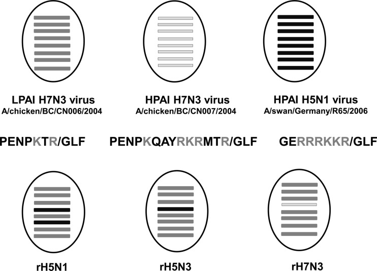
To further identify viral determinants that contribute to AIV transmission and virulence, we used reverse genetics to generate two reassortant viruses that possess the HA gene from the HPAI H5N1 isolate A/swan/Germany/R65/2006 (Germany/R65). The HA of this virus has the polybasic HA0 cleavage site PQGERRRKKR/GLF. One reassortant (rH5N1) possesses the HA and NA genes of Germany/R65 and the remaining six gene segments from the LPAI H7N3 isolate A/chicken/British Columbia/CN006/2004 (BC/CN006), while the second reassortant (rH5N3) possesses the HA of Germany/R65 and the remaining seven gene segments of BC/CN006. Since rH5N1 and rH5N3 viruses contained identical polybasic HA0 cleavage sites, we predicted that both should be highly pathogenic for chickens. As it turned out, rH5N1 had an HPAI phenotype, while rH5N3 did not. Passaging rH5N3 six times in chickens led to a progressive increase in virulence. Our findings support previous observations that suggest that the activities of the HA and NA proteins are functionally linked, and their balance in this case is essential for expression of the highly pathogenic phenotype.
MATERIALS AND METHODS
Generation of recombinant viruses.
The recombinant viruses used in the present study were derived from the LP H7N3 avian influenza virus isolate A/chicken/British Columbia/CN006/2004 (BC/CN006), the HP H7N3 avian influenza virus isolate A/chicken/British Columbia/CN007/2004 (BC/CN007), and the HP H5N1 avian influenza virus isolate A/swan/Germany/R65/2006 (Germany/R65) (12). LPAI BC/CN006 was shown to be the immediate precursor of HPAI BC/CN007 (13). The GenBank accession numbers of the gene segments used to generate the recombinant viruses are KP055066 to KP055076. RNA was extracted from stock virus preparations that had been propagated in embryonated chicken eggs (ECE) using TriPure isolation reagent (Roche Diagnostic Corp., Indianapolis, IN) according to the manufacturer's instructions. The 8 viral gene segments were amplified by reverse transcription-PCR (RT-PCR) according to the protocol described by Hoffmann et al. (14) and then cloned into the influenza A virus eight-plasmid DNA transfection system pHW2000 (15). Prior to transfection, all expression plasmids were sequenced to confirm their identity. Cocultures of human 293T embryonic kidney and Madin-Darby canine kidney (MDCK) cells in six-well culture dishes were transfected with a DNA transfection mixture that consisted of 1 μg of each plasmid (8 μg of total DNA per well) plus 16 μl of TransIT-LT1 transfection reagent (Mirus Bio LLC, Madison, WI) in 2 ml of Opti-MEM medium (Life Technologies, Burlington, Ontario, Canada). At 6 h after transfection, the transfection medium was replaced with RPMI medium, and the cultures were monitored daily for cytopathic effect. Rescued viruses were amplified in T-75 flasks of MDCK cells.
Plaque assay.
Plaque assays were performed in MDCK cells that were grown to 95 to 100% confluence in 24- or 48-well Corning tissue culture plates. Virus containing material was adsorbed onto the cells for 1 h at 37°C; the inoculum was then removed, and 2× Dulbecco modified Eagle medium (DMEM) mixed with 3% carboxymethyl cellulose (Sigma, Oakville, Ontario, Canada) at 1:1 ratio was applied as an overlay. After 48 to 72 h of incubation, the cells were fixed with 10% formalin in phosphate-buffered saline (PBS) and permeabilized with 10% acetone in PBS. Viral plaques were immunostained by sequential incubation with influenza A virus nucleoprotein monoclonal antibody F28 (16), horseradish peroxidase-conjugated goat anti-mouse secondary antibody (Jackson ImmunoResearch, Inc., West Grove, PA), and True Blue peroxidase substrate (KPL, Gaithersburg, MD).
Sequencing of influenza virus genes.
All eight genes from the rescued viruses were sequenced to confirm their identities. The gene segments were amplified in a one-step RT-PCR using a universal primer set (14) and a high-fidelity RT-PCR kit (Invitrogen; SuperScript III One-Step RT-PCR System with Platinum TaqHigh Fidelity). RT-PCR amplicons were cloned into pCR4-Topo (Invitrogen) and used to transform OneShot TOP10 or Mach-T competent E. coli. The resulting plasmids were sequenced using BigDye Terminator chemistry version 3.1 (Life Technologies) and an Applied Biosystems 3130xl genetic analyzer.
In vitro characterization of viruses.
Multistep growth kinetics of each virus was determined in MDCK and quail fibrosarcoma QT-35 cells. Cells grown in six-well culture dishes were inoculated with each recombinant reassortant virus at a multiplicity of infection (MOI) of 0.0001 or 0.00001. The virus inoculum was allowed to adsorb for 1 h; the residual inoculum was then removed, the cell monolayers were washed three times with PBS, and 3 ml of fresh infection medium without trypsin was added. Culture supernatants were sampled at selected time points postinoculation for plaque titration.
In vivo characterization of viruses.
The pathogenic phenotypes of the viruses generated were assessed in 4- to 6-week-old White Leghorn chickens (Gallus gallus domesticus). Briefly, 10 specific-pathogen-free (SPF) chickens were inoculated intravenously with 0.1 ml of virus stock that had been diluted 1:10 in sterile PBS. Virus stocks had a minimum titer of 512 hemagglutination units/25 μl of allantoic fluid. Birds were observed daily for a total of 10 days and given a clinical score, as described in the OIE Manual of Diagnostic Tests and Vaccines for Terrestrial Animals (17). The intravenous pathogenicity index (IVPI) is the mean score per bird per observation over the 10-day period. A maximum score of 3.00 means that all birds died by 24 h postinoculation. Viruses with an IVPI of >1.2 are considered highly pathogenic. In an attempt to determine the 50% lethal dose (LD50), groups of five White Leghorn chickens were inoculated intranasally with 0.2 ml of 10-fold serial dilutions of virus.
Serial passaging of recombinant reassortant H5N3 virus in chickens.
Serial passaging of rH5N3 began with a cloacal swab specimen that was taken at 10 days postinoculation (dpi) from a single bird (Ck#544) belonging to a group of five chickens that were intranasally inoculated with 40,000 PFU of rH5N3 as part of an experiment to determine the LD50. The cloacal swab material was treated with an antibiotic cocktail consisting of streptomycin, vancomycin, gentamicin, and nystatin prior to inoculating into the allantoic cavity of 9-day-old ECE. The virus in the allantoic fluid harvested at 2 dpi, considered passage 1 (P1), was titrated on MDCK cells prior to intranasally inoculating the next group of five chickens with a dose of 40,000 PFU/bird. A 10% tissue homogenate was prepared using lung collected at 6 dpi from one of these birds (Ck#577), and the resulting virus isolated in ECE was considered passage 2 (P2). The allantoic fluid containing the P2 virus which had a HA titer of 1:128 was diluted 1:100 in PBS and 0.5 ml/bird used to intranasally inoculate five chickens. Brain tissue collected from a single bird (Ck#585) at 6 dpi was used to inoculate ECE, from which P3 virus was derived. The allantoic fluid containing this virus was diluted 1:100, and 0.5 ml/bird was used to intranasally inoculate an additional five chickens. P4 virus was derived from ECE inoculated with brain homogenate of Ck#589 harvested at 5 dpi. The allantoic fluid containing the P4 virus was diluted 1:100 in PBS and used to intranasally inoculate five chickens. Brain tissue collected from Ck#636 at 5 dpi was used to isolate P5 virus in ECE. Allantoic fluid containing P5 virus was diluted 1:100 in PBS, and 0.5 ml/bird was used to intranasally inoculate an additional five chickens. P6 virus was derived from brain collected from Ck#640 at 4 dpi. RNA was extracted from rH5N3 isolates P0 to P6 using a Qiagen RNeasy minikit (Qiagen, Toronto, Ontario, Canada), and sequences of the eight viral gene segments were confirmed as described above.
Serology.
Serum samples collected at 0, 7, 10, and 20 dpi were tested for influenza A virus-specific nucleoprotein antibodies using a competitive enzyme-linked immunosorbent assay described previously (16). These sera were also tested for anti-HA antibodies by a hemagglutination inhibition (HI) test using 4 hemagglutination units of A/teal/Germany/Wv632/2005 (H5N1) antigen and a 0.5% (vol/vol) suspension of chicken erythrocytes.
Neuraminidase inhibition assay.
Oseltamivir resistance of P0 rH5N3 and P6 rH5N3 viruses was assessed by using the NA-Star (Life Technologies) chemiluminescent neuraminidase inhibition assay according to the manufacturer's instructions. Briefly, Triton X-100 was added to 0.5 ml of infectious allantoic fluid that contained equivalent hemagglutination units of each virus to give a final concentration of 1% (vol/vol). The virus sample was diluted in NA-Star assay buffer, and 25 μl was added to wells containing 25 μl of serially diluted (10 half-log dilutions) oseltamivir. The plate was incubated for 20 min at 37°C, after which 10 μl of NA-Star substrate was added. The plate was then incubated for a further 30 min at room temperature. The chemiluminescent signal was read, and the averaged signal intensity was exported for nonlinear curve fitting and 50% inhibitory concentration (IC50) calculation.
NA enzyme kinetic assays.
The NA enzyme activities of P0 rH5N3 and P6 rH5N3 viruses were determined by using a fluorometric assay (18). Briefly, both virus stocks were adjusted to a concentration of 106 PFU/ml in minimal essential medium containing 0.1% (wt/vol) bovine serum albumin. Then, 5 μl of each virus preparation was incubated in triplicate at 37°C with the fluorogenic substrate MUNANA [2′-(4-methylumbelliferyl)-α-d-N-acetylneuraminic acid] at concentrations that ranged from 0 to 2,000 μM. Reactions were performed in a round-bottom 96-well white plate. Fluorescence was monitored every 90 s for 60 min at 37°C using excitation and emission wavelengths of 360 and 460 nm, respectively. The Michaelis constant (Km) and the maximum velocity (Vm) were calculated by using Prism software (GraphPad5) and fitting the data to Michaelis-Menten equations using nonlinear regression. An extra sum of square F-test was used to compare individual values to Mx10, and a P value was determined. Each experiment was performed three times.
Virus elution assay.
The ability of the NA to elute virus from chicken erythrocytes was assessed according to the procedure described by Castrucci and Kawaoka (19). Briefly, 50 μl of 2-fold serial dilutions of virus preparations adjusted to a HA titer of 1:128 were made in calcium saline (6.8 mM CaCl2–154 mM NaCl in 20 mM borate buffer [pH 7.2]) and incubated with 50 μl of 0.5% chicken erythrocytes in V-bottom microtiter plates at 4°C for 1 h. The microtiter plates were then incubated at 37°C, and changes in HA titers were recorded hourly.
RNA extraction and qRT-PCR.
Total RNA was extracted from 1 ml of clarified oropharyngeal and cloacal swab samples using the MagMax-96/AI/ND Viral RNA Isolation kit (Ambion, Austin, TX) according to the manufacturer's protocol. To quantify the amount of viral RNA in each swab, a quantitative RT-PCR (qRT-PCR) specific for the influenza A virus M1 gene was performed as previously described (20). Standard curves were generated for each run using serial dilutions of full-length in vitro-transcribed influenza A virus segment 7. The nucleic acid copy number in each specimen was extrapolated from the standard curve.
Histopathology and immunohistochemistry.
Tissue samples (brain, trachea, lung, liver, spleen, cecal tonsil, pectoral muscle, kidney, heart, pancreas, jejunum, proventriculus, bursa of Fabricius, and thymus) were collected from each bird and fixed in 10% neutral buffered formalin for a minimum of 48 h, embedded in paraffin, sectioned, and stained with hematoxylin and eosin (H&E). Tissues were screened, and a subset was selected for microscopic examination and immunohistochemical testing. Microscopic lesions were assessed semiquantitatively as mild, moderate, or severe. For immunohistochemistry, paraffin-embedded tissue sections were quenched for 10 min in aqueous 3% H2O2 and then pretreated with proteinase K for 15 min. Mouse monoclonal antibody F26NP9, specific for influenza A nucleoprotein (16), was used at a 1:10,000 dilution for 1 h. Influenza antigen in tissue sections was then visualized using a horseradish peroxidase-labeled polymer (Envision+ system anti-mouse, Dako, USA) that reacted with the chromogen diaminobenzidine (DAB). The sections were counterstained with Gill's hematoxylin.
Generation of N3 mutants.
Based on the sequence of the NA gene segment for rH5N3 P0, a mutant gene sequence was synthesized at GenScript USA, Inc. (Piscataway, NJ), and then cloned into pHW2000 and used to generate a N3 mutant in an rH5N3 P0 background. This NA mutant, NA-3-R293K, contained a lysine in the place of an arginine at amino acid residue 293.
Statistical analysis.
Statistically significant differences between experimental groups were determined using Student t test and analysis of variance (ANOVA) with the SYSTAT 10 software package. Multiple comparison testing using the Bonferroni t test was performed following ANOVA. P values of <0.05 were considered statistically significant.
RESULTS
Evaluation of the biological characteristics of recombinant viruses.
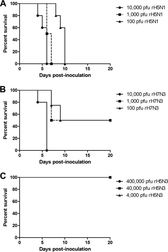
The rescued recombinant viruses rH7N3 (the HA gene derived from BC/CN007 and the remaining gene segments derived from BC/CN006), rH5N1 (the HA and NA genes from Germany/R65 and the remaining gene segments from BC/CN006), and rH5N3 (the HA gene from Germany/R65 and the remaining genes segments from BC/CN006) were sequenced to confirm their identities prior to evaluating their in vitro and in vivo biological characteristics (Fig. 1). Since rH5N1 and rH5N3 contain identical polybasic HA0 cleavage sites, we predicted that both viruses should be highly pathogenic for chickens. IVPI testing of rH7N3, rH5N1 and rH5N3 carried out in 4- to 6-week-old chickens produced scores of 3.0, 2.3, and 0.0, respectively. The LD50 of each recombinant was estimated by intranasally administering 10-fold serial dilutions of virus to groups of five chickens. Figure 2 shows the kill curves for each recombinant virus. Although rH7N3 and rH5N1 each produced 100% mortality within 6 days of intranasally inoculating 10,000 PFU virus/bird, chickens inoculated with 400,000 PFU of rH5N3 showed no clinical signs over a 20-day observation period, despite the fact that they replicated and shed virus via the oropharyngeal and cloacal routes and seroconverted by 7 dpi.
FIG 1.
Generation of recombinant reassortant viruses. Three parent viruses were used to generate the reassortant viruses that were used in the present study. LPAI H7N3 A/chicken/BC/CN006/2004 and HPAI H7N3 A/chicken/BC/CN007/2004 can be considered isogenic with the exception of the HA gene, which in the case of A/chicken/BC/CN007/2004 contains a 21-nucleotide/7-amino-acid insert in its cleavage site that is derived from the matrix gene. The HA cleavage sites are indicated in bold letters, and the basic amino acid residues are highlighted.
FIG 2.
Survival of White Leghorn chickens following intranasal inoculation with reassortant viruses. Four- to six-week-old chickens were inoculated intranasally with 0.2 ml of an inoculum containing various PFU of the respective reassortant viruses and mortality was monitored over a period of 20 days. (A) Five chickens each were inoculated with 10,000, 1,000, or 100 PFU of rH5N1. (B) Five chickens each were inoculated with 10,000, 1,000, or 100 PFU of rH7N3. (C) Five chickens each were inoculated with 400,000, 40,000, or 4,000 PFU of rH5N3.
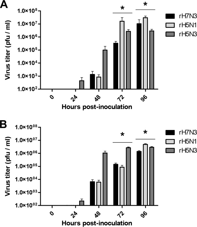
To determine whether the observed differences in pathogenicity could be correlated with growth characteristics in vitro, the replication kinetics of rH7N3, rH5N1, and rH5N3 were compared in MDCK and QT-35 cells. Confluent monolayers of MDCK and QT-35 cells were inoculated with each recombinant virus at an MOI of 0.00001 in the absence of exogenous trypsin. Culture supernatants were sampled immediately following the 3× PBS wash that followed virus adsorption and at 24, 48, 72, and 96 h postinoculation and titrated on MDCK cells. All three viruses grew in both cell lines in the absence of exogenously added trypsin with minor differences in the magnitude of replication among the three (Fig. 3). An accelerated production of rH5N3 was noted at earlier time points in MDCK and QT-35 cells.
FIG 3.
Multicycle replication of reassortant viruses in MDCK and QT-35 cells. MDCK and QT-35 cell monolayers were inoculated with each virus at an MOI of 0.00001. After the inoculum had adsorbed for 1 h, the cell monolayers were washed three times with PBS, and infection medium was added. Culture supernatants were collected at the indicated time points and titrated on MDCK cells. (A) MDCK cells; (B) QT-35 cells. *, P < 0.05 based on one-way ANOVA. The Bonferroni adjusted probabilities were 0.002 for rH5N3 versus rH7N3 at 72 h postinoculation in MDCK cells and 0.006 for rH5N3 versus rH5N1 at 96 h postinoculation in MDCK cells. The Bonferroni adjusted probabilities were 0.004 for rH5N3 versus rH5N1 at 72 h postinoculation in QT-35 cells, 0.005 for rH5N3 versus rH7N3 at 72 h postinoculation in QT-35 cells, and 0.003 for rH5N1 versus rH7N3 at 96 h postinoculation in QT-35 cells.
Serial passaging of rH5N3 in chickens.
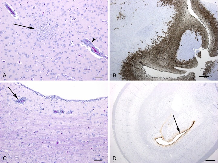
Despite the possession of a polybasic HA0 cleavage site and replication characteristics in chickens and cell culture that were comparable to those of highly virulent rH5N1 and rH7N3 viruses, rH5N3's display of a nonvirulent phenotype suggested that additional viral determinants were required for expression of high pathogenicity. To assess whether rH5N3 was capable of evolving into a phenotypically HPAI virus, it was serially passaged six times in chickens. A total of five chickens were intranasally inoculated with each virus passage as described in Materials and Methods, and virus was reisolated from a cloacal swab at P1, lung tissue at P2, and brain tissue at P3 through P6. A progressive increase in virulence, as evidenced by clinical signs and mortality, was observed from P0 to P6. Repeat of the IVPI on P0 rH5N3 confirmed the score of 0. IVPI results for P2 rH5N3, P4 rH5N3, and P6 rH5N3 viruses were 1.55, 2.55, and 2.71, respectively (Fig. 4), showing that after only two passages in chickens we could obtain a HPAI virus. Histopathological and immunohistochemical analysis of tissues collected from rH5N3 P0- and rH5N3 P6-infected chickens at 1, 2, and 3 dpi revealed distinct differences (Table 1). Viral antigen could be detected within the spleen as early as 1 dpi in the P6 rH5N3 birds, whereas no viral antigen was detected in any tissue analyzed in the P0 rH5N3-inoculated birds. By 2 dpi, the most significant differences between the two groups were observed within the brain. In the P0 rH5N3 group no significant lesions were observed, except for one very small focus of mild inflammation in one bird, and no viral antigen was detected in any of the brains at this time point. In the P6 rH5N3 group, 3/3 animals had mild lesions in the brain with viral antigen detected in all three brains. By 3 dpi, moderate brain lesions, including gliosis, necrosis, meningitis, and perivascular cuffing with mononuclear cells, could be observed multifocally throughout the brain in all of the P6 rH5N3-inoculated birds (Fig. 5A). Abundant viral antigen could be detected extensively in the brain, including the ependymal epithelial cells, neurons, and neuropil (Fig. 5B). In contrast, 2/3 P0 rH5N3-inoculated birds did not have lesions, and one bird had only mild lesions characterized by perivascular cuffing in the periventricular area (Fig. 5C). In one of the birds there was a prominent focus of immunostaining that was primarily limited to ependymal cells (Fig. 5D). In the other two birds immunostaining was negative or very weak (<5 cells). In general, lesions observed in the heart, spleen and lung were similar between the two groups, but overall more viral antigen could be detected in the P6 rH5N3 group for all three tissues. Significant lesions of the cecal tonsil and pancreas were only present in the P6 rH5N3 group (Table 1).
FIG 4.
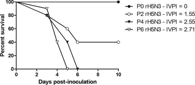
Survival of White Leghorn chickens following intranasal inoculation with P0 to P6 rH5N3 virus. The percent survival of chickens following intranasal inoculation of each passaged virus was determined. Intravenous pathogenicity indices (IVPI) are indicated for P0, P2, P4, and P6 rH5N3 viruses.
TABLE 1.
Immunohistochemical detection of influenza A viral antigen in P0 rH5N3- and P6 rH5N3-inoculated birdsa
| Tissue | 1 dpi |
2 dpi |
3 dpi |
|||
|---|---|---|---|---|---|---|
| P0 rH5N3 | P6 rH5N3 | P0 rH5N3 | P6 rH5N3 | P0 rH5N3 | P6 rH5N3 | |
| Brain | – | – | – | 1+ | 1+ | 3+ |
| Lung | – | – | 1+ | 2+ | wk+ | 1+ |
| Spleen | – | wk+ | wk+ | 2+ | wk+ | 1+ |
| Cecal tonsil | – | – | – | 2+ | wk+ | wk+ |
| Heart | – | – | 1+ | 2+ | 1+ | 3+ |
| Pancreas | – | – | wk+ | – | – | 2+ |
–, No immunostaining; wk+, weak immunostaining (<20 cells); 1+, mild immunostaining (<25% of the section); 2+, moderate immunostaining (25 to 50% of the section); 3+, abundant immunostaining (51 to 75% of the section). dpi, days postinoculation.
FIG 5.
Comparison of histopathology and immunohistochemistry findings in brains of P6 rH5N3 (A and B)- and P0 rH5N3 (C and D)-inoculated birds at 3 dpi. (A) Perivascular cuffing with mononuclear inflammatory cells (arrowhead) was observed throughout the brain. Note area of gliosis and necrosis (arrow). Bar, 50 μm; H&E stain. (B) Extensive positive immunostaining for influenza A virus antigen. Bar, 500 μm. (C) Perivascular cuffing was observed only in the periventricular area of one bird (arrow). Bar, 50 μm; H&E stain. (D) Detection of viral antigen was limited to the ependymal cells lining the ventricles. Bar, 500 μm.
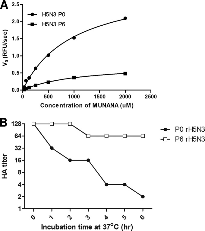
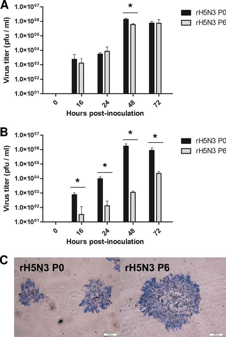
To determine the differences between the virus isolates obtained through passaging in chickens at the genotype level, whole-genome sequencing of P0 through P6 rH5N3 viruses revealed a total of 12 nonsynonymous mutations involving all eight gene segments (Table 2). The arginine-to-lysine substitution at amino acid position 293 (R293K; R292K for N2 numbering) of the NA protein was of particular interest to us since this amino acid, along with R118, D151, R152, R225, E278, R370, and Y405, forms the catalytic site. The R293K substitution has been associated with decreased enzyme activity, resistance to oseltamivir, and compromised fitness and transmissibility in ferrets (21, 22). Comparison of the neuraminidase activities of P0 rH5N3 and P6 rH5N3 revealed that although the Km did not significantly differ between the two viruses (Km P0 rH5N3 = 995.9 μM ± 59.04; Km P6 rH5N3 = 1,113 μM ± 48.89; P = 0.5612), there was a 4-fold reduction in the Vmax of P6 rH5N3 relative to P0 rH5N3 (Vmax P0 rH5N3 = 3.115 relative fluorescent units/s ± 0.8894; Vmax P6 rH5N3 = 0.7626 relative fluorescent units/s ± 0.01673; P < 0.0001). This finding was supported by the results of the chicken erythrocyte elution assay (Fig. 6) in which a progressive reduction in HA titer was observed in P0 rH5N3 relative to P6 rH5N3 over a 6-h period. Oseltamivir resistance was also confirmed for P6 rH5N3, which had an IC50 of 2726.25 nM compared to an IC50 of 1.81 nM for P0 rH5N3 determined by neuraminidase inhibition assay. To further analyze whether the differences in pathogenicity between P0 and P6 rH5N3 viruses were also evident in vitro, multistep growth kinetics for P0 rH5N3 and P6 rH5N3 were assessed on MDCK and QT-35 cells (Fig. 7). The replication kinetics of both viruses were comparable in QT-35 cells with the exception of 48 h postinoculation, at which point there was a statistically significant difference between P0 rH5N3 and P6 rH5N3 titers (Fig. 7A). P6 rH5N3 replication in MDCK cells was significantly impaired compared to that of P0 rH5N3 at all time points assayed (Fig. 7B). Overall, this indicates that the changes in P6 rH5N3 resulted in a relative loss of replication efficiency in mammalian cells compared to avian cells. Although there did not appear to be any difference in the replication kinetics between P0 and P6 rH5N3 viruses in QT-35 cells, there was a significant difference (P < 0.001) in the size of the plaques formed by the two viruses. The mean plaque size for P0 rH5N3 was 377 ± 55 μm (n = 33), whereas the mean plaque size for P6 rH5N3 was 631 ± 78 μm (n = 26), suggesting more efficient cell-to-cell spread of P6 rH5N3.
TABLE 2.
Amino acid substitutions in rH5N3 genes after six serial passages in White Leghorn chickensa
| Virus passage | Substitution in rH5N3 genes |
|||||||||||
|---|---|---|---|---|---|---|---|---|---|---|---|---|
| PB2 (aa 466) | PB1 (aa 633) | PA (aa 263) | HA (aa 387) | NP (aa 371) | NA |
M1 (aa 97) | NS1 (aa 193) | NEP |
||||
| aa 82 | aa 293 | aa 329 | aa 36 | aa 52 | ||||||||
| P0 | D | S | T | N | M | E | R | A | V | R | E | M |
| P1 | D | S | T | N | T | G | R | A | V | R | E | M |
| P2 | D | R | T | N | T | G | K | A | V | R | E | T |
| P3 | D | R | T | D | T | G | K | T | V | R | E | T |
| P4 | D | R | T | D | T | G | K | T | I | R | E | T |
| P5 | N | R | K | D | T | G | K | T | I | Q | K | T |
| P6 | N | R | K | D | T | G | K | T | I | Q | K | T |
aa, amino acid.
FIG 6.
Comparison of the neuraminidase activities of rH5N3 P0 versus rH5N3 P6. (A) Neuraminidase enzyme kinetic curves for rH5N3 P0 and rH5N3 P6 expressed in relative fluorescent units per second as a function of MUNANA concentration. (B) Elution of rH5N3 P0 versus rH5N3 P6 from chicken erythrocytes. rH5N3 P0 and rH5N3 P6 were adjusted to a HA titer of 1:128, and 2-fold serial dilutions were incubated with equal volumes of 0.5% (vol/vol) chicken erythrocyte suspensions in V-bottom microtiter plates at 4°C for 1 h. The microtiter plates were then incubated at 37°C, and the reduction in HA titers was recorded every hour over a period of 6 h.
FIG 7.
Comparison of multicycle replication of rH5N3 P0 and rH5N3 P6 in MDCK and QT-35 cells. MDCK and QT-35 cell monolayers were inoculated with each virus at an MOI of 0.0001. After the inoculum had adsorbed for 1 h the cell monolayers were washed three times with PBS, and infection medium was added. Culture supernatants were collected at the indicated time points and titrated on MDCK cells. (A) QT-35 cells; (B) MDCK cells. (C) Representative viral plaques formed by rH5N3 P0 and rH5N3 P6 in QT-35 cells. *, P < 0.05 based on the Student t test.
Transmissibility of P0 rH5N3 versus P6 rH5N3.
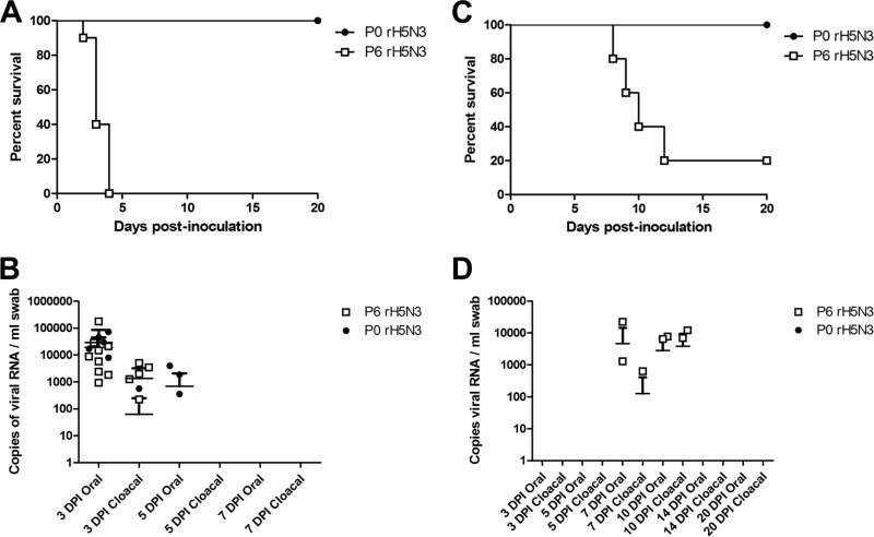
To compare the transmissibility of P0 rH5N3 and P6 rH5N3 viruses, 10 SPF chickens, housed in separate containment cubicles, were oronasally inoculated with 4.0 × 10⁴ PFU of each virus. After 24 h, five naive contact chickens were placed in each cubicle and allowed to commingle with the inoculated birds for the remainder of the experiment. All P0 rH5N3-inoculated birds and their respective contacts survived the entire 20-day observation period. In contrast, all of the P6 rH5N3-inoculated birds died by 5 dpi, and only one of the five contact birds had survived at the end of the 20-day observation period (Fig. 8A and C). While the levels of oropharyneal and cloacal virus shedding in P0 rH5N3- and P6 rH5N3-inoculated birds were similar, only the P6 rH5N3 contacts shed virus at detectable levels just prior to death. Although all P0 rH5N3-inoculated birds tested positive for NP and HI antibodies at the end of the 20-day observation period (data not shown), the respective contact birds did not, indicating that P0 rH5N3 did not transmit bird to bird.
FIG 8.
Survival of White Leghorn chickens and transmission of virus to naive contact birds following intranasal inoculation with P0 rH5N3 versus P6 rH5N3. (A) Ten chickens, each in separate isolation cubicles, were intranasally inoculated with 40,000 PFU of either P0 rH5N3 or P6 rH5N3, and survival was monitored over a period of 20 days. (B and D) Oropharyngeal and cloacal swabs from P0 rH5N3- and P6 rH5N3-inoculated birds (B) or from P0 rH5N3 and P6 rH5N3 contact birds (D) were tested by qRT-PCR to assess virus shedding. (C) One day following inoculation, five naive contact birds were placed in each isolation cubicle and allowed to commingle with the infected birds. The survival of the contact birds was monitored for a period of 19 days.
Amino acid substitution R293K in the NA protein contributes to the highly pathogenic phenotype of P6 rH5N3.
Since rH5N1, which had an IVPI of 2.30, and P0 rH5N3, which had an IVPI of 0, differed only by their NA genes, we hypothesized that the increased virulence of P6 rH5N3 could be attributed to the amino acid substitutions that had occurred in the N3 protein and, more specifically, to the single R293K amino acid substitution in the catalytic site of the NA. To test this, a mutant of P0 rH5N3 was generated that contained the modified NA gene NA-3-R293K. IVPI testing of the NA-3-R293K mutant virus resulted in a score of 2.44, suggesting that this single amino acid substitution significantly contributed to the increases in IVPI scores associated with P2 rH5N3 (1.55), P4 rH5N3 (2.55), and P6 rH5N3 (2.71) viruses. To assess transmissibility of this mutant virus, 10 SPF chickens were intranasally inoculated with 4.0 × 10⁴ PFU of virus and, 24 h later, 5 naive contact chickens were placed in the animal cubicle and allowed to commingle with the inoculated birds for the remainder of the experiment. Four of the inoculated birds died or had to be euthanized at 6 dpi. Clinical signs in those birds were depression, ataxia, and green feces. All of the surviving inoculated birds tested positive for NP antibodies at 16 dpi, and five of the six had positive H5 HI titers. Only one of the five contact birds had virus-positive oropharyngeal and cloacal swabs. This bird developed NP antibodies at 6 days after contact and was euthanized at 8 days after contact with severe depression and hemorrhages on the feet. The remaining contact birds were all seronegative at 15 days postcontact. Altogether, these findings show that although the R293K substitution contributed significantly to virulence by increasing the IVPI, it only partially enhanced the contact transmission characteristics of the virus. Hence, in addition to the interplay between HA and NA, other determinants were necessary for virus transmissibility.
DISCUSSION
In this study a reassortant H5N3 virus that possessed the HA gene from H5N1 HPAI Germany/R65 and the remaining gene segments from H7N3 LPAI BC/CN006 was generated using reverse genetics. Based on the presence of a polybasic HA0 cleavage site, we predicted that this rH5N3 virus should exhibit a highly pathogenic phenotype in chickens. Despite the fact that this virus replicated efficiently in cell culture in the absence of trypsin and could infect and replicate in chickens, as well as induce seroconversion, it was not pathogenic in this host. By serially passaging this rH5N3 virus in chickens, a progressive increase in virulence was observed. P6 rH5N3 produced an IVPI of 2.71 and was highly transmissible to naive contact birds. This change in phenotype was associated with a corresponding change in genotype. Changes involving the NA of rH5N3 were of particular interest since the rH5N1 virus which possessed the HA and NA genes of Germany/R65 and the remaining genes of BC/CN006 was highly pathogenic in chickens, suggesting that the NA plays an important role in expression of the highly pathogenic phenotype.
Although the HA0 cleavage site of H5 and H7 subtype viruses is considered the dominant virulence determinant of HPAIV, virulence is nevertheless considered to be a polygenic trait. Early studies by Rott et al. (23) showed that the pathogenicity of a HPAI H7N1 virus for chickens could be attenuated after reassortment with equine, swine, or human influenza A viruses even when the reassortants retained the HA and NA of the parent virus. A follow-up study by Webster et al. (24) demonstrated that not all reassortants that contained the HA and M genes of HPAIV A/chicken/Pennsylvania/1370/83 (H5N2) and the remaining gene segments from wild duck, shorebird, and domestic poultry viruses resulted in a HPAI phenotype. These researchers found that a disproportionate number of reassortant viruses that contained gene constellations of shorebird or avirulent H5N2 live bird market origin were highly pathogenic in chickens, suggesting that not all gene constellations were capable of complementing the HA gene from chicken/Penn (H5N2). Field isolates and laboratory generated viruses have further confirmed that a polybasic HA0 cleavage site is necessary but not sufficient to confer a HPAI phenotype. To illustrate, a H5N2 virus isolated from a broiler flock and two live bird markets in Texas in 2004 was phenotypically low pathogenic, although possessing the HA0 cleavage site PQRKKR/GLF and hence genotypically HPAIV (7). Discordant results between molecular and in vivo tests used to characterize virus pathogenicity were retrospectively recognized for other H5 field virus isolates (8, 9), as well as non-H5/H7 viruses that had been engineered to contain polybasic HA0 cleavage sites (25, 26, 27). For example Stech et al. (26) showed that inserting a polybasic cleavage site identical to that of HPAI A/chicken/Italy/8/98 (H5N2) into the HA of the LPAI A/duck/Ukraine/1/63 (H3N8) virus was not sufficient to immediately transform it into a highly pathogenic virus, despite the fact that it was able to replicate in tissue culture in the absence of exogenous trypsin. This implied that the evolution of highly pathogenic H5/H7 viruses from low-pathogenic precursors might also involve other changes in addition to a polybasic HA0 cleavage site. In contrast, Veits et al. (27) reported that avian influenza viruses with H2, H4, H8, and H14 hemagglutinins could support a HPAI phenotype after acquiring a polybasic HA0 cleavage site. However, this approach has yielded different results for H6 viruses. Although the insertion of a polybasic cleavage site in the H6N1 virus A/mallard/Sweden/81/02 (28) supported a HPAI phenotype in vivo, the insertion of a polybasic cleavage site in the H6N2 A/turkey/Germany/R617/07 virus did not (27), indicating that other “cryptic” virulence determinants are also involved in expression of the highly pathogenic phenotype in vivo. The involvement of such cryptic virulence determinants has been supported by results from a number of studies (8, 25, 29).
rH7N3, rH5N1, and rH5N3 all possess identical internal gene segments and differ only in their HA and NA genes. While rH7N3 and rH5N1 were highly pathogenic in chickens, rH5N3 was not, implying that the interplay between HA and NA was at the root of the difference in pathogenicity between rH5N1 and rH5N3 viruses. Although it has been well established that HA-NA functional balance impacts virus replication efficiency, the results of the present study suggest that HA-NA balance can also influence expression of the HPAI phenotype. HA and NA both recognize sialic acid-containing receptors on target cells with HA binding to sialic acid receptors required to initiate virus infection and cleavage of sialic acid from receptors by NA required to facilitate the release of progeny virus from infected cells. The interaction between HA receptor binding and NA receptor destroying activities has been reported to affect virus replication efficiency in eggs and cell culture (30, 31). HA-NA balance can be disturbed by reassortment, transmission to a new host, or treatment with neuraminidase inhibitors. Reassortants expressing avian H3, H4, H10, and H13 subtypes in combination with a human N1 replicated poorly in ECE compared to the parental viruses and were associated with the formation of virus aggregates (32). Serial passaging of these reassortants in ECE resulted in high-yielding nonaggregating variants which contained compensatory mutations in the HA that resulted in decreased affinity for sialic acid containing receptors (33). Amino acid substitutions involving conserved residues associated with the enzymatic active site of NA can affect its catalytic functions. This in turn has been reported to compromise virus replication in some animal models (21, 22). The R293K substitution found in P2-P6 rH5N3 viruses in this study was associated with a progressive increase in pathogenicity, as determined by IVPI, a reduction in neuraminidase activity and resistance to the neuraminidase inhibitor oseltamivir. This association of enhanced pathogenicity was confirmed by introducing the R293K substitution into the NA of P0 rH5N3, which resulted in an IVPI increase from 0 to 2.44. It should be noted that the NA of Germany/R65 (GenBank accession DQ464355) possesses a 20-amino-acid deletion in its stalk (the deletion of amino acids 49 to 68). NA stalk deletions have been associated with increased host range, changes in the ability of the virus to replicate in vivo, reduced NA enzymatic activity, and in some instances increased virus virulence (34, 35, 36). Influenza A viruses with shorter NA stalks were found to have a decreased ability to elute from red blood cells (19, 34, 37). This is thought to be due to their reduced ability to access sialic acid substrates properly, possibly because the enzyme active site is located too close to the viral envelope. We speculate that the R293K substitution in the NA of rH5N3 might compensate for the stalk deletion found in the NA of Germany/R65, thus restoring HA-NA balance and contributing to increased pathogenicity. In this regard, the finding that rH5N3 P6 formed significantly larger plaques in QT-35 cells compared to rH5N3 P0, suggests more efficient cell-to-cell spread. This could be linked to increased “stickiness” of the virus imparted by the R293K substitution, which may in turn have other effects on traits such as transmissibility.
To date, one naturally occurring H5N3 HPAI virus has been recognized. A/tern/South Africa/1961 (H5N3) was responsible for an explosive die-off common terns (Sterna hirundo) along the coast of the Cape Province of South Africa during April of 1961 (38). The HA (GenBank accession U20460.1) of this virus has the HA0 cleavage site PQRETRRQKR/GLF and has a wild-type K293 in its NA (GenBank accession AY207524.1) but no deletion involving the NA stalk. A comprehensive analysis of NA sequences with a focus on stalk deletions was recently reported by Li et al. (39). Of the 58 natural H5N3 virus isolates for which NA sequences were analyzed, 3 or roughly 5% had a deletion in the stalk and some of these were responsible for outbreaks in poultry. In addition to reduced NA enzymatic activity (34), a shortened NA stalk has been associated with adaptation to gallinaceous poultry (36). A matching reduction in HA affinity for receptor binding often accompanies NA stalk deletions/reduced enzymatic activity in order for efficient virus replication to be maintained (30, 40). In this regard additional glycosylation sites on HA1 have been reported to compensate for the decreased NA activity that is associated with a truncated NA stalk (30, 41). It is worth noting that the HA1 of Germany/R65 has four glycosylation sites, while the HA1 of BC/CN006 and BC/CN007 have only three. This difference in HA1 glycosylation of the H5 of Germany/R65 when paired with the N3 from BC/CN006 may have predisposed rH5N3 to an initial HA-NA imbalance which was overcome by the R293K substitution involving the catalytic site of the NA. The reason for why a NA stalk deletion was not observed is not known, but could involve unknown constraints for this particular gene constellation. Although the 11 other amino acid substitutions that were found in rH5N3 P6 were not experimentally assessed for their ability to contribute to increased virulence and transmissibility, a search of the literature failed to identify any of them as having associations with increased virulence for poultry.
Although the subject of the research described here, a reassortant between a HPAI Eurasian and a LPAI North American lineage virus, may at one time have been considered unlikely to occur in nature, this very event did occur in 2014. In late November 2014, a novel reassortant HPAI H5N2 virus that contained gene segments related to Eurasian H5N8 was detected in a domestic turkey and broiler breeder flock in British Columbia, Canada (42). Shortly thereafter, Eurasian HPAI H5N8 and reassortant HPAI H5N2 viruses were isolated from wild birds in Washington, USA (43). A second novel reassortant HPAI H5N1 virus was subsequently isolated from a hunter-killed green-winged teal in Washington, USA (44). The gene constellation of this reassortant differed from the earlier H5N2 reassortant. A nearly identical HPAI H5N1 was later isolated from a backyard chicken flock near Chilliwack, British Columbia, in February 2015. Despite the fact that the reassortant HPAI H5N2 has become dominant causing widespread outbreaks in several midwestern states in the United States, novel reassortant viruses possessing unique phenotypes may still be generated.
In summary, we show that following reassortment not all gene constellations are immediately capable of complementing a HA with a polybasic cleavage site and that HA-NA balance is important in the expression of the HPAI phenotype. Furthermore, since it may only take a few passages in chickens for a genotypically HPAI virus to become phenotypically HPAI, all H5 and H7 viruses with a polybasic HA0 cleavage site should be treated as HPAI. Lastly, unexpected phenotypic traits such as resistance to antiviral drugs may also arise following reassortment and passaging in domestic poultry which can have significant public health implications.
ACKNOWLEDGMENTS
We thank Timm Harder, Friedrich-Loeffler-Institut, for providing the virus isolates A/swan/Germany/R65/2006 (H5N1) and A/teal/Germany/Wv632/2005 (H5N1) used in this study. We are grateful to Yan Li, National Microbiology Laboratory, Public Health Agency of Canada, for performing the NA-Star chemiluminescent neuraminidase inhibition assay. We especially thank Kurtis Swekla, Maggie Forbes, Jaime Bernstein, Kory Nakamura, Kevin Tierney, and Marlee Phair for their service and dedication during the animal work for this project.
This work was supported by funding provided by the Canadian Poultry Research Council and the Canadian Food Inspection Agency.
REFERENCES
- 1.Tong S, Li Y, Rivailler P, Conrardy C, Castillo DA, Chen LM, Recuenco S, Ellison JA, Davis CT, York IA, Turmelle AS, Moran D, Rogers S, Shi M, Tao Y, Weil MR, Tang K, Rowe LA, Sammons S, Xu X, Frace M, Lindblade KA, Cox NJ, Anderson LJ, Rupprecht CE, Donis RO. 2012. A distinct lineage of influenza A virus from bats. Proc Natl Acad Sci U S A 109:4269–4274. doi: 10.1073/pnas.1116200109. [DOI] [PMC free article] [PubMed] [Google Scholar]
- 2.Tong S, Zhu X, Li Y, Shi M, Zhang J, Bourgeois M, Yang H, Chen X, Recuenco S, Gomez J, Chen LM, Johnson A, Tao Y, Dreyfus C, Yu W, McBride R, Carney PJ, Gilbert AT, Chang J, Guo Z, Davis CT, Paulson JC, Stevens J, Rupprecht CE, Holmes EC, Wilson IA, Donis RO. 2013. New world bats harbor diverse influenza A viruses. PLoS Pathog 9:e1003657. doi: 10.1371/journal.ppat.1003657. [DOI] [PMC free article] [PubMed] [Google Scholar]
- 3.Li KS, Guan Y, Wang J, Smith GJ, Xu KM, Duan L, Rahardjo AP, Puthavathana P, Buranthal C, Nguyen TD, Estoepangestie ATS, Chaisingh A, Auewarakul P, HTlong Hanh NTH, Webby RJ, Poon LLM, Chen H, Shortridge KF, Yuen KY, Webster RG, Peris JSM. 2004. Genesis of a highly pathogenic and potentially pandemic H5n1 influenza virus in eastern Asia. Nature 430:209–213. doi: 10.1038/nature02746. [DOI] [PubMed] [Google Scholar]
- 4.Stieneke-Grober A, Vey M, Angliker H, Shaw E, Thomas G, Roberts C, Klenk H-D, Garten W. 1992. Influenza virus hemagglutinin with multibasic cleavage site is activated by furin, a subtilism-like endoprotease. EMBO J 11:2407–2414. [DOI] [PMC free article] [PubMed] [Google Scholar]
- 5.Wiley DC, Skehel JJ. 1987. The structure and function of the hemagglutinin membrane glycoprotein of influenza virus. Annu Rev Biochem 56:365–394. doi: 10.1146/annurev.bi.56.070187.002053. [DOI] [PubMed] [Google Scholar]
- 6.Abdelwhab E-SM, Veits J, Mettenleiter TC. 2013. Genetic changes that accompanied shifts of low pathogenic avian influenza viruses toward higher pathogenicity in poultry. Virulence 4:1–12. doi: 10.4161/viru.22788. [DOI] [PMC free article] [PubMed] [Google Scholar]
- 7.Lee CW, Swayne DE, Linares JA, Linares JA, Senne DA, Suarez DL. 2005. H5N2 avian influenza outbreak in Texas in 2004: the first highly pathogenic strain in the United States in 20 years? J Virol 79:11412–11421. doi: 10.1128/JVI.79.17.11412-11421.2005. [DOI] [PMC free article] [PubMed] [Google Scholar]
- 8.Londt BZ, Banks J, Alexander DJ. 2007. Highly pathogenic avian influenza viruses with low virulence for chickens in in vivo tests. Avian Path. 36:347–350. doi: 10.1080/03079450701589134. [DOI] [PubMed] [Google Scholar]
- 9.Lee C-CD, Zhu H, Huang P-Y, Peng L, Chang Y-C, Yip C-H, Li Y-T, Cheung C-L, Compans R, Yang C, Smith DK, Lam TT-Y, King C-C. 2014. Emergence and evolution of avian H5N2 influenza viruses in chickens in Taiwan. J Virol 88:5677–5686. doi: 10.1128/JVI.00139-14. [DOI] [PMC free article] [PubMed] [Google Scholar]
- 10.Wasilenko JL, Lee CW, Sarmento L, Spackman E, Kapczynski DR, Suarez DL, Pantin-Jackwood MJ. 2008. NP, PB1, and PB2 viral genes contribute to altered replication of H5N1 avian influenza viruses in chickens. J Virol 82:4544–4553. doi: 10.1128/JVI.02642-07. [DOI] [PMC free article] [PubMed] [Google Scholar]
- 11.Song J, Huapeng F, Xu J, Zhao D, Shi J, Li Y, Deng G, Jiang Y, Li X, Zhu P, Guan Y, Bu Z, Kawaoka Y, Chen H. 2011. The PA protein directly contributes to the virulence of H5N1 avian influenza viruses in domestic ducks. J Virol 85:2180–2188. doi: 10.1128/JVI.01975-10. [DOI] [PMC free article] [PubMed] [Google Scholar]
- 12.Weber S, Harder T, Starick E, Beer M, Werner O, Hoffmann B, Mettenleiter TC, Mundt E. 2007. Molecular analysis of highly pathogenic avian influenza virus of subtype H5N1 isolated from wild birds and mammals in northern Germany. J Gen Virol 88:554–558. doi: 10.1099/vir.0.82300-0. [DOI] [PubMed] [Google Scholar]
- 13.Pasick J, Handel K, Robinson J, Copps J, Ridd D, Hills K, Kehler H, Cottam-Birt C, Neufeld J, Berhane Y, Czub S. 2005. Intersegmental recombination between the haemagglutinin and matrix genes was responsible for the emergence of a highly pathogenic H7N3 avian influenza virus in British Columbia. J Gen Virol 86:727–731. doi: 10.1099/vir.0.80478-0. [DOI] [PubMed] [Google Scholar]
- 14.Hoffmann E, Stech J, Guan Y, Webster RG, Perez DR. 2001. Universal primer set for the full-length amplification of all influenza A viruses. Arch Virol 146:2275–2289. doi: 10.1007/s007050170002. [DOI] [PubMed] [Google Scholar]
- 15.Hoffmann E, Neumann G, Kawaoka Y, Hobom G, Webster RG. 2000. A DNA transfection system for generation of influenza A virus from eight plasmids. Proc Natl Acad Sci U S A 97:2020–2028. [DOI] [PMC free article] [PubMed] [Google Scholar]
- 16.Yang M, Berhane Y, Salo T, Li M, Hole KK, Clavijo A. 2008. Development and application of monoclonal antibodies against avian influenza virus nucleoprotein. J Virol Methods 147:265–274. doi: 10.1016/j.jviromet.2007.09.016. [DOI] [PubMed] [Google Scholar]
- 17.Swayne D. 2012. Avian influenza, p 436–452. In Manual of diagnostic tests and vaccines for terrestrial animals, vol 1, 7th ed Office International des Epizooties, Paris, France. [Google Scholar]
- 18.Marathe BM, Lévêque V, Klumpp K, Webster RG, Govorkova EA. 2013. Determination of neuraminidase kinetic constants using whole influenza virus preparations and correction for spectroscopic interference by a fluorogenic substrate. PLoS One 8:e71401. doi: 10.1371/journal.pone.0071401. [DOI] [PMC free article] [PubMed] [Google Scholar]
- 19.Castrucci MR, Kawaoka Y. 1993. Biologic importance of neuraminidase stalk length in influenza A virus. J Virol 67:759–764. [DOI] [PMC free article] [PubMed] [Google Scholar]
- 20.Spackman E, Senne DA, Myers TJ, Bulaga LL, Garber LP, Perdue ML, Lohman K, Daum LT, Suarez DL. 2002. Development of a real-time reverse transcriptase PCR assay for type A influenza virus and the avian H5 and H7 hemagglutinin subtypes. J Clin Microbiol 40:3256–3260. doi: 10.1128/JCM.40.9.3256-3260.2002. [DOI] [PMC free article] [PubMed] [Google Scholar]
- 21.Yen H-L, Herlocher ML, Hoffmann E, Matrosovich MN, Monto AS, Webster RG, Govorkova EA. 2005. Neuraminidase inhibitor-resistant influenza viruses may differ substantially in fitness and transmissibility. Antimicrob Agents Chemother 49:4075–4084. doi: 10.1128/AAC.49.10.4075-4084.2005. [DOI] [PMC free article] [PubMed] [Google Scholar]
- 22.Yen H-L, Hoffmann E, Taylor G, Scholtissek C, Monto AS, Webster RG, Govorkova E. 2006. Importance of neuraminidase active-site residues to the neuraminidase inhibitor resistance of influenza viruses. J Virol 80:8787–8795. doi: 10.1128/JVI.00477-06. [DOI] [PMC free article] [PubMed] [Google Scholar]
- 23.Rott R, Orlich M, Scholtissek C. 1976. Attenuation of pathogenicity of fowl plague virus by recombination with other influenza A viruses non-pathogenic for fowl: nonexclusive dependence of pathogenicity on hemagglutinin and neuraminidase of the virus. J Virol 19:54–60. [DOI] [PMC free article] [PubMed] [Google Scholar]
- 24.Webster RG, Kawaoka Y, Bean WJ. 1989. What is the potential of avirulent influenza viruses to complement a cleavable hemagglutinin and generate virulent strains? Virology 171:484–492. doi: 10.1016/0042-6822(89)90618-1. [DOI] [PubMed] [Google Scholar]
- 25.Gohrbandt S, Veits J, Breithaupt A, Hundt J, Teifke JP, Stech O, Mettenleiter TC, Stech J. 2011. H9 avian influenza reassortant with engineered polybasic cleavage site displays a highly pathogenic phenotype in chicken. J Gen Virol 92:1843–1853. doi: 10.1099/vir.0.031591-0. [DOI] [PubMed] [Google Scholar]
- 26.Stech O, Veits J, Weber S, Deckers D, Schröer D, Vahlenkamp TW, Breithaupt A, Teifke J, Mettenleiter TC, Stech J. 2009. Acquisition of a polybasic hemagglutinin cleavage site by a low-pathogenic avian influenza virus is not sufficient for immediate transformation into a highly pathogenic strain. J Virol 83:5864–5868. doi: 10.1128/JVI.02649-08. [DOI] [PMC free article] [PubMed] [Google Scholar]
- 27.Veits J, Weber S, Stech O, Breithaupt A, Gräber M, Gohrbandt S, Bogs J, Hundt J, Teifke JP, Mettenleiter TC, Stech J. 2012. Avian influenza hemagglutinins H2, H4, H8, and H14 support a highly pathogenic phenotype. Proc Natl Acad Sci U S A 109:2579–2584. doi: 10.1073/pnas.1109397109. [DOI] [PMC free article] [PubMed] [Google Scholar]
- 28.Munster VJ, Schrauwen EJA, de Wit E, van den Brand JMA, Bestebroer TM, Herfst S, Rimmelzwaan GF, Osterhaus ADME, Fouchier RAM. 2010. Insertion of a multibasic cleavage site motif into the hemagglutinin of a low-pathogenic avian influenza H6N1 virus induces a highly pathogenic phenotype. J Virol 84:7953–7960. doi: 10.1128/JVI.00449-10. [DOI] [PMC free article] [PubMed] [Google Scholar]
- 29.Bogs J, Veits J, Gohrbandt S, Hundt J, Stech O, Breithaupt A, Teifke JP, Mettenleiter TC, Stech J. 2010. Highly pathogenic H5N1 influenza viruses carry virulence determinants beyond the polybasic hemagglutinin cleavage site. PLoS One 5:e11826. doi: 10.1371/journal.pone.0011826. [DOI] [PMC free article] [PubMed] [Google Scholar]
- 30.Mitnaul LJ, Matrosovich MN, Castrucci MR, Tuzikov AB, Bovin NV, Kobasa D, Kawaoka Y. 2000. Balanced hemagglutinin and neuraminidase activities are critical for efficient replication of influenza A virus. J Virol 74:6015–6020. doi: 10.1128/JVI.74.13.6015-6020.2000. [DOI] [PMC free article] [PubMed] [Google Scholar]
- 31.Wagner R, Matrosovich M, Klenk H-S. 2002. Functional balance between haemagglutinin and neuraminidase in influenza virus infections. Rev Med Virol 12:159–166. doi: 10.1002/rmv.352. [DOI] [PubMed] [Google Scholar]
- 32.Rudneva IA, Kovaleva VP, Varich NL, Farashyan VR, Gubareva LV, Yamnikova SS, Popova IA, Presnova VP, Kaverin NV. 1993. Influenza A virus reassortants with surface glycoprotein genes of the avian parent virus: effects of HA and NA gene combinations on virus aggregation. Arch Virol 133:437–450. doi: 10.1007/BF01313781. [DOI] [PubMed] [Google Scholar]
- 33.Rudneva IA, Sklyanskaya EI, Barulina OS, Yamnikova SS, Kovaleva VP, Tsvetkova IV, Kaverin NV. 1996. Phenotypic expression of HA-NA combinations in human-avian influenza A virus reassortants. Arch Virol 141:1091–1099. doi: 10.1007/BF01718612. [DOI] [PubMed] [Google Scholar]
- 34.Hossain MJ, Hickman D, Perez DR. 2008. Evidence of expanded host range and mammalian-associated genetic changes in a duck H9N2 influenza virus following adaptation in quail and chickens. PLoS One 3:e3170. doi: 10.1371/journal.pone.0003170. [DOI] [PMC free article] [PubMed] [Google Scholar]
- 35.Munier S, Larcher T, Cormier-Aline F, Soubieux D, Su B, Guigand L, Labrosse B, Cherel Y, Quéré P, Marc D, Naffakh N. 2010. A genetically engineered waterfowl influenza virus with a deletion in the stalk of the neuraminidase has increased virulence for chickens. J Virol 84:940–952. doi: 10.1128/JVI.01581-09. [DOI] [PMC free article] [PubMed] [Google Scholar]
- 36.Els MC, Air GM, Murti KG, Webster RG, Laver WG. 1985. An 18-amino acid deletion in an influenza neuraminidase. Virology 142:241–247. doi: 10.1016/0042-6822(85)90332-0. [DOI] [PubMed] [Google Scholar]
- 37.Matsuoka Y, Swayne DE, Thomas C, Rameix-Welti MA, Naffakh N, Cwarnes Altholz M, Donis R, Subbarao K. 2009. Neuraminidase stalk length and additional glycosylation of the hemagglutinin influence the virulence of influenza H5N1 viruses for mice. J Virol 83:4704–4708. doi: 10.1128/JVI.01987-08. [DOI] [PMC free article] [PubMed] [Google Scholar]
- 38.Becker WB. 1966. The isolation and classification of tern virus: influenza virus A/Tern/South Africa/1961. J Hyg (Lond) 64:309–320. [DOI] [PMC free article] [PubMed] [Google Scholar]
- 39.Li J, Hzu Dohna Cardona CJ, Miller J, Carpenter T. 2011. Emergence and genetic variation of neuraminidase stalk deletions in avian influenza viruses. PLoS One 6:e14722. doi: 10.1371/journal.pone.0014722. [DOI] [PMC free article] [PubMed] [Google Scholar]
- 40.Wagner R, Wolff T, Herwig A, Pleschka S, Klenk HD. 2000. Interdependence of hemagglutinin glycosylation and neuraminidase as regulators of influenza virus growth: a study by reverse genetics. J Virol 74:6316–6323. doi: 10.1128/JVI.74.14.6316-6323.2000. [DOI] [PMC free article] [PubMed] [Google Scholar]
- 41.Baigent SJ, McCauley JW. 2001. Glycosylation of haemagglutinin and stalk-length of neuraminidase combine to regulate the growth of avian influenza viruses in tissue culture. Virus Res 79:177–185. doi: 10.1016/S0168-1702(01)00272-6. [DOI] [PubMed] [Google Scholar]
- 42.Pasick J, Berhane Y, Joseph T, Bowes V, Hisanaga T, Handel K, Alexandersen S. 2015. Reassortant highly pathogenic influenza A H5N2 virus containing gene segments related to Eurasian H5N8 in British Columbia, Canada, 2014. Sci Rep 5:9484. doi: 10.1038/srep09484. [DOI] [PMC free article] [PubMed] [Google Scholar]
- 43.Ip HS, Torchetti MK, Crespo R, Kohrs P, DeBruyn P, Mansfield KG, Baszler T, Badcoe L, Bodenstein B, Shearn-Bochsler V, Killian ML, Pedersen JC, Hines N, Gidlewski T, DeLiberto T, Sleeman JM. 2014. 2015. Novel Eurasian highly pathogenic avian influenza A H5 viruses in wild birds, Washington, USA, 2014. Emerg Infect Dis 21:886–890. doi: 10.3201/eid2105.142020. [DOI] [PMC free article] [PubMed] [Google Scholar]
- 44.Torchetti MK, Killian ML, Dusek RJ, Pedersen JC, Hines N, Bodenstein B, White CL, Ip HS. 2015. Novel H5 clade 2.3.4.4 reassortant (H5N1) virus from a green-winged teal in Washington, U S A. Genome Announc 3:e00195-15. doi: 10.1128/genomeA.00195-15. [DOI] [PMC free article] [PubMed] [Google Scholar]


