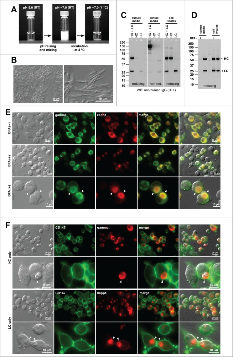
Figure 2.

Human IgG2κ (mAb-B) forms needle-shaped aggregates at neutral pH in vitro and induces Russell bodies in the ER. (A) Glass vial containing mAb-B solution at 70 mg/ml at pH 5.0 (left) and after pH shift to pH ∼7.0 followed by a brief mixing (middle). The vial shown in the middle panel was subsequently kept at 4°C for 24 hr (right). (B) DIC micrographs of the clouded mAb-B solution after pH raising. (C) Western blotting results for the cell culture media and cell lysates harvested on Day-7 post transfection. The expression constructs used for transfection are shown at the top of each lane. (D) Western blotting results on the cell culture media and cell lysates after 24 hr of BFA or null treatment from Day-2 to Day-3 post transfection. The cell viabilities after the 24 hr BFA and mock treatment were 94.6% and 96.9%, respectively. (E) Fluorescent micrographs of HEK293 cells co-transfected with HC and LC constructs. Cells at steady-state are shown in the first row. Cells under BFA treatment are shown in the second and third rows. Green, anti-gamma staining. Red, anti-kappa staining. RBs are pointed by arrowheads in the bottom row. (F) Fluorescent micrographs of HEK293 cells transfected with HC construct (first 2 rows) or with LC construct (bottom 2 rows). Green, anti-CD147 staining. Red, anti-gamma or anti-kappa staining. RBs are pointed by arrowheads in the second and fourth rows.
