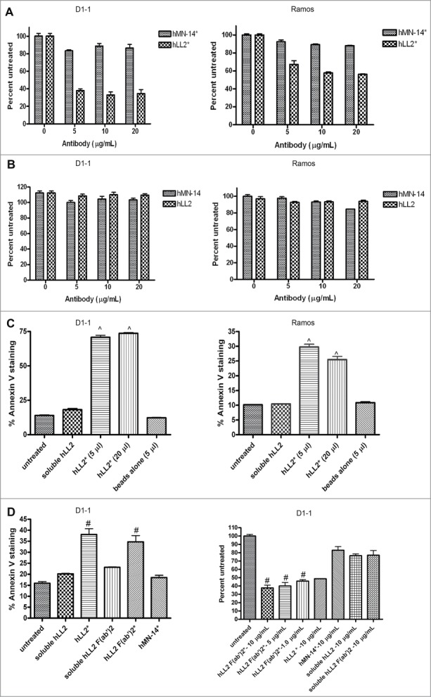
Figure 1.

Evaluation of growth-inhibition and apoptosis in D1–1 and Ramos cells. Cell viability determined by the MTS assay after 4-day incubation for (A) the Dried-I format of epratuzumab (hLL2*) or labetuzumab (hMN-14*) and (B) the Wet-I format of epratuzumab (hLL2) or labetuzumab (hMN-14). Apoptosis as determine by Annexin V staining (C) following the indicated treatments of D1–1 and Ramos cells for 24 and 48 h, respectively. (D) Plate-immobilized F(ab’)2 of epratuzumab (hLL2 F(ab’)2*) effectively induced apoptosis (left panel) and inhibited proliferation (right panel) in D1–1 cells as determined by the annexin V assay at 24 h and the MTS assay after 4 days, respectively. Error bars represent standard deviation (SD), where n = 3. Significant differences compared to untreated or nonspecific antibody are indicated with ^ (P < 0.005) and # (P < 0.05).
