Abstract
Monoclonal B lymphocytosis (MBL) is defined as the presence of a clonal B-cell population in the peripheral blood with fewer than 5 × 10⁹/L B-cells and no other signs of a lymphoproliferative disorder. The majority of cases of MBL have the immunophenotype of chronic lymphocytic leukemia (CLL). MBL can be categorized as either low count or high count based on whether the B-cell count is above or below 0.5 × 10⁹/L. Low-count MBL can be detected in ∼5% of adults over the age of 40 years when assessed using standard-sensitivity flow cytometry assays. A number of biological and genetic characteristics distinguish low-count from high-count MBL. Whereas low-count MBL rarely progresses to CLL, high-count MBL progresses to CLL requiring therapy at a rate of 1% to 2% per year. High-count MBL is distinguished from Rai 0 CLL based on whether the B-cell count is above or below 5 × 10⁹/L. Although individuals with both high-count MBL and CLL Rai stage 0 are at increased risk of infections and second cancers, the risk of progression requiring treatment and the potential to shorten life expectancy are greater for CLL. This review highlights challenging questions regarding the classification, risk stratification, management, and supportive care of patients with MBL and CLL.
Introduction
Chronic lymphocytic leukemia (CLL) is a clonal lymphoproliferative disorder characterized by >5 × 10⁹/L peripheral B-lymphocytes coexpressing CD5, CD19, and CD23 and a weak expression of CD20, CD79b, and surface immunoglobulin (sIg).1 When such a population is detected in enlarged lymph nodes of patients without peripheral lymphocytes, the term small lymphocytic lymphoma (SLL) is used, indicating a clinical variant of the same histopathological and molecular entity.2
The possibility of a precursor state to CLL was first identified in the early 1990s when a series of cross-sectional population-based studies was conducted in the United States to determine the health risks of living near hazardous waste sites.3,4 Using a 2-color panel (CD19 and CD5), 11 out of 1926 (0.6%) individuals older than 40 years were found to have a clonal population of CD5+CD19+ B cells, an immunophenotype classically associated with CLL. However, none of them met the diagnostic criteria for CLL or SLL. In particular, none had an absolute lymphocyte count (ALC) >5000/µL, as originally required by the diagnostic criteria for CLL.5 This phenomenon, later categorized monoclonal B-cell lymphocytosis (MBL), opened a new chapter in the field of B-cell lymphoproliferative disorders, suggesting that a precursor state of these lymphoid malignancies may occur at high prevalence in the general population.
Evaluation of lymphocytosis
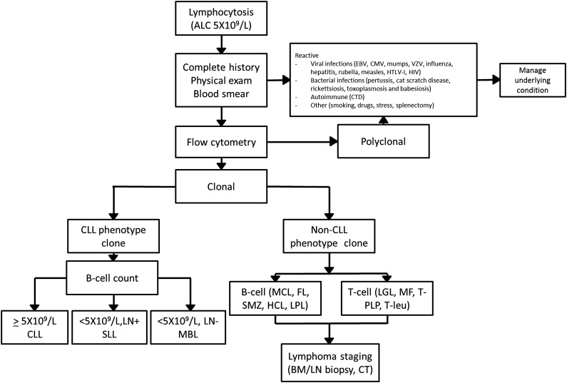
Lymphocytosis is a laboratory finding frequently encountered by the general internist and/or hematologist. An ALC ≥5 × 10⁹/L has been suggested as the threshold in need of further investigation to identify infectious, autoimmune, or neoplastic etiology.6,7 A general approach to the workup of lymphocytosis is suggested in Figure 1.
Figure 1.
General approach to the workup of lymphocytosis. BM, bone marrow; CMV, cytomegalovirus; CTD, connective tissue disease; EBV, Epstein-Barr virus; FL, follicular lymphoma; HCL, hairy cell leukemia; HTLV, human T-lymphotropic virus; LGL, large-granular leukemia; LN, lymph nodes; LPL, lymphoplasmacytic lymphoma; MCL, mantle cell lymphoma; MF, mycosis fungoides; T-PLP, T prolymphocytic leukemia; SMZ, splenic marginal zone lymphoma; T-leu, T-cell leukemia; VZV, varicella zoster virus.
A complete history and physical examination should represent the first step of such an evaluation, aimed at identifying causes of reactive (polyclonal) lymphocytosis. The most common cause of reactive lymphocytosis is viral infections, including hepatitis infection and HIV infection. Autoimmune conditions (particularly connective tissue diseases), smoking, hypersensitivity reactions, acute stress, and splenectomy can also induce polyclonal lymphocytosis.8,9
If the clinical and laboratory evaluation point toward a neoplastic origin, clonality should be evaluated through flow cytometry. A variety of clonal B-cell disorders can be identified based on surface protein markers with such analysis (Table 1). The management of clonal disorders of CLL phenotype is the focus of the remainder of this review. The detection of clonal B cells with a non-CLL phenotype (non-CLL MBL) or T-cell monoclonal lymphocytosis should warrant further testing, including computed tomography (CT) imaging, bone marrow biopsy, and molecular and genetic studies according to the suspected lymphoproliferative disorder.10,11
Table 1.
Immunophenotype of common clonal B-cell disorders
| CD5 | CD19 | CD20 | CD23 | CD10 | CD103 | Dual CD11c/22 | sIg | CD200 | Genetic defects | |
|---|---|---|---|---|---|---|---|---|---|---|
| CLL | + | + | Dim | + | − | − | − | Dim | + | — |
| MCL* | + | + | Bright | Dim/− | − | − | − | Bright | − | t(11;14) |
| FL | − | + | + | +/− | +/− | − | − | + | − | t(14;18) |
| MZL† | − | + | Bright | − | − | − | + | + | − | 7q− |
| HCL | − | Bright | Bright | − | − | + | Bright | + | + | — |
| LPL‡ | +/− | + | + | +/− | − | − | − | Dim | + | — |
It is important to look at the flow cytometry histograms to determine the intensity of expression and whether the staining is “all, none, or partial.” The immunophenotype profile of classic CLL is dim sIg and dim CD20; CD5 and CD23 expression (not partial expression for either) is critical. Some degree of immunophenotype overlap among CLL, marginal zone lymphoma, and lymphoplasmacytic lymphoma exists. If the diagnosis is uncertain based on peripheral blood flow cytometry, lymph node biopsy should be pursued.
CD23 is usually negative, but ∼20% of cases will have partial CD23 expression.
CD5 is positive in 10% to 20% of cases, and it may be bright or partial in these situations.
20% of cases are CD5+; CD23 is usually negative, but some cases will have partial CD23 expression.
FL, follicular lymphoma; HCL, hairy cell leukemia; LPL, lymphoplasmacytic lymphoma; MZL, marginal zone lymphoma; sIg, surface immunoglobulin
Classifying patients with clonal cells of CLL phenotype
Definition and prevalence of MBL
In 2005, the International Familial CLL Consortium proposed the term “monoclonal B lymphocytosis” to define the presence of CLL-phenotype cells in the peripheral blood in the absence of other features of CLL or SLL. The initially proposed diagnostic criteria for CLL phenotype MBL are as follows12:
Documentation of a clonal B-cell population in peripheral blood (light-chain restriction [abnormal κ/λ ratio or low sIg in >25% B cells] or heavy-chain monoclonal IGHV rearrangement).
B-cell count <5 × 10⁹/L.
Presence of CLL phenotype (CD5, CD19, CD23 positive; CD20 and sIg dim [reduced]).
No evidence of lymphoma, infection, or autoimmune conditions.
This entity was later acknowledged by the International Working Group of CLL, which in 2008 revised the 1996 National Cancer Institute–sponsored Working Group diagnostic criteria for CLL and SLL to include MBL. These revisions also redefined the threshold to diagnose CLL based on the absolute B-lymphocyte count rather than the ALC.1 The prevalence of MBL observed in the initial reports was revised after subsequent studies using more sensitive flow cytometry evaluation strategies. Two population studies were conducted in Europe in 2002 and 2004, analyzing either individuals referred to the hospital for nonhematologic conditions or in primary health facilities for routine evaluation. Using a 4-color flow cytometry approach and acquiring 200 000 events, both studies reported a prevalence of MBL of ∼3.5% in individuals older than 40 years.13,14 A subsequent population study from Italy, employing a 5-color panel and 500 000 acquisitions, found a prevalence of 6.7% among healthy individuals older than 40 years.15 A more sensitive technique was finally used, with an 8-color panel and 5 000 000 acquisitions, and reported a prevalence of 12% in healthy subjects of the same age.16 These studies indicated that large increases in assay sensitivity resulted in only small increases in the prevalence of MBL, indicating that MBL is not a universal phenomenon and only impacts a subset of the population.17,18
Risk factors for developing MBL
The findings above imply that MBL is not a physiological event that occurs in all individuals with increasing age but a specific condition affecting select patients in whom at least some predisposing risk factors have been identified (Table 2). A genetic predisposition for MBL is suggested by family studies. CLL has one of the strongest inherited predispositions among lymphoid malignancies. Familial CLL is defined as a pedigree with at least 2 first-degree relatives with CLL and characterizes about one-tenth of patients with CLL. The prevalence of MBL among unaffected relatives in such familial pedigrees is two- to threefold higher (∼15% among individuals older than 40 years) than that of the general population.19-21 Relatives of patients with sporadic CLL also have an increased prevalence of MBL, with a prevalence of 15.6% among relatives older than 60 years.22 Single-nucleotide polymorphisms in over 20 normal genes have now been found to be associated with familial CLL.23,24 Of interest, early studies of MBL demonstrated that at least 6 of these single-nucleotide polymorphisms known to confer an increased risk of developing CLL also relate to the risk of developing MBL in a large case-control series, supporting the concept of a genetic basis for MBL.25
Table 2.
Risk factors for MBL onset and progression to CLL requiring therapy
| Risk factors for MBL onset | Risk factors for MBL progression to CLL |
|---|---|
| Family history of CLL | CD38 positivity |
| Genetic polymorphisms* | Unmutated IGHV |
| Age | Deletion 17p |
| Infections† | Elevated B-cell count |
More than 20 single-nucleotide polymorphisms associated with the development of CLL have been reported.110 At least 6 of these have also been confirmed as risk factors for MBL (rs17483466, rs13397985, rs757978, rs872071, rs2456449, and rs735665),25 whereas the association of the others with MBL remains under investigation.
As outlined above, the frequency of MBL in the general population progressively increases with age. Although present in only 0.2% to 0.3% of individuals younger than 40 years, its frequency is 3.5% to 6.7% in those aged 40 to 60 years and 5% to 9% among individuals older than 60 years when assessed using standard-sensitivity assays.14,15 When assessed using the most sensitive assay techniques, this prevalence can be >20% in healthy individuals older than 60 years and as high as 75% in subjects older than 90 years.16 Several studies point to a link between MBL and infection. One recent study identified MBL in ∼30% of hepatitis C virus–infected patients.26 Population-based studies have also reported an increased risk of CLL among patients with pneumonia.26 Conversely, a lower incidence of MBL has been reported among patients vaccinated for influenza or pneumonia.27,28 Studies aimed to determine whether specific antigenic stimuli can lead to the development of MBL are ongoing and may shed light on its pathogenesis and natural history.
Evidence supporting current classification and remaining classification challenges
Although on a theoretic level, classifying patients based on the presence of peripheral blood ALC and/or enlarged lymph nodes may seem simple (eg, B-cell count <5 × 10⁹/L and no nodes: MBL; B-cell count <5 × 10⁹/L with enlarged nodes: SLL; B-cell count >5 × 10⁹/L: CLL), the clinical circumstance is often far more nuanced, as will be discussed below.
Low- vs high-count MBL
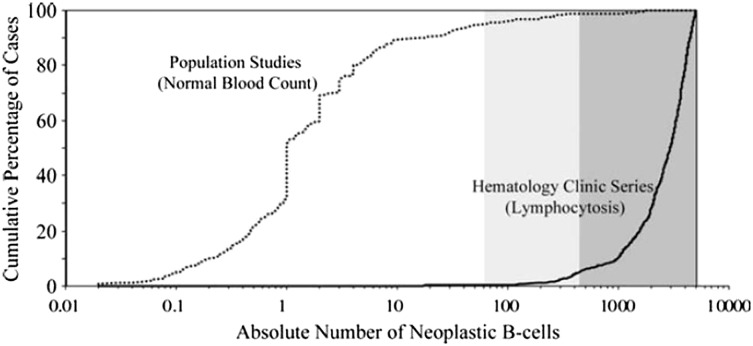
In 2010, a cumulative analysis of MBL series worldwide was performed and revealed the distribution of MBL cases followed a bimodal B-cell count distribution (Figure 2).29 Notable differences in the size of the clonal B-cell population between cases of MBL identified in population screening studies and clinical series were observed. Among cases identified in population screening studies, the overwhelming majority of patients had clonal B-cell counts between 0.1 and 10 clonal B cells/µL (median, 1/µL). In contrast, among cases identified in clinical cohorts (typically undergoing evaluation of low-level lymphocytosis), the clonal B-cell count was between 0.5 and 5 × 10⁹/L (median, 2.9 × 10⁹/L). Strikingly, little overlap was observed between the population and clinical cohorts, with very few cases lying in the middle. These results led to the descriptive segregation of MBL into 2 groups (high-count MBL and low-count MBL) based on the size of the B-cell clone (<0.5 × 10⁹/L clonal B cells: low count MBL; ≥0.5 × 10⁹/L clonal B cells: high count MBL).29-31
Figure 2.
Distribution of clonal CLL-like B-cell count in published studies. Two main entities can be identified: low-count MBL, usually detected in general population studies, and high-count MBL, usually detected in series of patients referred for lymphocytosis. Reprinted with permission from Rawstron et al.29
Biological characteristics of CLL, such as immunoglobulin repertoire, cytogenetic abnormalities, and, more recently, molecular mutations, were subsequently investigated in both low-count and high-count MBL, shedding light on their differential biology. Specific immunoglobulin gene rearrangements, frequently observed in patients with CLL, such as IGHV 4-34, 3-23, and 1-69, are underrepresented or absent in subjects with low-count MBL.32 By contrast, their frequency in high-count MBL is comparable to that observed in CLL.15 The frequency of IGHV-mutated cases is also significantly higher in individuals with low-count MBL than in subjects with high-count MBL or CLL.33 Moreover, whereas at least 25% of patients with CLL and 22% of subjects with high-count MBL demonstrate stereotyped complementarity determining region 3 sequences, these are present in <5% of individuals with low-count MBL.15,34 Cases of low-count MBL are also enriched with genetic abnormalities typically associated with more favorable prognosis in CLL, such as deletion 13q.35 By contrast, the distribution of genetic abnormalities as identified by fluorescence in situ hybridization (FISH) analysis in high-count MBL is comparable to that observed in CLL.16,36-38 High-throughput sequencing and new biological markers, such as noncoding RNA, may help to further characterize the biological differences of these 2 conditions.39,40 Novel mutations, such as NOTCH1 and SF3B1, described in 10% and 15% of patients with CLL, respectively, appear to be extremely rare in MBL, including high-count MBL.41,42 Additional gene mutations in patients with MBL continue to be discovered.43 Collectively, these findings suggest a potential stepwise evolution from high-count MBL to CLL through a progressive acquisition of high-risk genetic abnormalities but indicate that relatively few individuals with low-count MBL transition to high-count MBL.
High-count MBL vs CLL Rai stage 0
The threshold selected to distinguish high-count MBL from CLL (5 × 10⁹ B cells/L) was arbitrary. Indeed, changing from an ALC-based criteria to a B-cell count–based criteria reclassified many patients who would have previously been considered Rai stage 0 CLL into the high-count MBL category.44,45 Initial analyses suggested, however, that the reclassification had clinical relevance, with different likelihoods of progression to require therapy for those in the high-count MBL and Rai 0 CLL categories.30,31,36 Nonetheless, as outlined above, many of the biological characteristics of high-count MBL are similar to Rai stage 0 CLL.46 These facts raised the question of whether high-count MBL should or should not remain an entity separate from Rai 0 CLL and, if a separate entity, what threshold should be used to segregate the 2 conditions. Because the designation of CLL and MBL are clinical diagnoses, a number of groups advocated that the distinction should be based on the clinical implications for patients, such as having an impact on survival. To answer this question, investigators from the Mayo Clinic evaluated what B-cell threshold best predicted overall survival among patients whose only manifestation of disease was a circulating clone of CLL phenotype (eg, no lymphadenopathy, organomegaly, or cytopenias). This analysis revealed that a B-cell threshold of between 10 and 11 × 109/L B cells was the best predictor of overall survival and predicted survival independent of other traditional molecular biomarkers.37 Three subsequent independent studies confirmed that a B-cell threshold of ∼10 × 109/L was the best predictor of survival.37,38,47,48 Notably, 10 × 109/L was also the B-cell threshold associated with changes in T-cell function, suggesting a potential biological underpinning for the association of this threshold with clinical outcomes.49 As can be easily intuited, higher B-cell counts are also associated with a shorter time to progression and a shorter time to first treatment.29,30
Nodal MBL
Accurate clinical assessment of lymphadenopathy is essential when evaluating individuals with MBL, because the presence of lymphadenopathy would suggest the diagnosis of SLL rather than MBL. Moreover, development of lymphadenopathy appears to be a common pattern of progression among individuals with MBL.29-31 This has raised the question of whether all individuals with MBL should be staged with a CT scan at time of diagnosis to rule out lymphadenopathy in nonpalpable lymph node regions. In one of the few studies evaluating this issue to date, 29 out of 62 individuals with clinically identified MBL (42%) had lymphadenopathy identified on CT imaging.50 Notably, however, after a median follow-up of 35 months, the rate of progression among cases of MBL with lymphadenopathy identified on CT imaging was only 6.9% (2/29 patients), which was no different than in cases of MBL without lymphadenopathy on CT imaging (rate of progression, 9%; 3/33 patients).50 These findings support the recommendation not to perform CT imaging in patients with MBL.51
It should also be noted that lymph node biopsy specimens used for the staging of other malignancies are often incidentally found to have a diffuse or focal infiltration of CLL-like cells in normal-sized or slightly enlarged lymph nodes.52 For example, up to 1.6% of patients undergoing sentinel lymph node biopsy for breast cancer show concomitant lymphoma, mostly with a CLL phenotype.53 How best to classify such patients (eg, SLL or MBL) when the circulating lymphocyte count is normal is unknown. Limited evidence suggests that patients in this circumstance with any lymph nodes ≥1.5 cm are at greater risk of progression/treatment.52 Based on this information, our current approach is to classify individuals with the incidental discovery of a CLL phenotype infiltrate in a normal-sized lymph node (eg, <1.5 cm), normal blood counts, and no other nodes >1.5 cm in size as nodal MBL and those with any enlarged lymph nodes (≥1.5 cm) as SLL. More studies evaluating how best to classify such patients are needed.
Other challenging situations and unanswered questions
Bone marrow involvement.
It is not uncommon to find a bone marrow infiltrate of CLL B cells in patients staged for other hematologic malignancies in the absence of other diagnostic criteria for either CLL or SLL. Of interest, most individuals with MBL who have a bone marrow biopsy at time of diagnosis have a concomitant bone marrow infiltration, with a median of 20% of marrow cellularity composed of CLL B cells.30,54 Although higher percentages of CLL infiltrate have been described,48,55 the percentage involvement is not clearly linked to the likelihood of clinical progression. Accordingly, it should be kept in mind that the diagnostic criteria for MBL do not include bone marrow features12 and, in the absence of other criteria, percent marrow infiltration does not change the diagnosis from MBL to CLL or SLL.1,56
Tissue involvement.
Clonal CLL-like cells can be detected in up to 0.4% of prostatic tissues at the time of prostatectomy57,58 and in 1.9% of liver biopsy specimens59 in the absence of meeting any other diagnostic criteria for CLL and SLL. Progression to first therapy has been only rarely reported in these cases, suggesting the concept of “tissue MBL.”56,60,61 Whether the coexistence of such a clone influences the outcome of the primary malignancy, as has been observed in CLL,62 is currently unknown. The extent of infiltration in such cases is also likely important, where extensive tissue replacement or infiltration is more consistent with a diagnosis of SLL.
Autoimmune disease.
According to the original guidelines, the presence of a concomitant autoimmune process excludes a diagnosis of MBL and was considered CLL.12 It has also long been recognized, however, that very small CLL-like clones can be found in individuals with immune thrombocytopenia and that many such patients may never develop any clinical manifestations of a lymphoproliferative disorder.63 In other patients with this profile, immune thrombocytopenia can occur many years before patients meet the criteria for CLL.63 How to best categorize patients with autoimmune conditions and very small clonal B-cell populations in blood or bone marrow remains challenging. Clinically, our approach is to consider individuals with autoimmune cytopenia who have very small B-cell clones (eg, normal lymphocyte counts and no lymphadenopathy) as “MBL with autoimmune cytopenia” rather than classifying them as CLL. Guidelines may need to be revised as new clinical/biological evidence becomes available.
Stem cell and blood donor issues.
Allogeneic stem cell transplantation (SCT) is an appropriate therapeutic option for selected high-risk patients with CLL,64,65 with matched related donors being the preferred source of stem cells. As outlined previously, MBL is more frequent in first-degree relatives of patients with both familial and sporadic CLL, raising the question of whether screening for MBL should be pursued in potential related stem cell donors. Indeed cases of MBL transfer from the donor to recipient during SCT have been described from both related and unrelated donors.66-68
Similar questions related to blood donation have also arisen. In 2007, studies using low-sensitivity flow cytometry indicated a prevalence of MBL of 0.14% in blood-bank donors.69 More recent studies using higher-sensitivity assays indicate a prevalence of MBL in blood donors of 7.1%.70 These findings are notable, given historical epidemiologic studies suggesting that blood transfusion is associated with an increased risk of developing CLL.71,72 Subsequent studies, however, refuted this finding, and the association in some historic studies may have been related to transfusion-associated hepatitis C.73 At present, there is no consensus as to whether routine screening for MBL among SCT donors should be pursued. Our approach is to screen all related donors with flow cytometry, independent of whether or not lymphocytosis is present, to identify cases of occult MBL.
Clinical implications and management of MBL
Clinical implications of low-count MBL
In 2009, using prediagnostic samples from the Prostate, Lung, Colon and Ovary Cancer Screening Trial study, investigators demonstrated that MBL could be observed in the peripheral blood of virtually all patients later diagnosed with CLL.74 This demonstrated for the first time that MBL is a precursor to CLL and that at least some cases progress. No information on the size of the B-cell clone was available in this study. However, based on the percent of lymphocytes that were clonal B cells, the vast majority of cases likely represented high-count MBL.
A few studies have prospectively examined the natural history of low-count MBL.75,76 The most robust data to date come from a prospective study of 1779 healthy adults from the Val Borbera valley in Northern Italy, of whom 138 had low-count MBL on screening (96 CLL-phenotype MBL, 21 atypical CLL-phenotype MBL, 20 CD5-negative–phenotype MBL). After a median follow-up of 34 months, a small clonal B-cell population persisted in 90% of those with CLL-phenotype MBL, but no patients progressed to CLL, SLL, or other lymphoid malignancy.65 Life expectancy for subjects with low-count MBL was also no different than in the general population.76 This evidence supports the current consensus that low-count MBL is at low risk of progression, has no clear clinical implications, and requires no specific clinical follow-up.51
Clinical implications of high-count MBL
A number of studies evaluating the natural history of high-count MBL indicate that the risk of progression to CLL or SLL requiring treatment is between 1% and 2% per year.30,36-38,51,77-79 As a consequence, an annual complete blood count and periodic lymph node examination are advised for individuals with high-count MBL.51
As a group, the overall survival of individuals with high-count MBL does not differ significantly from age- and sex-matched controls in the general population.38,80,81 A number of preliminary studies have suggested, however, that the prognostic parameters used in CLL (eg, IGHV mutational status, FISH, and CD38) have utility for stratifying outcome among individuals with high-count MBL30,31,78 and may identify a subset in whom the presence of MBL impacts overall survival.80
In contrast to individuals with low-count MBL,76 subjects with high-count MBL appear to have a significantly higher risk of hospitalization due to serious infections82 as well as a higher risk of hematologic83,84 and nonhematologic cancers.62,85 Given the prevalence of MBL, these findings could have public health implications, and more research in this area is needed.
Finally, although studies indicate that the diagnosis of CLL can precipitate substantial anxiety and adversely impact quality of life, data regarding the impact of an MBL diagnosis on quality of life are lacking.86
Clinical implications and management of early-stage CLL and SLL
Prognosis
Patients with CLL have a substantial shorter life expectancy compared with age- and sex-matched populations.87 A number of basic demographic and clinical characteristics, such as age, sex, and performance status, along with Rai and Binet staging systems, have been the foundation of risk stratification for the last 40 years.88,89 Although useful, most patients with CLL are now diagnosed with early-stage disease, where these parameters do not predict outcome for individual patients. Over the last 2 decades, a number of molecular biomarkers have been found to predict shorter survival. Inferior outcomes have been reported for patients with unmutated IGHV,90 positive CD38, ZAP70, or CD49,91-93 elevated serum β-2 microglobulin levels,94 and unfavorable genetic abnormalities such as deletion 11q and 17p on FISH testing95 or TP53 mutations on sequencing. The recent studies using next-generation sequencing have also identified a number of recurrent mutations such as NOTCH1, SF3B1, DDX3X, and ATM associated with clinical outcome.96
Several recent efforts have attempted to integrate the results of multiple prognostic markers into a single risk score.97,98 Although useful at the population level, most of these tools are not sufficiently robust to predict the outcome of individual CLL patients.
A comprehensive prognostic model was recently developed by the German CLL Study group. After analysis of 23 different prognostic markers in a cohort of ∼2000 patients participating in prospective clinical trials, the investigators identified 8 factors with independent prognostic value: age, sex, performance status, IGHV mutational status, FISH (deletion 17p13, deletion 11q23), serum β-2 microglobulin, and serum thymidine kinase. Each item was assigned a weighted score based on its hazard ratio for survival in the multivariate analysis. These values were then summed into a single risk score, which stratified patients into 4 risk groups with median 5-year survivals ranging from 95% to 18%.99 The prognostic index was then validated in an independent cohort of 700 newly diagnosed patients and was the first prognostic tool with sufficient accuracy to allow prediction of outcome in individual patients. Although a significant step forward, the cohorts in which this index was developed and validated are younger than the majority of real-world CLL patients. The index also did not account for the impact of comorbid health conditions, and incorporation of this information into future prognostic tools may further improve accuracy.
Building from the results of these prior efforts, a collaborative, international effort to develop an International Prognostic Index for patients with CLL is currently ongoing.
Indications for treatment
Phase 3 trials evaluating the benefit of early administration of chlorambucil compared with observation for asymptomatic early-stage CLL indicated a lack of clinical benefit for early treatment and established observation as the standard of care for early stage patients.100 According to the 2008 guidelines, the acceptable indications for treatment in patients with CLL are anemia (hemoglobin <11 g/dL) or thrombocytopenia (platelet count <100 × 109/L), progressive or symptomatic lymphadenopathy or organomegaly, and severe constitutional symptoms.1 Several recent studies have assessed the role of early treatment of select high-risk patients. Recent phase 3 trials comparing fludarabine to observation101 or fludarabine, cyclophosphamide, and rituximab to observation102 in early-stage patients at high risk of progression have again found no advantage of early treatment. These studies have further solidified active monitoring as the standard of care for asymptomatic early-stage patients outside of clinical trials regardless of the findings of prognostic testing. Whether the recent introduction of highly active, targeted, oral small-molecule inhibitors with a generally favorable toxicity profile (eg, ibrutinib and idelalisib) will change this paradigm is unknown. The German CLL Study Group is currently conducting a phase 3 study comparing ibrutinib with observation in patients with early-stage CLL at high-risk for progression. This study will provide important insights regarding the value of such an approach.
Supportive care
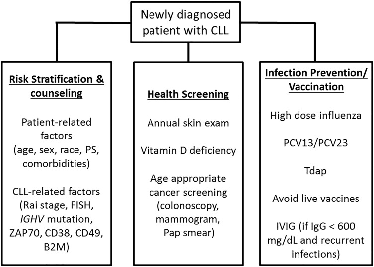
Patients with early-stage disease are at risk of a number of CLL-related complications, including nonhematologic cancers and infection. Second cancers, particularly skin cancers, are more frequent among CLL patients than in the general population.62,85 In addition to increased incidence, skin cancers may behave more aggressively in patients with CLL.103 Patients with CLL should be counseled regarding use of sun block and protective clothing and should undergo an annual skin examination. Sun avoidance can increase the risk of vitamin D deficiency, a condition that already affects 40% to 50% of the general US population. Given this fact and the association between vitamin D deficiency and CLL progression in several studies,104,105 it is reasonable to periodically assess vitamin D levels in patients with CLL. Given their apparent increased risk of other nonhematologic cancers, patients with CLL and high-count MBL should also adhere to age-appropriate cancer screening, such as colonoscopy, mammograms, and cervical cancer screening. Given their increased risk of infection, it is recommended that patients with CLL be kept up to date with influenza, pneumococcal,106 and tetanus, diphtheria, acellular pertussis vaccines despite the fact they are less likely to mount an immune response to vaccines (Figure 3). Live attenuated vaccines should be avoided.107
Figure 3.
Management of early-stage CLL and SLL. B2M, β-2-microglobulin; IgG, immunoglobulin G; IVIG, intravenous immunoglobulins; MX, mammogram; PCV, pneumococcus vaccine; PS, performance status; Tdap, tetanus diphtheria, acellular pertussis.
Finally, it must be noted that a family history of CLL is a strong risk factor for the development of CLL.108 In particular, first-degree relatives have a risk of developing CLL that is 2 to 7 times greater than the general population.109 This obviously represents a stressor among relatives of CLL patients, particularly in reference to children. Although guidelines about surveillance and counseling do not exist, periodic complete blood count and physical examination may be advised. Prospective observational studies may provide further hints in the future.
Acknowledgments
T.S. is a Clinical Scholar of the Leukemia and Lymphoma Society.
Authorship
Contribution: P.S. and T.D.S. conceived the study and wrote the paper.
Conflict-of-interest disclosure: T.D.S. has received research support from Genentech, Pharmacyclics/Janssen, Celgene, GlaxoSmithKline, Cephalon, Hospira, and Polyphenon E International. P.S. declares no competing financial interests.
Correspondence: Tait D. Shanafelt, Mayo Clinic, 200 First St SW, Rochester, MN 55905; e-mail: shanafelt.tait@mayo.edu.
References
- 1.Hallek M, Cheson BD, Catovsky D, et al. International Workshop on Chronic Lymphocytic Leukemia. Guidelines for the diagnosis and treatment of chronic lymphocytic leukemia: a report from the International Workshop on Chronic Lymphocytic Leukemia updating the National Cancer Institute-Working Group 1996 guidelines. Blood. 2008;111(12):5446–5456. doi: 10.1182/blood-2007-06-093906. [DOI] [PMC free article] [PubMed] [Google Scholar]
- 2.Santos FP, O’Brien S. Small lymphocytic lymphoma and chronic lymphocytic leukemia: are they the same disease? Cancer J. 2012;18(5):396–403. doi: 10.1097/PPO.0b013e31826cda2d. [DOI] [PubMed] [Google Scholar]
- 3.Vogt RF, Shim YK, Middleton DC, et al. Monoclonal B-cell lymphocytosis as a biomarker in environmental health studies. Br J Haematol. 2007;139(5):690–700. doi: 10.1111/j.1365-2141.2007.06861.x. [DOI] [PubMed] [Google Scholar]
- 4.Shim YK, Vogt RF, Middleton D, et al. Prevalence and natural history of monoclonal and polyclonal B-cell lymphocytosis in a residential adult population. Cytometry B Clin Cytom. 2007;72(5):344–353. doi: 10.1002/cyto.b.20174. [DOI] [PubMed] [Google Scholar]
- 5.Cheson BD, Bennett JM, Grever M, et al. National Cancer Institute-sponsored Working Group guidelines for chronic lymphocytic leukemia: revised guidelines for diagnosis and treatment. Blood. 1996;87(12):4990-4997. [PubMed]
- 6.Carney D. Peripheral blood lymphocytosis—what is the threshold for further investigation? Leuk Lymphoma. 2008;49(9):1659–1661. doi: 10.1080/10428190802389843. [DOI] [PubMed] [Google Scholar]
- 7.Andrews JM, Cruser DL, Myers JB, Fernelius CA, Holm MT, Waldner DL. Using peripheral smear review, age and absolute lymphocyte count as predictors of abnormal peripheral blood lymphocytoses diagnosed by flow cytometry. Leuk Lymphoma. 2008;49(9):1731–1737. doi: 10.1080/10428190802251787. [DOI] [PubMed] [Google Scholar]
- 8.Teggatz JR, Parkin J, Peterson L. Transient atypical lymphocytosis in patients with emergency medical conditions. Arch Pathol Lab Med. 1987;111(8):712-714. [PubMed]
- 9.Lesesve JF, Troussard X. Persistent polyclonal B-cell lymphocytosis. Blood. 2011;118(25):6485. [DOI] [PubMed]
- 10.Wilcox RA. Cutaneous T-cell lymphoma: 2014 update on diagnosis, risk-stratification, and management. Am J Hematol. 2014;89(8):837–851. doi: 10.1002/ajh.23756. [DOI] [PubMed] [Google Scholar]
- 11.Karube K, Scarfò L, Campo E, Ghia P. Monoclonal B cell lymphocytosis and “in situ” lymphoma. Semin Cancer Biol. 2014;24:3–14. doi: 10.1016/j.semcancer.2013.08.003. [DOI] [PubMed] [Google Scholar]
- 12.Marti GE, Rawstron AC, Ghia P, et al. International Familial CLL Consortium. Diagnostic criteria for monoclonal B-cell lymphocytosis. Br J Haematol. 2005;130(3):325–332. doi: 10.1111/j.1365-2141.2005.05550.x. [DOI] [PubMed] [Google Scholar]
- 13.Ghia P, Prato G, Scielzo C, et al. Monoclonal CD5+ and CD5- B-lymphocyte expansions are frequent in the peripheral blood of the elderly. Blood. 2004;103(6):2337–2342. doi: 10.1182/blood-2003-09-3277. [DOI] [PubMed] [Google Scholar]
- 14.Rawstron AC, Green MJ, Kuzmicki A, et al. Monoclonal B lymphocytes with the characteristics of “indolent” chronic lymphocytic leukemia are present in 3.5% of adults with normal blood counts. Blood. 2002;100(2):635-639. [DOI] [PubMed]
- 15.Dagklis A, Fazi C, Sala C, et al. The immunoglobulin gene repertoire of low-count chronic lymphocytic leukemia (CLL)-like monoclonal B lymphocytosis is different from CLL: diagnostic implications for clinical monitoring. Blood. 2009;114(1):26–32. doi: 10.1182/blood-2008-09-176933. [DOI] [PubMed] [Google Scholar]
- 16.Nieto WG, Almeida J, Romero A, et al. Primary Health Care Group of Salamanca for the Study of MBL. Increased frequency (12%) of circulating chronic lymphocytic leukemia-like B-cell clones in healthy subjects using a highly sensitive multicolor flow cytometry approach. Blood. 2009;114(1):33–37. doi: 10.1182/blood-2009-01-197368. [DOI] [PubMed] [Google Scholar]
- 17.Scarfò L, Dagklis A, Scielzo C, Fazi C, Ghia P. CLL-like monoclonal B-cell lymphocytosis: are we all bound to have it? Semin Cancer Biol. 2010;20(6):384–390. doi: 10.1016/j.semcancer.2010.08.005. [DOI] [PubMed] [Google Scholar]
- 18.Almeida J, Nieto WG, Teodosio C, et al. Primary Health Care Group of Salamanca for the Study of MBL. CLL-like B-lymphocytes are systematically present at very low numbers in peripheral blood of healthy adults. Leukemia. 2011;25(4):718–722. doi: 10.1038/leu.2010.305. [DOI] [PubMed] [Google Scholar]
- 19.Goldin LR, Lanasa MC, Slager SL, et al. Common occurrence of monoclonal B-cell lymphocytosis among members of high-risk CLL families. Br J Haematol. 2010;151(2):152–158. doi: 10.1111/j.1365-2141.2010.08339.x. [DOI] [PMC free article] [PubMed] [Google Scholar]
- 20.Marti GE, Carter P, Abbasi F, et al. B-cell monoclonal lymphocytosis and B-cell abnormalities in the setting of familial B-cell chronic lymphocytic leukemia. Cytometry B Clin Cytom. 2003;52(1):1–12. doi: 10.1002/cyto.b.10013. [DOI] [PubMed] [Google Scholar]
- 21.Rawstron AC, Yuille MR, Fuller J, et al. Inherited predisposition to CLL is detectable as subclinical monoclonal B-lymphocyte expansion. Blood. 2002;100(7):2289–2290. doi: 10.1182/blood-2002-03-0892. [DOI] [PubMed] [Google Scholar]
- 22.Matos DM, Ismael SJ, Scrideli CA, de Oliveira FM, Rego EM, Falcão RP. Monoclonal B-cell lymphocytosis in first-degree relatives of patients with sporadic (non-familial) chronic lymphocytic leukaemia. Br J Haematol. 2009;147(3):339–346. doi: 10.1111/j.1365-2141.2009.07861.x. [DOI] [PubMed] [Google Scholar]
- 23.Sellick GS, Goldin LR, Wild RW, et al. A high-density SNP genome-wide linkage search of 206 families identifies susceptibility loci for chronic lymphocytic leukemia. Blood. 2007;110(9):3326–3333. doi: 10.1182/blood-2007-05-091561. [DOI] [PMC free article] [PubMed] [Google Scholar]
- 24.Slager SL, Rabe KG, Achenbach SJ, et al. Genome-wide association study identifies a novel susceptibility locus at 6p21.3 among familial CLL. Blood. 2011;117(6):1911–1916. doi: 10.1182/blood-2010-09-308205. [DOI] [PMC free article] [PubMed] [Google Scholar]
- 25.Crowther-Swanepoel D, Corre T, Lloyd A, et al. Inherited genetic susceptibility to monoclonal B-cell lymphocytosis. Blood. 2010;116(26):5957–5960. doi: 10.1182/blood-2010-07-294975. [DOI] [PubMed] [Google Scholar]
- 26.Anderson LA, Landgren O, Engels EA. Common community acquired infections and subsequent risk of chronic lymphocytic leukaemia. Br J Haematol. 2009;147(4):444–449. doi: 10.1111/j.1365-2141.2009.07849.x. [DOI] [PMC free article] [PubMed] [Google Scholar]
- 27.Fazi C, Dagklis A, Cottini F, et al. Monoclonal B cell lymphocytosis in hepatitis C virus infected individuals. Cytometry B Clin Cytom. 2010;78(Suppl 1):S61–S68. doi: 10.1002/cyto.b.20545. [DOI] [PubMed] [Google Scholar]
- 28.Casabonne D, Almeida J, Nieto WG, et al. Primary Health Care Group of Salamanca for the Study of MBL. Common infectious agents and monoclonal B-cell lymphocytosis: a cross-sectional epidemiological study among healthy adults. PLoS ONE. 2012;7(12):e52808. doi: 10.1371/journal.pone.0052808. [DOI] [PMC free article] [PubMed] [Google Scholar]
- 29.Rawstron AC, Shanafelt T, Lanasa MC, et al. Different biology and clinical outcome according to the absolute numbers of clonal B-cells in monoclonal B-cell lymphocytosis (MBL). Cytometry B Clin Cytom. 2010;78(suppl 1):S19–S23. doi: 10.1002/cyto.b.20533. [DOI] [PMC free article] [PubMed] [Google Scholar]
- 30.Rossi D, Sozzi E, Puma A, et al. The prognosis of clinical monoclonal B cell lymphocytosis differs from prognosis of Rai 0 chronic lymphocytic leukaemia and is recapitulated by biological risk factors. Br J Haematol. 2009;146(1):64–75. doi: 10.1111/j.1365-2141.2009.07711.x. [DOI] [PubMed] [Google Scholar]
- 31.Shanafelt TD, Kay NE, Rabe KG, et al. Brief report: natural history of individuals with clinically recognized monoclonal B-cell lymphocytosis compared with patients with Rai 0 chronic lymphocytic leukemia. J Clin Oncol. 2009;27(24):3959–3963. doi: 10.1200/JCO.2008.21.2704. [DOI] [PMC free article] [PubMed] [Google Scholar]
- 32.Dagklis A, Fazi C, Scarfo L, Apollonio B, Ghia P. Monoclonal B lymphocytosis in the general population. Leuk Lymphoma. 2009;50(3):490–492. doi: 10.1080/10428190902763475. [DOI] [PubMed] [Google Scholar]
- 33.Henriques A, Rodríguez-Caballero A, Nieto WG, et al. Combined patterns of IGHV repertoire and cytogenetic/molecular alterations in monoclonal B lymphocytosis versus chronic lymphocytic leukemia. PLoS ONE. 2013;8(7):e67751. doi: 10.1371/journal.pone.0067751. [DOI] [PMC free article] [PubMed] [Google Scholar]
- 34.Vardi A, Dagklis A, Scarfò L, et al. Immunogenetics shows that not all MBL are equal: the larger the clone, the more similar to CLL. Blood. 2013;121(22):4521–4528. doi: 10.1182/blood-2012-12-471698. [DOI] [PubMed] [Google Scholar]
- 35.Ghia P, Fazi C, Pecciarini L, et al. CLL-like MBL in the general population persist over time, without clinical progression, though carrying the same cytogenetic abnormalities of CLL. Blood. 2010;116(21):1012–1013. doi: 10.1182/blood-2011-05-357251. [DOI] [PubMed] [Google Scholar]
- 36.Rawstron AC, Bennett FL, O’Connor SJ, et al. Monoclonal B-cell lymphocytosis and chronic lymphocytic leukemia. N Engl J Med. 2008;359(6):575–583. doi: 10.1056/NEJMoa075290. [DOI] [PubMed] [Google Scholar]
- 37.Shanafelt TD, Kay NE, Jenkins G, et al. B-cell count and survival: differentiating chronic lymphocytic leukemia from monoclonal B-cell lymphocytosis based on clinical outcome. Blood. 2009;113(18):4188–4196. doi: 10.1182/blood-2008-09-176149. [DOI] [PMC free article] [PubMed] [Google Scholar]
- 38.Molica S, Mauro FR, Giannarelli D, et al. Differentiating chronic lymphocytic leukemia from monoclonal B-lymphocytosis according to clinical outcome: on behalf of the GIMEMA chronic lymphoproliferative diseases working group. Haematologica. 2011;96(2):277–283. doi: 10.3324/haematol.2010.030189. [DOI] [PMC free article] [PubMed] [Google Scholar]
- 39.Ferrajoli A, Shanafelt TD, Ivan C, et al. Prognostic value of miR-155 in individuals with monoclonal B-cell lymphocytosis and patients with B chronic lymphocytic leukemia. Blood. 2013;122(11):1891–1899. doi: 10.1182/blood-2013-01-478222. [DOI] [PMC free article] [PubMed] [Google Scholar]
- 40.Lionetti M, Fabris S, Cutrona G, et al. High-throughput sequencing for the identification of NOTCH1 mutations in early stage chronic lymphocytic leukaemia: biological and clinical implications. Br J Haematol. 2014;165(5):629–639. doi: 10.1111/bjh.12800. [DOI] [PubMed] [Google Scholar]
- 41.Rasi S, Monti S, Spina V, Foà R, Gaidano G, Rossi D. Analysis of NOTCH1 mutations in monoclonal B-cell lymphocytosis. Haematologica. 2012;97(1):153–154. doi: 10.3324/haematol.2011.053090. [DOI] [PMC free article] [PubMed] [Google Scholar]
- 42.Greco M, Capello D, Bruscaggin A, et al. Analysis of SF3B1 mutations in monoclonal B-cell lymphocytosis. Hematol Oncol. 2013;31(1):54–55. doi: 10.1002/hon.2013. [DOI] [PubMed] [Google Scholar]
- 43.Ojha J, Secreto C, Rabe K, et al. Monoclonal B-cell lymphocytosis is characterized by mutations in CLL putative driver genes and clonal heterogeneity many years before disease progression. Leukemia. 2014;28(12):2395–2398. doi: 10.1038/leu.2014.226. [DOI] [PMC free article] [PubMed] [Google Scholar]
- 44.Norman AD, Call TG, Hanson CA, et al. Incidence of chronic lymphocytic leukemia and monoclonal B-cell lymphocytosis in Olmsted county, 2000-2010: impact of the 2008 International Workshop on CLL Guidelines. Cancer Res. 2013;73(8) [Google Scholar]
- 45.Mulligan CS, Thomas ME, Mulligan SP. Lymphocytes, B lymphocytes, and clonal CLL cells: observations on the impact of the new diagnostic criteria in the 2008 Guidelines for Chronic Lymphocytic Leukemia (CLL). Blood. 2009;113(25):6496–6497, author reply 6497-6498. doi: 10.1182/blood-2008-07-166710. [DOI] [PubMed] [Google Scholar]
- 46.Morabito F, Mosca L, Cutrona G, et al. Clinical monoclonal B lymphocytosis versus Rai 0 chronic lymphocytic leukemia: A comparison of cellular, cytogenetic, molecular, and clinical features. Clin Cancer Res. 2013;19(21):5890-5900. [DOI] [PubMed]
- 47.Scarfò L, Zibellini S, Tedeschi A, et al. Impact of B-cell count and imaging screening in cMBL: any need to revise the current guidelines? Leukemia. 2012;26(7):1703–1707. doi: 10.1038/leu.2012.20. [DOI] [PubMed] [Google Scholar]
- 48.Oliveira AC, Fernandez de Sevilla A, Domingo A, et al. Prospective study of prognostic factors in asymptomatic patients with B-cell chronic lymphocytic leukemia-like lymphocytosis: the cut-off of 11 x 10/L monoclonal lymphocytes better identifies subgroups with different outcomes. Ann Hematol. 2014;94(4):627–632. doi: 10.1007/s00277-014-2263-1. [DOI] [PubMed] [Google Scholar]
- 49.te Raa GD, Tonino SH, Remmerswaal EB, et al. Chronic lymphocytic leukemia specific T-cell subset alterations are clone-size dependent and not present in monoclonal B lymphocytosis. Leuk Lymphoma. 2012;53(11):2321–2325. doi: 10.3109/10428194.2012.698277. [DOI] [PubMed] [Google Scholar]
- 50.Gentile M, Cutrona G, Fabris S, et al. Total body computed tomography scan in the initial work-up of Binet stage A chronic lymphocytic leukemia patients: results of the prospective, multicenter O-CLL1-GISL study. Am J Hematol. 2013;88(7):539–544. doi: 10.1002/ajh.23448. [DOI] [PubMed] [Google Scholar]
- 51.Shanafelt TD, Ghia P, Lanasa MC, Landgren O, Rawstron AC. Monoclonal B-cell lymphocytosis (MBL): biology, natural history and clinical management. Leukemia. 2010;24(3):512–520. doi: 10.1038/leu.2009.287. [DOI] [PMC free article] [PubMed] [Google Scholar]
- 52.Gibson SE, Swerdlow SH, Ferry JA, et al. Reassessment of small lymphocytic lymphoma in the era of monoclonal B-cell lymphocytosis. Haematologica. 2011;96(8):1144–1152. doi: 10.3324/haematol.2011.042333. [DOI] [PMC free article] [PubMed] [Google Scholar]
- 53.Fox JP, Grignol VP, Gustafson J, et al. Incidental lymphoma during sentinel lymph node biopsy for breast cancer [abstract]. J Clin Oncol. 2010;28(15) Abstract e11083. [Google Scholar]
- 54.Herrick JL, Shanafelt TD, Kay NE, VanDyke DL, Morice WG, Hanson CA. Monoclonal B-cell lymphocytosis (MBL): a bone marrow study of an indolent form of chronic lymphocytic leukemia (CLL). Mod Pathol. 2008;21:256a. [Google Scholar]
- 55.Randen U, Tierens AM, Tjønnfjord GE, Delabie J. Bone marrow histology in monoclonal B-cell lymphocytosis shows various B-cell infiltration patterns. Am J Clin Pathol. 2013;139(3):390–395. doi: 10.1309/AJCPPHSUQM8XBJH7. [DOI] [PubMed] [Google Scholar]
- 56.Rawstron AC. Occult B-cell lymphoproliferative disorders. Histopathology. 2011;58(1):81–89. doi: 10.1111/j.1365-2559.2010.03702.x. [DOI] [PubMed] [Google Scholar]
- 57.Winstanley AM, Sandison A, Bott SR, Dogan A, Parkinson MC. Incidental findings in pelvic lymph nodes at radical prostatectomy. J Clin Pathol. 2002;55(8):623-626. [DOI] [PMC free article] [PubMed]
- 58.Chu PG, Huang Q, Weiss LM. Incidental and concurrent malignant lymphomas discovered at the time of prostatectomy and prostate biopsy: a study of 29 cases. Am Journal Surg Pathol. 2005;29(5):693-699. [DOI] [PubMed]
- 59.Liu CL, Fan ST, Lo CM, et al. Hepatic resection for incidentaloma. J Gastrointest Surg. 2004;8(7):785-793. [DOI] [PubMed]
- 60.He H, Cheng L, Weiss LM, Chu PG. Clinical outcome of incidental pelvic node malignant B-cell lymphomas discovered at the time of radical prostatectomy. Leuk Lymphoma. 2007;48(10):1976–1980. doi: 10.1080/10428190701584007. [DOI] [PubMed] [Google Scholar]
- 61.Fend F, Cabecadas J, Gaulard P, et al. Early lesions in lymphoid neoplasia: conclusions based on the Workshop of the XV meeting of the European Association of Hematopathology and the Society of Hematopathology, in Uppsala, Sweden. J Hematop. 2012;5(3):169-199. [DOI] [PMC free article] [PubMed]
- 62.Solomon BM, Rabe KG, Slager SL, Brewer JD, Cerhan JR, Shanafelt TD. Overall and cancer-specific survival of patients with breast, colon, kidney, and lung cancers with and without chronic lymphocytic leukemia: a SEER population-based study. J Clin Oncol. 2013;31(7):930–937. doi: 10.1200/JCO.2012.43.4449. [DOI] [PubMed] [Google Scholar]
- 63.Cines DB, Bussel JB, Liebman HA, Luning Prak ET. The ITP syndrome: pathogenic and clinical diversity. Blood. 2009;113(26):6511–6521. doi: 10.1182/blood-2009-01-129155. [DOI] [PMC free article] [PubMed] [Google Scholar]
- 64.Dreger P, Schetelig J, Andersen N, et al. European Research Initiative on CLL (ERIC) and the European Society for Blood and Marrow Transplantation (EBMT) Managing high-risk CLL during transition to a new treatment era: stem cell transplantation or novel agents? Blood. 2014;124(26):3841–3849. doi: 10.1182/blood-2014-07-586826. [DOI] [PMC free article] [PubMed] [Google Scholar]
- 65.Dreger P, Corradini P, Kimby E, et al. Chronic Leukemia Working Party of the EBMT. Indications for allogeneic stem cell transplantation in chronic lymphocytic leukemia: the EBMT transplant consensus. Leukemia. 2007;21(1):12–17. doi: 10.1038/sj.leu.2404441. [DOI] [PubMed] [Google Scholar]
- 66.Perz JB, Ritgen M, Moos M, Ho AD, Kneba M, Dreger P. Occurrence of donor-derived CLL 8 years after sibling donor SCT for CML. Bone Marrow Transplant. 2008;42(10):687–688. doi: 10.1038/bmt.2008.230. [DOI] [PubMed] [Google Scholar]
- 67.Hardy NM, Grady C, Pentz R, et al. Bioethical considerations of monoclonal B-cell lymphocytosis: donor transfer after haematopoietic stem cell transplantation. Br J Haematol. 2007;139(5):824–831. doi: 10.1111/j.1365-2141.2007.06862.x. [DOI] [PubMed] [Google Scholar]
- 68.Ferrand C, Garnache-Ottou F, Collonge-Rame MA, et al. Systematic donor blood qualification by flow cytometry would have been able to avoid CLL-type MBL transmission after unrelated hematopoietic stem cell transplantation. Eur J Haematol. 2012;88(3):269–272. doi: 10.1111/j.1600-0609.2011.01741.x. [DOI] [PubMed] [Google Scholar]
- 69.Rachel JM, Zucker ML, Fox CM, et al. Monoclonal B-cell lymphocytosis in blood donors. Br J Haematol. 2007;139(5):832–836. doi: 10.1111/j.1365-2141.2007.06870.x. [DOI] [PubMed] [Google Scholar]
- 70.Shim YK, Rachel JM, Ghia P, et al. Monoclonal B-cell lymphocytosis in healthy blood donors: an unexpectedly common finding. Blood. 2014;123(9):1319–1326. doi: 10.1182/blood-2013-08-523704. [DOI] [PMC free article] [PubMed] [Google Scholar]
- 71.Brandt L, Brandt J, Olsson H, Anderson H, Moller T. Blood transfusion as a risk factor for non-Hodgkin lymphoma. Br J Cancer. 1996;73(9):1148-1151. [DOI] [PMC free article] [PubMed]
- 72.Cerhan JR, Wallace RB, Dick F, et al. Blood transfusions and risk of non-Hodgkin's lymphoma subtypes and chronic lymphocytic leukemia. Cancer Epidemiol Biomarkers Prev. 2001;10(4):361-368. [PubMed]
- 73.Slager SL, Benavente Y, Blair A, et al. Medical history, lifestyle, family history, and occupational risk factors for chronic lymphocytic leukemia/small lymphocytic lymphoma: the InterLymph Non-Hodgkin Lymphoma Subtypes Project. J Natl Cancer Inst Monogr. 2014;2014(48):41–51. doi: 10.1093/jncimonographs/lgu001. [DOI] [PMC free article] [PubMed] [Google Scholar]
- 74.Landgren O, Albitar M, Ma W, et al. B-cell clones as early markers for chronic lymphocytic leukemia. N Engl J Med. 2009;360(7):659–667. doi: 10.1056/NEJMoa0806122. [DOI] [PMC free article] [PubMed] [Google Scholar]
- 75.Fazi C, Scarfò L, Pecciarini L, et al. General population low-count CLL-like MBL persists over time without clinical progression, although carrying the same cytogenetic abnormalities of CLL. Blood. 2011;118(25):6618–6625. doi: 10.1182/blood-2011-05-357251. [DOI] [PubMed] [Google Scholar]
- 76.Bennett FL, Dalal S, Jack AS, Hillmen P, Rawstron AC. Five-year follow-up of monoclonal B-cell lymphocytosis (MBL) in individuals with a normal blood count: expansion of the abnormal B-cell compartment but no progressive disease [abstract]. Blood. 2009 114(22). Abstract 30. [Google Scholar]
- 77.Fung SS, Hillier KL, Leger CS, et al. Clinical progression and outcome of patients with monoclonal B-cell lymphocytosis. Leuk Lymphoma. 2007;48(6):1087–1091. doi: 10.1080/10428190701321277. [DOI] [PubMed] [Google Scholar]
- 78.Kern W, Bacher U, Haferlach C, et al. Monoclonal B-cell lymphocytosis is closely related to chronic lymphocytic leukaemia and may be better classified as early-stage CLL. Br J Haematol. 2012;157(1):86–96. doi: 10.1111/j.1365-2141.2011.09010.x. [DOI] [PubMed] [Google Scholar]
- 79.Xu W, Li JY, Wu YJ, et al. Clinical features and outcome of Chinese patients with monoclonal B-cell lymphocytosis. Leuk Res. 2009;33(12):1619–1622. doi: 10.1016/j.leukres.2009.01.029. [DOI] [PubMed] [Google Scholar]
- 80.Shanafelt TD, Kay NE, Rabe KG, et al. Survival of patients with clinically identified monoclonal B-cell lymphocytosis (MBL) relative to the age- and sex-matched general population. Leukemia. 2012;26(2):373–376. doi: 10.1038/leu.2011.211. [DOI] [PubMed] [Google Scholar]
- 81.Molica S, Levato D, Dattilo A. Natural history of early chronic lymphocytic leukemia. A single institution study with emphasis on the impact of disease-progression on overall survival. Haematologica. 1999;84(12):1094-1099. [PubMed]
- 82.Moreira J, Rabe KG, Cerhan JR, et al. Infectious complications among individuals with clinical monoclonal B-cell lymphocytosis (MBL): a cohort study of newly diagnosed cases compared to controls. Leukemia. 2013;27(1):136–141. doi: 10.1038/leu.2012.187. [DOI] [PubMed] [Google Scholar]
- 83.Rodríguez-Caballero A, Henriques A, Criado I, et al. Subjects with chronic lymphocytic leukaemia-like B-cell clones with stereotyped B-cell receptors frequently show MDS-associated phenotypes on myeloid cells. Br J Haematol. 2015;168(2):258–267. doi: 10.1111/bjh.13127. [DOI] [PubMed] [Google Scholar]
- 84.Sanchez ML, Almeida J, Gonzalez D, et al. Incidence and clinicobiologic characteristics of leukemic B-cell chronic lymphoproliferative disorders with more than one B-cell clone. Blood. 2003;102(8):2994–3002. doi: 10.1182/blood-2003-01-0045. [DOI] [PubMed] [Google Scholar]
- 85.Mansfield AS, Rabe KG, Slager SL, et al. Skin cancer surveillance and malignancies of the skin in a community-dwelling cohort of patients with newly diagnosed chronic lymphocytic leukemia. J Oncol Pract. 2014;10(1):e1-e4. [DOI] [PMC free article] [PubMed]
- 86.Shanafelt TD, Bowen D, Venkat C, et al. Quality of life in chronic lymphocytic leukemia: an international survey of 1482 patients. Br J Haematol. 2007;139(2):255–264. doi: 10.1111/j.1365-2141.2007.06791.x. [DOI] [PubMed] [Google Scholar]
- 87.Shanafelt TD, Rabe KG, Kay NE, et al. Age at diagnosis and the utility of prognostic testing in patients with chronic lymphocytic leukemia. Cancer. 2010;116(20):4777–4787. doi: 10.1002/cncr.25292. [DOI] [PMC free article] [PubMed] [Google Scholar]
- 88.Rai KR, Sawitsky A, Cronkite EP, Chanana AD, Levy RN, Pasternack BS. Clinical staging of chronic lymphocytic leukemia. Blood. 1975;46(2):219-234. [DOI] [PubMed]
- 89.Binet JL, Auquier A, Dighiero G, et al. A new prognostic classification of chronic lymphocytic leukemia derived from a multivariate survival analysis. Cancer. 1981;48(1):198-206. [DOI] [PubMed]
- 90.Hamblin TJ, Davis Z, Gardiner A, Oscier DG, Stevenson FK. Unmutated Ig V(H) genes are associated with a more aggressive form of chronic lymphocytic leukemia. Blood. 1999;94(6):1848-1854. [PubMed]
- 91.Hamblin TJ, Orchard JA, Ibbotson RE, et al. CD38 expression and immunoglobulin variable region mutations are independent prognostic variables in chronic lymphocytic leukemia, but CD38 expression may vary during the course of the disease. Blood. 2002;99(3):1023-1029. [DOI] [PubMed]
- 92.Rassenti LZ, Huynh L, Toy TL, et al. ZAP-70 compared with immunoglobulin heavy-chain gene mutation status as a predictor of disease progression in chronic lymphocytic leukemia. N Engl J Med. 2004;351(9):893–901. doi: 10.1056/NEJMoa040857. [DOI] [PubMed] [Google Scholar]
- 93.Gattei V, Bulian P, Del Principe MI, et al. Relevance of CD49d protein expression as overall survival and progressive disease prognosticator in chronic lymphocytic leukemia. Blood. 2008;111(2):865–873. doi: 10.1182/blood-2007-05-092486. [DOI] [PubMed] [Google Scholar]
- 94.Hallek M, Wanders L, Ostwald M, et al. Serum beta(2)-microglobulin and serum thymidine kinase are independent predictors of progression-free survival in chronic lymphocytic leukemia and immunocytoma. Leuk Lymphoma. 1996;22(5-6):439–447. doi: 10.3109/10428199609054782. [DOI] [PubMed] [Google Scholar]
- 95.Döhner H, Stilgenbauer S, Benner A, et al. Genomic aberrations and survival in chronic lymphocytic leukemia. N Engl J Med. 2000;343(26):1910–1916. doi: 10.1056/NEJM200012283432602. [DOI] [PubMed] [Google Scholar]
- 96.Braggio E, Kay NE, VanWier S, et al. Longitudinal genome-wide analysis of patients with chronic lymphocytic leukemia reveals complex evolution of clonal architecture at disease progression and at the time of relapse. Leukemia. 2012;26(7):1698–1701. doi: 10.1038/leu.2012.14. [DOI] [PMC free article] [PubMed] [Google Scholar]
- 97.Wierda WG, O’Brien S, Wang X, et al. Prognostic nomogram and index for overall survival in previously untreated patients with chronic lymphocytic leukemia. Blood. 2007;109(11):4679–4685. doi: 10.1182/blood-2005-12-051458. [DOI] [PubMed] [Google Scholar]
- 98.Wierda WG, O’Brien S, Wang X, et al. Multivariable model for time to first treatment in patients with chronic lymphocytic leukemia. J Clin Oncol. 2011;29(31):4088–4095. doi: 10.1200/JCO.2010.33.9002. [DOI] [PMC free article] [PubMed] [Google Scholar]
- 99.Pflug N, Bahlo J, Shanafelt TD, et al. Development of a comprehensive prognostic index for patients with chronic lymphocytic leukemia. Blood. 2014;124(1):49–62. doi: 10.1182/blood-2014-02-556399. [DOI] [PMC free article] [PubMed] [Google Scholar]
- 100.Dighiero G, Maloum K, Desablens B, et al. French Cooperative Group on Chronic Lymphocytic Leukemia. Chlorambucil in indolent chronic lymphocytic leukemia. N Engl J Med. 1998;338(21):1506–1514. doi: 10.1056/NEJM199805213382104. [DOI] [PubMed] [Google Scholar]
- 101.Bergmann MA, Busch R, Eichhorst B, et al. Overall survival in early stage chronic lymphocytic leukemia patients with treatment indication due to disease progression: follow-up data of the CLL1 Trial of the German CLL Study Group [abstract]. Blood. 2013;122(21). Abstract 4127. [Google Scholar]
- 102.Schweighofer CD, Cymbalista F, Muller C, et al. Early versus deferred treatment with combined fludarabine, cyclophosphamide and rituximab (FCR) improves event-free survival in patients with high-risk Binet stage A chronic lymphocytic leukemia: first results of a randomized German-French cooperative phase III trial [abstract]. Blood. 2013;122(21) Abstract 524. [Google Scholar]
- 103.Brewer JD, Shanafelt TD, Otley CC, et al. Chronic lymphocytic leukemia is associated with decreased survival of patients with malignant melanoma and Merkel cell carcinoma in a SEER population-based study. J Clin Oncol. 2012;30(8):843–849. doi: 10.1200/JCO.2011.34.9605. [DOI] [PubMed] [Google Scholar]
- 104.Shanafelt TD, Drake MT, Maurer MJ, et al. Vitamin D insufficiency and prognosis in chronic lymphocytic leukemia. Blood. 2011;117(5):1492–1498. doi: 10.1182/blood-2010-07-295683. [DOI] [PMC free article] [PubMed] [Google Scholar]
- 105.Molica S, Digiesi G, Antenucci A, et al. Vitamin D insufficiency predicts time to first treatment (TFT) in early chronic lymphocytic leukemia (CLL). Leuk Res. 2012;36(4):443–447. doi: 10.1016/j.leukres.2011.10.004. [DOI] [PubMed] [Google Scholar]
- 106.Pasiarski M, Rolinski J, Grywalska E, et al. Antibody and plasmablast response to 13-valent pneumococcal conjugate vaccine in chronic lymphocytic leukemia patients—preliminary report. PLoS ONE. 2014;9(12):e114966. doi: 10.1371/journal.pone.0114966. [DOI] [PMC free article] [PubMed] [Google Scholar]
- 107.Whitaker JA, Shanafelt TD, Poland GA, Kay NE. Room for improvement: immunizations for patients with monoclonal B-cell lymphocytosis or chronic lymphocytic leukemia. Clin Adv Hematol Oncol. 2014;12(7):440-450. [PubMed]
- 108.Goldin LR, Björkholm M, Kristinsson SY, Turesson I, Landgren O. Elevated risk of chronic lymphocytic leukemia and other indolent non-Hodgkin’s lymphomas among relatives of patients with chronic lymphocytic leukemia. Haematologica. 2009;94(5):647–653. doi: 10.3324/haematol.2008.003632. [DOI] [PMC free article] [PubMed] [Google Scholar]
- 109.Capalbo S, Trerotoli P, Ciancio A, Battista C, Serio G, Liso V. Increased risk of lymphoproliferative disorders in relatives of patients with B-cell chronic lymphocytic leukemia: relevance of the degree of familial linkage. Eur J Haematol. 2000;65(2):114-117. [DOI] [PubMed]
- 110.Di Bernardo MC, Crowther-Swanepoel D, Broderick P, et al. A genome-wide association study identifies six susceptibility loci for chronic lymphocytic leukemia. Nat Genet. 2008;40(10):1204–1210. doi: 10.1038/ng.219. [DOI] [PubMed] [Google Scholar]


