Abstract
Background:
A growing body of evidence suggests that many downstream pathologies of obesity are amplified or even initiated by molecular changes within the white adipose tissue (WAT). Such changes are the result of an excessive expansion of individual white adipocytes and could potentially be ameliorated via an increase in de novo adipocyte recruitment (adipogenesis). Mesoderm-specific transcript (MEST) is a protein with a putative yet unidentified enzymatic function and has previously been shown to correlate with adiposity and adipocyte size in mouse.
Objectives:
This study analysed WAT samples and employed a cell model of adipogenesis to characterise MEST expression and function in human.
Methods and Results:
MEST mRNA and protein levels increased during adipocyte differentiation of human multipotent adipose-derived stem cells. Further, obese individuals displayed significantly higher MEST levels in WAT compared with normal-weight subjects, and MEST was significantly correlated with adipocyte volume. In striking contrast to previous mouse studies, knockdown of MEST enhanced human adipocyte differentiation, most likely via a significant promotion of peroxisome proliferator-activated receptor signalling, glycolysis and fatty acid biosynthesis pathways at early stages. Correspondingly, overexpression of MEST impaired adipogenesis. We further found that silencing of MEST fully substitutes for the phosphodiesterase inhibitor 3-isobutyl-1-methylxanthine (IBMX) as an inducer of adipogenesis. Accordingly, phosphorylation of the pro-adipogenic transcription factors cyclic AMP responsive element binding protein (CREB) and activating transcription factor 1 (ATF1) were highly increased on MEST knockdown.
Conclusions:
Although we found a similar association between MEST and adiposity as previously described for mouse, our functional analyses suggest that MEST acts as an inhibitor of human adipogenesis, contrary to previous murine studies. We have further established a novel link between MEST and CREB/ATF1 that could be of general relevance in regulation of metabolism, in particular obesity-associated diseases.
Introduction
White adipose tissue (WAT) is the quantitatively most important energy storage in the vast majority of animal species.1 The proper function of its parenchymal cells—the white adipocytes—is a critical determinant for metabolic health. Indeed, many follow-up complications of obesity, most importantly insulin resistance and associated type 2 diabetes, have been shown to be amplified or even caused by white adipocyte dysfunction. Such detrimental changes can be related to the adipocytes' exhausted capacity for triglyceride storage (resulting in lipotoxicity) and also comprise a pro-inflammatory shift in the WAT secretory profile.2, 3 It has further been demonstrated that adipocyte necrosis is highly increased in the obese state.4 On the other hand, increasing the number of white adipocytes by pharmacological treatments, for example, via thiazolidinediones, results in marked amelioration of obesity-associated insulin resistance.5, 6
Physiologically, the differentiation of WAT-residing precursor cells (stem cells and preadipocytes) into mature adipocytes is triggered (or restrained) by a large repertoire of hormones and nutritionally derived substances that act on a large array of pro- and anti-adipogenic genes.7 The plethora of molecules generated by enzymes that are involved in lipid and carbohydrate metabolism constitutes a critical regulatory layer in adipogenesis, such as, for example, arachidonic acid, which is implicated in the generation of endogenous ligands for the nuclear receptor peroxisome proliferator-activated receptor-γ (PPARγ),8 an indispensable factor for adipocyte development,9 or steroids such as cortisol,10 which acts as a ligand of the glucocorticoid receptor.
Despite the already detailed knowledge about transcriptional networks in adipogenesis,11 there are still several genes that are known to be expressed in the adipocyte lineage, which are predicted (based on amino acid sequence and/or secondary structure) to have enzymatic activity but for which a particular enzymatic reaction has not been identified. One such protein is mesoderm-specific transcript (MEST, PEG1), harbouring an α/β hydrolase fold that is present on a large number of enzymes including lipases, esterases, amidases, epoxide hydrolases, dehalogenases and hydroxynitrile lyases.12 Mest was originally identified as highly abundant in the mesoderm and its derivatives,13 and was subsequently found to be an imprinted gene that is expressed solely from the paternal allele in mouse14 and human,15 at least in early developmental stages. When comparing gene expression patterns between wild-type and genetically obese (leptin deficient) mice, Soukas et al.16 for the first time revealed a strong upregulation of Mest in periuterine WAT in the obese state. This finding was corroborated and extended by several other studies, implying a potential relevance for Mest in murine WAT physiology. First, Mest was also shown to be increased in parametrial WAT of wild-type mice exposed to a 10-week high-fat diet.17 Second, Koza et al.18 profiled inguinal WAT of C57BL/6J mice on a high-fat diet. Despite the animal's identical genetic background, this study found a substantial variation in body weight gain between individual mice, with Mest being the most dynamically regulated transcript between the groups with highest and lowest weight gains.18 Third, the Mest induction in WAT was found to be promoted by diets high in unsaturated and saturated fats, and this induction was prevented when high-fat diet-fed mice were simultaneously exposed to cold stress (4 °C, preventing high-fat diet-induced body weight gain).19 Altogether, the levels of Mest in various mouse WAT depots appear to be closely linked to the accumulated fat mass (that is, adiposity). In line with this observation, transgenic mice overexpressing Mest in the adipogenic lineage display an enlargement of adipocyte size,17 whereas global Mest knockout mice exhibit reduced adiposity.19 These effects are likely cell autonomous, as Mest overexpression promoted, whereas Mest knockdown impaired adipocyte differentiation of 3T3-L1 preadipocytes.17, 20
In the present study, we have aimed to analyse, for the first time, possible functions of MEST in human adipocyte development. As previously described for mouse,17, 18, 19, 21 MEST expression was upregulated during human adipocyte differentiation, increased in human WAT in the obese state and significantly correlated with adipocyte volume. However, knockdown of MEST during human adipocyte differentiation evoked an unexpected increase in lipid accumulation and adipocyte marker gene expression, and microarray analysis revealed a significant promotion of PPAR signaling and glycolysis pathways. In line with these results, overexpression of MEST reduced human adipocyte differentiation. Interestingly, knockdown of MEST could fully substitute the phosphodiesterase inhibitor 3-isobutyl-1-methylxanthine (IBMX) as an inducer of adipogenesis and correspondingly promoted the phosphorylation of cyclic AMP responsive element binding protein 1 (CREB) and activating transcription factor 1 (ATF1), revealing a previously unknown inhibitory function for MEST in the control of these transcriptional regulators.
Material and methods
Cell culture experiments
Human multipotent adipose-derived stem (hMADS) cells were established from a 2.6-year-old female donor (umbilical adipose tissue; hMADS-1 cells) and a 4-month-old male donor (prepubic adipose tissue; hMADS-3 cells), respectively.22 hMADS cells were used for experiments between passages 15 and 30. Proliferation medium consisted of Dulbecco's modified Eagle's medium (Lonza, Basel, Switzerland) supplemented with 10% fetal bovine serum (Lonza), 10 mM HEPES (Life Technologies, Karlsruhe, Germany), 2 mM L-glutamine (Life Technologies), 100 μg ml−1 Normocin (InvivoGen, Toulouse, France), and 2.5 ng ml−1 recombinant human fibroblast growth factor 2 (FGF2; Sigma, St Louis, MO, USA). For experiments, cells were seeded and grown to confluence (designated d-2), when the medium was changed to proliferation medium without human FGF2. Adipocyte differentiation was induced 2 days later (d0) by a chemically defined medium. Unless indicated otherwise, this medium consisted of Dulbecco's modified Eagle's medium/Ham's F12 (1:1, Lonza), 5 mM HEPES, 2 mM L-glutamine, 100 μg ml−1 Normocin and the pro-adipogenic components Insulin (10 nM), apo-transferrin (10 μg ml−1), triiodothyronine (0.2 nM; all Sigma), rosiglitazone (100 nM; Cayman Chemical Technology, Ann Arbor, MI, USA), and for the first 3 days IBMX (100 μM; Sigma) and dexamethasone (1 μM; Sigma). Medium was replaced every 2–3 days. Transfection of cells with small interfering RNAs (siRNAs) was performed at d-2 using HiPerFect transfection reagent (Qiagen, Hilden, Germany) according to the manufacturer's instructions, with a final siRNA concentration of 10 nM. Two distinct siRNAs (1-sense: 5′-UGACUAAGGUUGACAUAAUUU-3′, 1-antisense: 5′-AUUAUGUCAACCUUAGUCAUU-3′ 2-sense: 5′-CAAAAGAGGUCCUGGCCAUUU-3′, 2-antisense: 5′-AUGGCCAGGACCUCUUUUGUU-3′) were designed using the Dharmacon/GE Healthcare siDESIGN center and applied as equimolar pool to silence MEST; both siRNAs targeted all six RefSeq mRNA transcript variants. For control transfections, miRIDIAN microRNA Mimic Negative Control 1 (GE Dharmacon, Lafayette, CO, USA, CN-001000-01) was used. Medium containing transfection solution was replaced after 2 days (d0). MEST overexpression was performed with lentiviral particles manufactured by GeneCopoeia (Rockville, MD, USA); the construct was based on the pReceiver-Lv105 vector containing the MEST coding sequence (NM_002402.2). A similar vector containing the enhanced green fluorescent protein coding sequence was used as control. Cells were transduced at d-2 using a multiplicity of infection of 10.
Analysis of lipid accumulation
Staining of intracellular lipids by Oil Red O was performed as described previously.23 To quantify intracellular triglyceride accumulation, cells were washed with phosphate-buffered saline (4 °C; Life Technologies) and scraped off the surface of 12-well plates using 300 μl phosphate-buffered saline per well. Homogenisation was performed by ultrasonication (20 s) using a Sonopuls UW2070 (Bandelin, Berlin, Germany). Fifteen microlitres of sample, as well as a dilution series of a glycerol standard (2 mM to 62.5 μM; Roth, Karlsruhe, Germany) were pipetted into wells of a 96-well plate before addition of Infinity Triglycerides Reagent (200 μl/well; Fisher Scientific, Hampton, NH, USA). Subsequently, reactions were incubated at 37 °C for 30 min and absorbance at 500 nm was recorded.
Preparation of adipocyte progenitors and mature adipocytes from human adipose tissue samples
The stromal vascular fraction and mature adipocytes were prepared from human subcutaneous adipose tissue obtained by liposuctions or plastic surgery as described.24 Patients with body mass index <30 were considered as non-obese and patients with body mass index ⩾30 were considered obese. Fluorescence-activated cell sorting of stromal vascular fraction was performed as described.25 Progenitor cells were sorted as CD45−CD31−/CD34+ cell population using the following antibodies: CD45 Pacific blue clone T29/33 (DakoCytomation, Glostrup, Denmark), and anti-CD34 clone 8G12 APC and anti-CD31 M89D3, fluorescein isothiocyanate (both from BD Biosciences, San Jose, CA, USA). Purity of progenitor fraction was 97.7±1.68%. Quantitative reverse-transcriptase PCR was performed as described.25
RNA isolation and analysis
Total RNA was obtained using TRIzol reagent (Life Technologies) according to the manufacturer's instructions. To analyse the expression of particular genes of interest, cDNA synthesis of 0.5–1 μg total RNA was performed using the QuantiTect Reverse Transcription Kit (Qiagen). Subsequently, quantitative real-time reverse transcription PCR was performed as described previously.26 Primer sequences are provided in Supplementary Table S1. Raw data storage and further analyses were performed with the quantitative PCR application27 using integrated AnalyzerMiner Cq and efficiency calculation algorithms.28 Global gene expression analysis of 15 539 distinct RefSeq mRNAs was performed by two-colour microarrays; metadata (experimental parameters and detailed procedures), raw data files and final (filtered and normalised) data are accessible via Gene Expression Omnibus (GSE63132).
Western blotting
Collection of total cellular protein, electrophoresis and blotting were performed as described previously.23 Membranes were blocked for 1 h in Tris-buffered saline supplemented with 0.05% Tween 20 and either 5% non-fat milk (Sigma) or 5% bovine serum albumin (GE Healthcare, Little Chalfont, UK). Subsequently, membranes were incubated overnight at 4 °C with primary antibodies. Detailed information on used antibodies is provided in Supplementary Table S2. After washing (3 × Tris-buffered saline supplemented with 0.05% Tween 20 for 10 min), membranes were incubated with horseradish peroxidase-conjugated secondary antibody for 2 h at room temperature, followed by washing as described above. Signals were detected by chemiluminescence using ECL Plus (Thermo Scientific, Waltham, MA, USA) and Lucent Blue X-ray films (Advansta, Menlo Park, CA, USA). Stripping of blots was performed using Restore Plus Western Blot Stripping Buffer (Thermo Scientific) according to the manufacturer's instructions, followed by re-blocking in Tris-buffered saline supplemented with 0.05% Tween 20 with 5% milk for 1 h. Detection of β-tubulin was performed as loading control. Densitometric analysis of bands was performed using ImageJ (http://imagej.nih.gov).
Glucose measurements
Glucose concentration in supernatants from cells was analysed using a BioProfile 100 Plus device (Nova Biomedical, Waltham, MA, USA) according to the manufacturer's instructions.
Enzyme immunoassays
Cells were lysed by incubation in 0.1 M HCl (275 μl per well of a six-well plate) for 20 min, then scraped off the wells and homogenised by repeated pipetting. Lysates were centrifuged (10 min/1000 g) and supernatants were used for acetylation and subsequent measurement of intracellular cAMP and cGMP concentrations by EIA (Cayman Chemical Technology) according to the manufacturer's instructions.
Statistical analysis
Data are presented as means±s.e.m. and were analysed by Student's unpaired t-test, unless indicated otherwise. Differences were considered as statistically significant when P<0.05.
Results
Endogenous expression of MEST in human adipocyte differentiation and adipose tissue
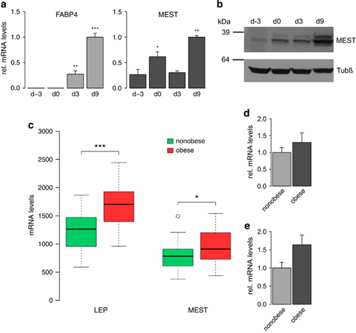
Exposure of 2-day post-confluent hMADS cells to a chemically defined adipogenic medium resulted in a strong upregulation of the adipocyte marker fatty acid-binding protein 4 within 9 days (Figure 1a, left panel). Similarly, MEST mRNA levels were high at day 9, but already showed a significant increase from day −3 to day 0, that is, when cells were still cultivated in growth medium (Figure 1a, right panel). This increase was also evident when MEST protein levels were analysed by western blotting, revealing only low amounts of MEST before the confluent stage, but already robust levels at the start of adipocyte differentiation, and a pronounced further increase as differentiation proceeded until day 9 (Figure 1b). To assess whether our in vitro adipogenesis model is reflecting the in vivo situation, we obtained data from a publicly available study in which gene expression profiles of human abdominal scWAT from 30 obese and 26 non-obese females had been compared.29 As expected, mRNA levels of the adipocytokine leptin were significantly higher in obese versus non-obese subjects (Figure 1c). Further, we also observed a significant elevation of MEST expression in the obese state (Figure 1c), thereby confirming previous results from mouse. In line with these results, we also found increased MEST levels in sorted (CD45−/CD31−/CD34+) adipocyte progenitors (Figure 1d) as well as in the mature adipocyte fraction (Figure 1e) of human scWAT. As previous studies have described that MEST overexpression results in enlarged adipocytes, we analyzed the relation of MEST and adipocyte size in human scWAT samples. Indeed, we found in 56 women a significant correlation between MEST mRNA and adipocyte volume (ρ=0.311; P=0.02) measured as described30 (Supplementary Figures S1a and b).
Figure 1.
Endogenous expression of MEST in human adipocyte differentiation and adipose tissue. (a and b) hMADS cells were grown to confluence (d-2) and stimulated to undergo adipocyte differentiation 2 days later (designated d0). (a) RNA was prepared at indicated time points and subjected to quantitative real-time reverse transcription PCR (RT-qPCR) for fatty acid-binding protein 4 (FABP4) and MEST. Expression levels were normalised to TBP and are presented relative to d9 (n=3). *P<0.05, **P<0.01, ***P<0.001 versus undifferentiated cells at d-3. (b) Protein lysates were prepared at indicated time points and analysed by western blotting for MEST and β-tubulin (Tubβ). (c) Abdominal scWAT biopsies from 26 non-obese and 30 obese female donors were analysed by microarray (Gene Expression Omnibus experiment GSE25401). Expression levels of leptin (LEP) and MEST are shown as boxplots. *P<0.05, ***P<0.001. (d and e) Expression of MEST was analysed by RT-qPCR in (d) adipocyte progenitor cells (CD45−/CD31−/CD34+) from scWAT biopsies of 10 non-obese and 8 obese subjects, and (e) mature adipocytes from scWAT biopsies of 4 non-obese and 6 obese subjects. MEST expression levels were normalised to 18S rRNA.
MEST suppresses human adipocyte differentiation
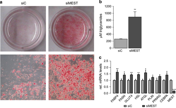
To analyse a possible function for MEST in human adipogenesis, we transfected hMADS cells with siRNAs to silence MEST (siMEST) and subsequently induced adipocyte differentiation. Interestingly, Oil Red O staining (Figure 2a) as well as triglyceride quantification (Figure 2b) revealed a markedly elevated intracellular lipid accumulation on MEST depletion at day 9 compared with control (siC)-transfected cells. Quantitative real-time reverse transcription PCR showed corresponding increases in the expression of fatty acid-binding protein 4, fatty acid synthase and the glucose transporter GLUT4 (SLC2A4) (Figure 2c). Further, PPARγ and CCAAT/enhancer-binding protein-α, both key transcriptional regulators of the adipogenic differentiation programme,31, 32 were increased by MEST silencing, as were the mRNAs of the lipid droplet-coating protein perilipin (PLIN1), adipose triglyceride lipase (PNPL2A) and hormone-sensitive lipase (LIPE) (Figure 2c). In line with these loss-of-function experiments, MEST overexpression resulted in diminished lipid accumulation and reduced expression of adipocyte markers (Supplementary Figure S2). As hMADS cells can differentiate into brown(-like) adipocytes,33 we also analysed uncoupling protein 1; however, uncoupling protein 1 mRNA levels were very low and not affected by changing the abundance of MEST (data not shown). Collectively, our results argue for a suppressive role of MEST in human adipogenesis.
Figure 2.
Silencing of MEST promotes human adipocyte differentiation. hMADS cells were transfected at confluence with siRNAs to target MEST (siMEST) or a control siRNA (siC) and adipocyte differentiation was induced 2 days later (d0). Cells were analysed at d9. (a) Triglyceride accumulation was visualised by Oil Red O staining. Upper panels: whole wells; lower panels: representative bright-field microscopy images (× 40 magnification). (b) Quantification of intracellular triglycerides (n=4). (c) Quantitative real-time reverse transcription PCR (RT-qPCR) analysis of adipocyte marker gene expression (normalised to TBP, relative to siC, n=3-6). (b and c) *P<0.05, **P<0.01, ***P<0.001 versus siC-transfected cells.
MEST silencing promotes PPAR signalling and glycolysis pathways
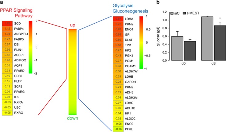
Previous studies in mouse 3T3-L1 preadipocytes have described pro-adipogenic effects of Mest.17, 20 Having observed the opposite in human, we decided to investigate the global impact of MEST silencing at the early stage of adipogenesis, that is, 3 days after induction of differentiation. Microarray analysis yielded a total of 2278 unique RefSeq mRNAs which were robustly detected (Supplementary Table S3). To obtain pathways that might be significantly addressed by MEST knockdown, we subjected this data set to gene set enrichment analysis.34 Interestingly, genes of the PPAR signalling pathway were found to be significantly (false discovery rate q-value <10−4) enriched among transcripts that were upregulated by MEST silencing (Figure 3a, left panel). The significant (q<10−4) induction of genes implicated in fatty acid, triacylglycerol and ketone body metabolism (derived from the reactome database, Supplementary Table S4) was an additional result supporting that adipocyte differentiation and, in particular, lipid biosynthesis were substantially promoted by MEST depletion even at this early stage of differentiation. Indeed, we found fatty acid synthase among the most highly upregulated transcripts (>2.5-fold higher abundance in siMEST-transfected versus siC-transfected cells; Supplementary Table S3). Further, genes of the glycolysis/gluconeogenesis pathway were also significantly (q=1.16 × 10⁻⁴) enriched among upregulated transcripts (Figure 3a, right panel). Correspondingly, glucose clearance from the differentiation medium was significantly increased in siMEST-transfected cells at day 3 (Figure 3b). Altogether, this suggests that MEST knockdown strongly promotes de novo triglyceride biosynthesis from glucose.
Figure 3.
MEST depletion induces PPAR signalling and glycolysis pathways. hMADS cells were transfected at confluence (d-2) with siRNAs targeting MEST (siMEST) or a control siRNA (siC). Adipocyte differentiation was induced 2 days later (d0). (a) RNA from cells at d3 was analysed by microarray to obtain a global view of mRNAs that are responsive to MEST depletion. A total of 2278 unique RefSeq mRNAs was sorted according to differences in expression between siMEST- and siC-transfected cells (middle heat map). Gene set enrichment analysis (GSEA preranked) revealed a significant enrichment of the KEGG pathways ‘PPAR signaling' (false discovery rate (FDR) q-value<0.0001) and ‘glycolysis/gluconeogenesis' (FDR q-value=0.0001) among upregulated transcripts. Expression of the transcripts assorted to both pathways are shown as heat maps (log2-transformed expression ratios (siMEST/siC). (b) Supernatants of hMADS cells at d0 and d3 were analysed for glucose concentration (n=5, *P<0.05).
MEST knockdown triggers early events in human adipogenesis
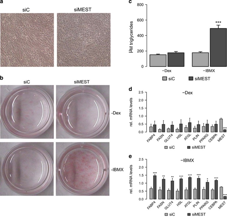
As transcriptomic analyses had revealed that MEST silencing evoked a substantial promotion of lipid biosynthesis pathways already at day 3 of adipocyte differentiation, we reasoned that MEST might be implicated in the early regulatory circuits of adipocyte differentiation. In further support of this hypothesis, the morphology of siMEST-transfected cells (compared to siC-transfected cells) at this time point was indicative of a more advanced differentiation stage, with a higher number of cells having changed their fibroblast-like shape into a more spherical shape (Figure 4a). As for the induction of adipocyte differentiation, various in vitro model systems (including hMADS cells) are usually exposed to a cocktail containing dexamethasone and IBMX for the first 2–3 days. We therefore asked whether knockdown of MEST might obviate the cell's need for one of these compounds. Although adipocyte differentiation was clearly suppressed for siC- as well as siMEST-transfected cells that were cultivated without dexamethasone (Figures 4b–d), we observed a strikingly efficient triglyceride accumulation (Figures 4b and c) as well as increased expression of adipocyte marker genes (Figure 4e) for MEST-depleted cells in the absence of IBMX.
Figure 4.
MEST depletion substitutes IBMX as inducer of human adipocyte differentiation. hMADS cells were transfected at confluence (d-2) with siRNAs targeting MEST (siMEST) or a control siRNA (siC). Adipocyte differentiation was induced 2 days later (d0). (a) Representative bright-field microscopy images (× 40 magnification) of cells at d3. (b–e) hMADS cells were stimulated to undergo adipocyte differentiation in suboptimal differentiation media lacking either dexamethasone (−Dex) or IBMX (-IBMX). (b) Triglyceride accumulation was visualised by Oil Red O staining at d9. (c) Quantification of intracellular triglycerides at d9 (n=4). (d and e) Quantitative real-time reverse transcription PCR (RT-qPCR) analysis of adipocyte marker gene expression (d9) for cells differentiated without Dex (d) or without IBMX (e). Gene expression was normalised to TBP and is presented relative to siC-transfected cells that were differentiated in complete differentiation medium (n=4). (c–e) *P<0.05, **P<0.01, ***P<0.001 versus siC-transfected cells exposed to the same differentiation cocktail.
MEST acts on the CREB/ATF1 signalling pathway
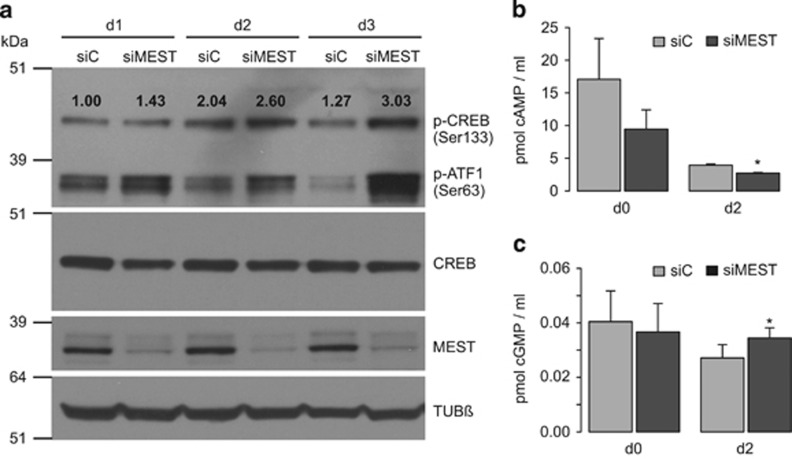
IBMX is a phosphodiesterase inhibitor, preventing the degradation of cAMP and cGMP. Both second messengers have been reported to trigger early events in adipocyte differentiation via activation of protein kinases A and G, respectively, which in turn leads to phosphorylation of the transcription factor CREB. As treatment of hMADS cells with IBMX could be fully substituted by MEST knockdown, we decided to analyse this signalling axis during the early stages of differentiation. Indeed, when exposed to complete differentiation medium, siMEST-transfected cells showed a substantial increase in phosphorylation of CREB and the related protein ATF1 compared with siC-transfected cells at days 1, 2 and 3 after induction of adipogenesis (Figure 5a). A similar, although less pronounced, effect was observed when cells were exposed to differentiation medium lacking IBMX (Supplementary Figure S3). We further measured intracellular cAMP and cGMP levels at the start of differentiation, as well as on day 2. Unexpectedly, cAMP levels were reduced due to MEST silencing (Figure 5b). In contrast, cGMP levels were increased by ~25% at day 2 (Figure 5c), yet the absolute levels of cGMP were found to be considerably lower than cAMP levels in differentiating hMADS cells. We thus propose that MEST acts on mediators which lie downstream of these second messengers, or that it affects a distinct signalling pathway, which ultimately regulates CREB/ATF1 phosphorylation.
Figure 5.
Effects of MEST depletion on phosphorylation of CREB/ATF1 and intracellular cAMP and cGMP levels. hMADS cells were transfected at confluence (d-2) with siRNAs targeting MEST (siMEST) or a control siRNA (siC). Adipocyte differentiation was induced 2 days later (d0) using complete differentiation medium. (a) Protein lysates of cells at d1, d2 and d3 were analysed by western blotting for phosphorylation of CREB and ATF1 (p-CREB and p-ATF-1), total CREB, MEST and β-Tubulin (Tubβ). Numbers above p-CREB bands reflect the results of densitometric analysis (ratio of p-CREB/CREB) and are presented relative to siC-transfected cells at d1. (b and c) Cells at d0 and d2 were analysed for intracellular levels of cAMP (b) and cGMP (c). Testing for significant differences in second messenger levels was performed by a paired t-test; *P<0.05.
Discussion
Throughout the past decades, excessive weight gain has become a global epidemic. Since 1980, the prevalence of obesity has almost doubled, with ~200 million male and ~300 million female adults worldwide being classified as obese in 2008.35 In contrast to earlier contention, which regarded adipose depots as a rather passive energy storage that expands as obesity develops, adipose tissue is nowadays considered a major endocrine organ with crucial implication in the pathology of obesity-associated diseases.2, 3 Indeed, as for the development of insulin resistance and preatherosclerotic changes, an ‘adipocentric view' has been proposed,36 placing detrimental changes in adipocyte physiology at the beginning of these afflictions. It should be noted that such weight gain-induced changes not only comprise an increase of adipocyte size37 (leading to necrosis at its extreme end4) but also a decrease in adipocyte differentiation.38 Thus, further elucidation of gene regulatory networks, which either promote or inhibit adipogenesis, is of utmost importance to better understand the molecular basis of adipose tissue malfunction in the obese state.
Proteins with predicted but not yet identified enzymatic function can be considered as particularly interesting candidates for further investigation. MEST is one such protein exhibiting an intriguingly high correlation with fat mass, which could be indicative of an involvement in lipid storage/mobilisation. In line with previous mouse studies,17, 18, 19 we also found MEST to be more abundant in WAT from obese as opposed to normal-weight humans (Figures 1c–e). MEST mRNA levels were further found to be significantly associated with adipocyte size (Supplementary Figure S1), albeit with less distinct correlation as leptin, the classical marker for adipocyte hypertrophy.
Although at least three studies in mouse have evolved the concept that Mest is a positive regulator of adipogenesis,17, 19, 20 our study suggests that MEST might have opposite effects in early steps of adipocyte differentiation in human. Similar to Jung et al.20 we have employed an RNA interference-based strategy to knock down MEST during adipocyte differentiation, yet have observed a pronounced upregulation of triglyceride accumulation and expression of adipocyte marker genes (Figure 2). These findings were corroborated by pathway analysis of global gene expression data, which revealed a significant upregulation of the PPAR signalling pathway, and genes involved in fatty acid, triacylglycerol and ketone body metabolism. Further, overexpression of MEST evoked a corresponding decrease in adipocyte differentiation (Supplementary Figure S2). Based on these results, it can be speculated that an increase of MEST in the obese state might participate in detrimental changes in WAT (and ultimately other tissues) by hampering de novo adipocyte recruitment. As for mouse cells, Jung et al.20 demonstrated that MEST inhibits the anti-adipogenic Wnt signalling pathway39, 40 via blocking maturation of the Wnt co-receptor low-density lipoprotein receptor-related protein 6.20 In contrast, although the Wnt signalling pathway was included in a collection of 186 gene sets (derived from the KEGG (Kyoto Encyclopedia of Genes and Genomes)) that we used as input for gene set enrichment analysis, we could not detect a significant upregulation of this pathway due to MEST silencing in differentiating hMADS cells. This was further supported by the fact that we observed a ~2-fold upregulation of the Wnt inhibitor dickkopf homolog 1(refs 41, 42) at this early stage of adipocyte differentiation (Supplementary Table S3). Altogether, we propose that species-specific regulatory circuits might be responsible for the diverging impact of MEST in human versus mouse adipogenesis as, for example, recently shown for the lim domain only 3 (LMO3) gene.43 Alternatively, we cannot rule out that the effect of MEST is critically dependent on the differentiation stage of adipocyte precursors, as we have employed mesenchymal stem cells that are multipotent at the clonal level, whereas others have used cells at the preadipocyte stage.
In addition to the pronounced upregulation during adipocyte differentiation, we also noted a preceding increase of MEST mRNA and protein as proliferating hMADS cells (d-3) reached the quiescent stage (d0, Figures 1a and b). In this phase of our experiments, FGF2 was withdrawn from the medium when hMADS cells had grown confluent (d-2). It can thus be hypothesised that MEST is under suppressive control of FGF2. Indeed, a recent study on human dermal fibroblasts (GSE48967) has revealed a significant downregulation of MEST on treatment with FGF2.44 Alternatively, it can be speculated that growth arrest due to contact inhibition might trigger pathways that unleash enhanced transcription of the MEST gene.
We also observed a significant promotion of the KEGG pathway ‘glycolysis and gluconeogensis' on MEST knockdown at day 3, concomitant with a depletion of glucose from the medium (Figure 3). In conjunction with the elevated lipid accumulation of hMADS-derived adipocytes at day 9, we propose that MEST silencing triggers de novo triglyceride biosynthesis from glucose. It will be of high interest to test whether the newly discovered relationship between MEST and glycolysis is also existent in other cell types, not least because an increase in glycolytic flux is a frequent characteristic in cancer.45 In this respect, it should be noted that a recent study has described increased CpG island methylation of the MEST promoter region (suggesting downregulation of MEST transcription) in drug-resistant ovarian cancer cell lines.46
Efficient adipocyte differentiation of quiescent human and mouse precursor cells has long been known to be induced by insulin, glucocorticoids and a cAMP-elevating agent.47, 48 As for the latter, it is important to keep in mind that the frequently used phosphodiesterase inhibitor IBMX also acts on cGMP-degrading phosphodiesterases, and that cGMP has pro-adipogenic effects comparable to cAMP.49 Through activation of protein kinases A/G, both second messengers trigger the phosphorylation of the downstream transcriptional effector CREB, which is necessary for adipocyte differentiation.50 Motivated by the fact that MEST knockdown promoted PPAR signalling and lipid biosynthesis pathways already at day 3 after induction, we decided to repeat experiments in suboptimal differentiation media lacking those pro-adipogenic components, which are only present at the very beginning of differentiation (Figure 4). Indeed, this approach revealed that MEST depletion could completely substitute IBMX (but not dexamethasone), thereby implying MEST in the regulatory pathway of cAMP/cGMP, protein kinases A/G and CREB. This hypothesis was principally confirmed as phosphorylation of CREB and the related transcription factor ATF1 was substantially increased by MEST knockdown (Figure 5a). However, we did not detect an elevation, but instead a reduction of cAMP due to MEST silencing in early adipogenesis (Figure 5b). Although cGMP levels were increased at day 2 (Figure 5c), this upregulation is unlikely to be the main cause for the promotion of CREB/ATF1 phosphorylation as (i) cAMP levels are decreased at the same time point and (ii) overall, cAMP levels were found to be >100-fold higher than cGMP levels in our cell model. We thus propose that invalidation of MEST promotes CREB/ATF1 phosphorylation via one or more distinct routes. For instance, Ras-mitogen activated protein kinase signalling,51, 52 Ca2+/calmodulin-dependent protein kinase53 and protein kinase C54 have been described to phosphorylate CREB. Future studies addressing the responsiveness of these pathways to MEST will therefore be of high interest to further explore the newly discovered link between MEST and CREB/ATF1. Keeping in mind the behavioural abnormalities of Mest knockout mice55 as well as the high importance of CREB in the central nervous system,56 this axis could also be of relevance for brain function.
In summary, we have used human samples and cells to confirm the high abundance of MEST in adipocytes and its association with fat mass, findings that had previously been obtained only in mouse experiments. Functional investigations have revealed an unexpected inhibitory role of MEST in human adipogenesis and, for the first time, have linked this protein to a pathway that triggers the phosphorylation of CREB, a crucial transcription factor not only in metabolism but also in cognitive abilities. Altogether, these results have laid a fruitful basis for further research that will ultimately elucidate the precise molecular activity of MEST, and that could eventually lead to the development of novel treatment strategies to ameliorate obesity-associated diseases.
Acknowledgments
We thank Christian Dani for providing hMADS cells, and Daniel Gütl, Lukas Fliedl and Regina Grillari-Voglauer for help with glucose measurements. This work was supported by the Austrian Science Fund (FWF, P25729-B19) and by the EU FP7 project DIABAT (HEALTH-F2-2011-278373).
The authors declare no conflict of interest.
Footnotes
Supplementary Information accompanies this paper on International Journal of Obesity website (http://www.nature.com/ijo)
Supplementary Material
References
- 1Gesta S, Tseng Y-H, Kahn CR. Developmental origin of fat: tracking obesity to its source. Cell 2007; 131: 242–256. [DOI] [PubMed] [Google Scholar]
- 2Virtue S, Vidal-Puig A. Adipose tissue expandability, lipotoxicity and the metabolic syndrome—an allostatic perspective. Biochim Biophys Acta 2010; 1801: 338–349. [DOI] [PubMed] [Google Scholar]
- 3Maury E, Brichard SM. Adipokine dysregulation, adipose tissue inflammation and metabolic syndrome. Mol Cell Endocrinol 2010; 314: 1–16. [DOI] [PubMed] [Google Scholar]
- 4Cinti S, Mitchell G, Barbatelli G, Murano I, Ceresi E, Faloia E et al. Adipocyte death defines macrophage localization and function in adipose tissue of obese mice and humans. J Lipid Res 2005; 46: 2347–2355. [DOI] [PubMed] [Google Scholar]
- 5Hallakou S, Doaré L, Foufelle F, Kergoat M, Guerre-Millo M, Berthault MF et al. Pioglitazone induces in vivo adipocyte differentiation in the obese Zucker fa/fa rat. Diabetes 1997; 46: 1393–1399. [DOI] [PubMed] [Google Scholar]
- 6Adams M, Montague CT, Prins JB, Holder JC, Smith SA, Sanders L et al. Activators of peroxisome proliferator-activated receptor gamma have depot-specific effects on human preadipocyte differentiation. J Clin Invest 1997; 100: 3149–3153. [DOI] [PMC free article] [PubMed] [Google Scholar]
- 7Cristancho AG, Lazar MA. Forming functional fat: a growing understanding of adipocyte differentiation. Nat Rev Mol Cell Biol 2011; 12: 722–734. [DOI] [PMC free article] [PubMed] [Google Scholar]
- 8Huang JT, Welch JS, Ricote M, Binder CJ, Willson TM, Kelly C et al. Interleukin-4-dependent production of PPAR-gamma ligands in macrophages by 12/15-lipoxygenase. Nature 1999; 400: 378–382. [DOI] [PubMed] [Google Scholar]
- 9Rosen ED, Sarraf P, Troy AE, Bradwin G, Moore K, Milstone DS et al. PPAR gamma is required for the differentiation of adipose tissue in vivo and in vitro. Mol Cell 1999; 4: 611–617. [DOI] [PubMed] [Google Scholar]
- 10Bujalska IJ, Kumar S, Hewison M, Stewart PM. Differentiation of adipose stromal cells: the roles of glucocorticoids and 11beta-hydroxysteroid dehydrogenase. Endocrinology 1999; 140: 3188–3196. [DOI] [PubMed] [Google Scholar]
- 11Siersbæk R, Mandrup S. Transcriptional networks controlling adipocyte differentiation. Cold Spring Harb Symp Quant Biol 2011; 76: 247–255. [DOI] [PubMed] [Google Scholar]
- 12Ollis DL, Cheah E, Cygler M, Dijkstra B, Frolow F, Franken SM et al. The alpha/beta hydrolase fold. Protein Eng 1992; 5: 197–211. [DOI] [PubMed] [Google Scholar]
- 13Sado T, Nakajima N, Tada M, Takagi N. A novel mesoderm-specific cdna isolated from a mouse embryonal carcinoma cell line. Dev Growth Differ 1993; 35: 551–560. [DOI] [PubMed] [Google Scholar]
- 14Kaneko-Ishino T, Kuroiwa Y, Miyoshi N, Kohda T, Suzuki R, Yokoyama M et al. Peg1/Mest imprinted gene on chromosome 6 identified by cDNA subtraction hybridization. Nat Genet 1995; 11: 52–59. [DOI] [PubMed] [Google Scholar]
- 15Kobayashi S, Kohda T, Miyoshi N, Kuroiwa Y, Aisaka K, Tsutsumi O et al. Human PEG1/MEST, an imprinted gene on chromosome 7. Hum Mol Genet 1997; 6: 781–786. [DOI] [PubMed] [Google Scholar]
- 16Soukas A, Cohen P, Socci ND, Friedman JM. Leptin-specific patterns of gene expression in white adipose tissue. Genes Dev 2000; 14: 963–980. [PMC free article] [PubMed] [Google Scholar]
- 17Takahashi M, Kamei Y, Ezaki O. Mest/Peg1 imprinted gene enlarges adipocytes and is a marker of adipocyte size. Am J Physiol Endocrinol Metab 2005; 288: E117–E124. [DOI] [PubMed] [Google Scholar]
- 18Koza RA, Nikonova L, Hogan J, Rim J-S, Mendoza T, Faulk C et al. Changes in gene expression foreshadow diet-induced obesity in genetically identical mice. PLoS Genet 2006; 2: e81. [DOI] [PMC free article] [PubMed] [Google Scholar]
- 19Nikonova L, Koza RA, Mendoza T, Chao P-M, Curley JP, Kozak LP. Mesoderm-specific transcript is associated with fat mass expansion in response to a positive energy balance. FASEB J 2008; 22: 3925–3937. [DOI] [PMC free article] [PubMed] [Google Scholar]
- 20Jung H, Lee SK, Jho E. Mest/Peg1 inhibits Wnt signalling through regulation of LRP6 glycosylation. Biochem J 2011; 436: 263–269. [DOI] [PubMed] [Google Scholar]
- 21Kadota Y, Yanagawa M, Nakaya T, Kawakami T, Sato M, Suzuki S. Gene expression of mesoderm-specific transcript is upregulated as preadipocytes differentiate to adipocytes in vitro. J Physiol Sci 2012; 62: 403–411. [DOI] [PMC free article] [PubMed] [Google Scholar]
- 22Rodriguez A-M, Pisani D, Dechesne CA, Turc-Carel C, Kurzenne J-Y, Wdziekonski B et al. Transplantation of a multipotent cell population from human adipose tissue induces dystrophin expression in the immunocompetent mdx mouse. J Exp Med 2005; 201: 1397–1405. [DOI] [PMC free article] [PubMed] [Google Scholar]
- 23Karbiener M, Pisani DF, Frontini A, Oberreiter LM, Lang E, Vegiopoulos A et al. MicroRNA-26 family is required for human adipogenesis and drives characteristics of brown adipocytes. Stem Cells 2014; 32: 1578–1590. [DOI] [PubMed] [Google Scholar]
- 24Pettersson AML, Stenson BM, Lorente-Cebrián S, Andersson DP, Mejhert N, Krätzel J et al. LXR is a negative regulator of glucose uptake in human adipocytes. Diabetologia 2013; 56: 2044–2054. [DOI] [PubMed] [Google Scholar]
- 25Gao H, Mejhert N, Fretz JA, Arner E, Lorente-Cebrián S, Ehrlund A et al. Early B cell factor 1 regulates adipocyte morphology and lipolysis in white adipose tissue. Cell Metab 2014; 19: 981–992. [DOI] [PMC free article] [PubMed] [Google Scholar]
- 26Karbiener M, Neuhold C, Opriessnig P, Prokesch A, Bogner-Strauss JG, Scheideler M. MicroRNA-30c promotes human adipocyte differentiation and co-represses PAI-1 and ALK2. RNA Biol 2011; 8: 850–860. [DOI] [PubMed] [Google Scholar]
- 27Pabinger S, Thallinger GG, Snajder R, Eichhorn H, Rader R, Trajanoski Z. QPCR: Application for real-time PCR data management and analysis. BMC Bioinformatics 2009; 10: 268. [DOI] [PMC free article] [PubMed] [Google Scholar]
- 28Zhao S, Fernald RD. Comprehensive algorithm for quantitative real-time polymerase chain reaction. J Comput Biol 2005; 12: 1047–1064. [DOI] [PMC free article] [PubMed] [Google Scholar]
- 29Arner E, Mejhert N, Kulyté A, Balwierz PJ, Pachkov M, Cormont M et al. Adipose tissue microRNAs as regulators of CCL2 production in human obesity. Diabetes 2012; 61: 1986–1993. [DOI] [PMC free article] [PubMed] [Google Scholar]
- 30Löfgren P, Hoffstedt J, Näslund E, Wirén M, Arner P. Prospective and controlled studies of the actions of insulin and catecholamine in fat cells of obese women following weight reduction. Diabetologia 2005; 48: 2334–2342. [DOI] [PubMed] [Google Scholar]
- 31Lefterova MI, Zhang Y, Steger DJ, Schupp M, Schug J, Cristancho A et al. PPARgamma and C/EBP factors orchestrate adipocyte biology via adjacent binding on a genome-wide scale. Genes Dev 2008; 22: 2941–2952. [DOI] [PMC free article] [PubMed] [Google Scholar]
- 32Schmidt SF, Jørgensen M, Chen Y, Nielsen R, Sandelin A, Mandrup S. Cross species comparison of C/EBPα and PPARγ profiles in mouse and human adipocytes reveals interdependent retention of binding sites. BMC Genomics 2011; 12: 152. [DOI] [PMC free article] [PubMed] [Google Scholar]
- 33Elabd C, Chiellini C, Carmona M, Galitzky J, Cochet O, Petersen R et al. Human multipotent adipose-derived stem cells differentiate into functional brown adipocytes. Stem Cells 2009; 27: 2753–2760. [DOI] [PubMed] [Google Scholar]
- 34Subramanian A, Tamayo P, Mootha VK, Mukherjee S, Ebert BL, Gillette MA et al. Gene set enrichment analysis: a knowledge-based approach for interpreting genome-wide expression profiles. Proc Natl Acad Sci USA 2005; 102: 15545–15550. [DOI] [PMC free article] [PubMed] [Google Scholar]
- 35WHO | Obesity and overweight. http://www.who.int/mediacentre/factsheets/fs311/en/# (accessed 10 June 2014).
- 36Naukkarinen J, Rissanen A, Kaprio J, Pietiläinen KH. Causes and consequences of obesity: the contribution of recent twin studies. Int J Obes (Lond) 2012; 36: 1017–1024. [DOI] [PubMed] [Google Scholar]
- 37Pietiläinen KH, Kannisto K, Korsheninnikova E, Rissanen A, Kaprio J, Ehrenborg E et al. Acquired obesity increases CD68 and tumor necrosis factor-alpha and decreases adiponectin gene expression in adipose tissue: a study in monozygotic twins. J Clin Endocrinol Metab 2006; 91: 2776–2781. [DOI] [PubMed] [Google Scholar]
- 38Pietiläinen KH, Naukkarinen J, Rissanen A, Saharinen J, Ellonen P, Keränen H et al. Global transcript profiles of fat in monozygotic twins discordant for BMI: pathways behind acquired obesity. PLoS Med 2008; 5: e51. [DOI] [PMC free article] [PubMed] [Google Scholar]
- 39Ross SE, Hemati N, Longo KA, Bennett CN, Lucas PC, Erickson RL et al. Inhibition of adipogenesis by Wnt signaling. Science 2000; 289: 950–953. [DOI] [PubMed] [Google Scholar]
- 40Longo KA, Wright WS, Kang S, Gerin I, Chiang S-H, Lucas PC et al. Wnt10b inhibits development of white and brown adipose tissues. J Biol Chem 2004; 279: 35503–35509. [DOI] [PubMed] [Google Scholar]
- 41Glinka A, Wu W, Delius H, Monaghan AP, Blumenstock C, Niehrs C. Dickkopf-1 is a member of a new family of secreted proteins and functions in head induction. Nature 1998; 391: 357–362. [DOI] [PubMed] [Google Scholar]
- 42Bafico A, Liu G, Yaniv A, Gazit A, Aaronson SA. Novel mechanism of Wnt signalling inhibition mediated by Dickkopf-1 interaction with LRP6/Arrow. Nat Cell Biol 2001; 3: 683–686. [DOI] [PubMed] [Google Scholar]
- 43Lindroos J, Husa J, Mitterer G, Haschemi A, Rauscher S, Haas R et al. Human but not mouse adipogenesis is critically dependent on LMO3. Cell Metab 2013; 18: 62–74. [DOI] [PMC free article] [PubMed] [Google Scholar]
- 44Kashpur O, LaPointe D, Ambady S, Ryder EF, Dominko T. FGF2-induced effects on transcriptome associated with regeneration competence in adult human fibroblasts. BMC Genomics 2013; 14: 656. [DOI] [PMC free article] [PubMed] [Google Scholar]
- 45Akram M. Mini-review on glycolysis and cancer. J Cancer Educ 2013; 28: 454–457. [DOI] [PubMed] [Google Scholar]
- 46Zeller C, Dai W, Steele NL, Siddiq A, Walley AJ, Wilhelm-Benartzi CSM et al. Candidate DNA methylation drivers of acquired cisplatin resistance in ovarian cancer identified by methylome and expression profiling. Oncogene 2012; 31: 4567–4576. [DOI] [PubMed] [Google Scholar]
- 47Russell TR, Ho R. Conversion of 3T3 fibroblasts into adipose cells: triggering of differentiation by prostaglandin F2alpha and 1-methyl-3-isobutyl xanthine. Proc Natl Acad Sci USA 1976; 73: 4516–4520. [DOI] [PMC free article] [PubMed] [Google Scholar]
- 48Hauner H, Entenmann G, Wabitsch M, Gaillard D, Ailhaud G, Negrel R et al. Promoting effect of glucocorticoids on the differentiation of human adipocyte precursor cells cultured in a chemically defined medium. J Clin Invest 1989; 84: 1663–1670. [DOI] [PMC free article] [PubMed] [Google Scholar]
- 49Hemmrich K, Gummersbach C, Paul NE, Goy D, Suschek CV, Kröncke K-D et al. Nitric oxide and downstream second messenger cGMP and cAMP enhance adipogenesis in primary human preadipocytes. Cytotherapy 2010; 12: 547–553. [DOI] [PubMed] [Google Scholar]
- 50Reusch JE, Colton LA, Klemm DJ. CREB activation induces adipogenesis in 3T3-L1 cells. Mol Cell Biol 2000; 20: 1008–1020. [DOI] [PMC free article] [PubMed] [Google Scholar]
- 51Ginty DD, Bonni A, Greenberg ME. Nerve growth factor activates a Ras-dependent protein kinase that stimulates c-fos transcription via phosphorylation of CREB. Cell 1994; 77: 713–725. [DOI] [PubMed] [Google Scholar]
- 52Klemm DJ, Roesler WJ, Boras T, Colton LA, Felder K, Reusch JE. Insulin stimulates cAMP-response element binding protein activity in HepG2 and 3T3-L1 cell lines. J Biol Chem 1998; 273: 917–923. [DOI] [PubMed] [Google Scholar]
- 53Sheng M, Thompson MA, Greenberg ME. CREB: a Ca(2+)-regulated transcription factor phosphorylated by calmodulin-dependent kinases. Science 1991; 252: 1427–1430. [DOI] [PubMed] [Google Scholar]
- 54Guo CM, Kasaraneni N, Sun K, Myatt L. Cross talk between PKC and CREB in the induction of COX-2 by PGF2α in human amnion fibroblasts. Endocrinology 2012; 153: 4938–4945. [DOI] [PMC free article] [PubMed] [Google Scholar]
- 55Lefebvre L, Viville S, Barton SC, Ishino F, Keverne EB, Surani MA. Abnormal maternal behaviour and growth retardation associated with loss of the imprinted gene Mest. Nat Genet 1998; 20: 163–169. [DOI] [PubMed] [Google Scholar]
- 56Carlezon WA, Duman RS, Nestler EJ. The many faces of CREB. Trends Neurosci 2005; 28: 436–445. [DOI] [PubMed] [Google Scholar]
Associated Data
This section collects any data citations, data availability statements, or supplementary materials included in this article.


