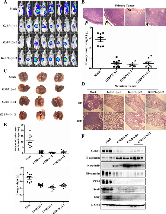
Figure 7. Downregulation of G3BP1 inhibits tumor metastasis in vivo.

A. BLI of the indicated cell lines. A total of 5.0 × 10⁵ 4T1-luc cells in 0.1 mL of PBS were orthotopically injected into the mammary fat pad of 6- to 8-week-old female BALB/c mice (n = 8 each). B. Representative images of primary tumor margins stained with hematoxylin and eosin (upper panel) (magnification, × 100). The arrows indicate the invasive front. The weight of the primary tumor from the indicated cell lines is shown in the lower panel. C. Representative lungs were harvested via necropsy after orthotopic injection (n = 3). D. Representative images of hematoxylin and eosin staining of lung sections (magnification, × 40 upper, × 100 lower). E. The number of metastatic nodules (upper panel) and the lung weight (lower panel) were used to evaluate metastasis to the lungs. The number of metastatic nodules and the lung weight are presented as the means±s.d. F. Tumors from mice sacrificed 41 days after injection of 4T1-luc cells (Mock, G3BP1(−)-1, G3BP1(−)-2 or G3BP1(−)-1/2 (n = 3 each)) were analyzed for the expression levels of EMT markers.
