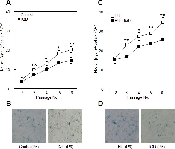
Figure 6. Effect of 1,5-isoquinolinediol (IQD) on formation of β-galactosidase.

A. Senescence was assessed using β-galactosidase staining after treatment with IQD. B. Lower panel shows representative field of view on microscopy at passage number 5. C. Under the senescence condition accelerated by hydroxyurea (HU), the number of β-galactosidase-positive cells was counted. D. Images showing representative β-galactosidase-positive cells. ns, not significant; *, p < 0.05; **, p < 0.01.
