CASE SUMMARY
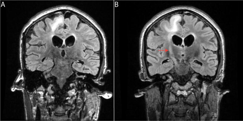
A 29-year-old gravida 3 para 2 woman with a history of penicillin-treated syphilis and ectopic pregnancy was transferred to this hospital for progressively worsening encephalopathy, left hemiparesis, and hemodynamic instability. Her symptoms had begun approximately 5.5 weeks earlier with left lower extremity weakness and falls. At that time, she was 30 weeks pregnant. The patient presented to an outside hospital where on neurologic exam she was found to have left lower extremity rigidity, hyperreflexia, and a positive Babinski sign. She was admitted and underwent MRI imaging of the brain and spine. These studies revealed no significant abnormality, and she was discharged home. Two weeks later she was admitted to another hospital for encephalopathy and worsening left-sided dysfunction involving both upper and lower extremities. Cerebrospinal fluid (CSF) analysis demonstrated a significantly elevated IgG concentration of 25 mg/dL with ≥ 2 oligoclonal bands (Table 1). Work-up for infectious and autoimmune causes was negative. In spite of treatment with high-dose intravenous methylprednisolone and plasma exchange therapy, she continued to show progressively worsening cognitive decline in addition to mood abnormalities, acute visual changes, and hallucinations. After undergoing uncomplicated spontaneous delivery of a preterm healthy infant at 34 weeks and 2 days, she developed abrupt further decline in cognitive function, tachycardia, low-grade fevers, and hemodynamic instability, which required transfer to the intensive care unit. A repeat brain MRI demonstrated generalized volume loss as well as multifocal cortical and subcortical T2 hyperintense lesions involving the right posterior frontal lobe, bilateral temporal lobes, and corpus callosum, with evidence of corticospinal tract degeneration on the right (Figure 1A). The clinical differential diagnosis was broad and included inflammatory, vascular, infectious, neoplastic, paraneoplastic, and metabolic etiologies. She was started on intravenous vancomycin/meropenem and transferred to this hospital 8 days postpartum.
Table 1.
Summary of Select Laboratory Studies
| CSF 1 (Day 16)* | CSF 2 (Day 18)* | CSF 3 (Day 42) | CSF 4 (Day 47) | ||
|---|---|---|---|---|---|
| Variable | Normal Range | Value | Value | Value | Value |
| Appearance | Clear | Clear | Clear | ||
| Red cell count (per mm3) | None | 50 | 0 | 1 | |
| White cell count (per mm3) | 0–5 | 0 | 2 | 3 | 4 |
| Lymphocytes (%) | 60–70 | 87 | 94 | ||
| Reactive lymphocytes (%) | 4 | ||||
| Monocytes (%) | < 30 | 13 | 2 | ||
| Neutrophils (%) | < 2 | ||||
| Protein (mg/dL) | 15–45 | 48 | 60 | 52 | 52 |
| Glucose (mg/dL) | 40–85 | 54 | 96 | 55 | 56 |
| Lactate (mg/dL) | < 35 | 2.1 | |||
| Xanthochromia | Negative | Negative | |||
| Oligoclonal bands | Absent | > 2 | > 5 | ||
| Albumin (mg/dL) | 13.4–23.7 | 10.6 | 15.2 | ||
| IgG (mg/dL) | 0.4–5.2 | 25 | 20.2 | ||
| IgG index | 0.23–0.64 | 7.9 | |||
| Cytology | Benign | Benign | Benign | Benign | |
| Flow cytometry | Benign | Benign | |||
| Anti-Measles IgG | < 1:64 | 1:1024 | |||
| Anti-Measles IgM | < 1:1 | < 1:1 | |||
| Additional studies performed at outside hospital | |||||
| Normal Range | Value | ||||
| Venereal disease research laboratory | Negative | Negative | |||
| Cryptococcal antigen | Negative | Negative | |||
| Herpes simplex virus 1 and 2 | Negative | Negative | |||
| Enterovirus | Negative | Negative | |||
| Humman immunodeficiency virus | Negative | Negative | |||
| Anti-Double stranded DNA | Negative | Negative | |||
| Anti-Ro (SSA) | Negative | Negative | |||
| Anti-La (SSB) | Negative | Negative | |||
| Anti-Ribonucleo-proteins | Negative | Negative | |||
| Anti-Smith | Negative | Negative | |||
| Anti-Chromatin | Negative | Negative | |||
| Anti-Ribosomal | Negative | Negative | |||
| Anti-Centromere | Negative | Negative | |||
| Anti-Scl70 | Negative | Negative | |||
| Anti-Jo1 | Negative | Negative | |||
| Anti-Hu | Negative | Negative | |||
| Anti-Yo | Negative | Negative | |||
| Anti-Ri | Negative | Negative | |||
| Anti-Tr | Negative | Negative | |||
| Anti-Amphiphysin | Negative | Negative | |||
| Anti-GAD-65 | Negative | Negative | |||
| Anti-Purkinge cell antibody 2 | Negative | Negative | |||
| Anti-Neuronal nuclear antibody 3 | Negative | Negative | |||
| Anti-NMDA receptor | Negative | Negative | |||
| Anti NMO | Negative | Negative | |||
| Anti TPO | Negative | Negative | |||
CSF studies 1 and 2 were performed at outside hospital
Figure 1.

MRI T2 FLAIR imaging studies obtained at 38 days (A) and 45 days (B) after symptom onset. There is generalized volume loss with associated multifocal cortical and subcortical T2 hyperintense lesions involving the right posterior frontal lobe, bilateral temporal lobes, and corpus callosum, with evidence of corticospinal tract degeneration on the right (A). Significant disease progression is noted on the later study, with worse multifocal white matter FLAIR abnormality involving the inferior right frontal lobe, posterior right parietal lobe, and bilateral temporal lobes (B). The arrow points to the corticospinal tract.
The patient was born and raised in India until the age of 3 when she immigrated to the United States. She worked at a nursing home and was married. Her 5-year-old daughter was healthy. Upon admission to this hospital, her vital signs showed a temperature of 37.9°C, heart rate of 75–140 beats per minute, respiratory rate of 21–48 breaths/minute, blood pressure of 105–180/52–100 mm Hg, and oxygen saturation of 98–100% at room air. On exam, her eyes were closed, and she did not respond to voice commands. Stiff posturing of all extremities was noted as well as flexion of the left upper extremity and bilateral lower extremity plantarflexion. Brainstem reflexes were intact. Her complete blood count showed mild anemia, with hemoglobin of 10.2 g/dL and hematocrit of 30%. A basic chemistry panel revealed hyperglycemia of 447 mg/dL. Liver function tests were remarkable for mildly elevated aspartate amino transferase of 55 U/L. An electroencephalogram (EEG) showed diffuse slowing and poor organization without epileptiform activity. CSF analysis revealed elevated IgG of 20.2 mg/dL without evidence of infection (Table 1). A brain MRI demonstrated significantly worsening multifocal white matter FLAIR abnormalities involving the inferior right frontal lobe, posterior right parietal lobe, and bilateral temporal lobes (Figure 1B). MR spectroscopy showed elevated choline and reduced N-acetyl aspartate within areas of white matter abnormality. She was started on ganciclovir, methylprednisolone, and intravenous immune globulin therapy. In spite of intensive care supportive measures, she continued to get progressively worse, with startle myoclonus, decorticate posturing, autonomic instability, and spontaneous periodic alternating conjugate horizontal gaze (“ping-pong gaze”), indicative of bilateral cortical damage with preserved brainstem function. A right parietal lobe brain biopsy was obtained as well as additional CSF studies.
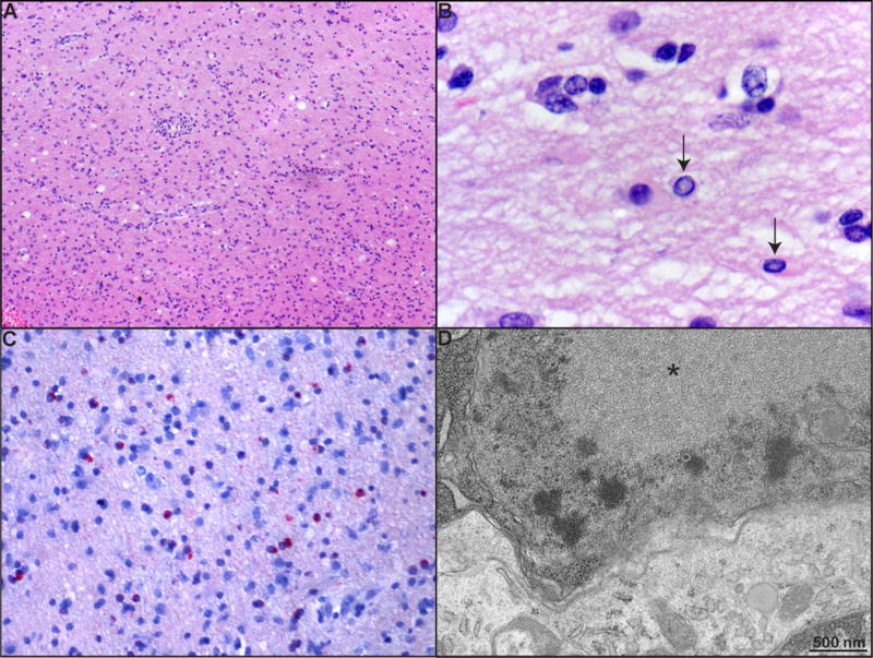
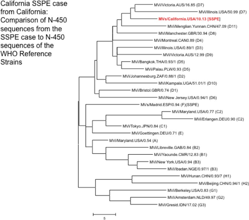
Hematoxylin and eosin (H&E)-stained sections of the brain tissue demonstrated hypercellular cortex and subcortical white matter with patchy white matter pallor, widespread microglial activation, perivascular and parenchymal lymphohistiocytic inflammation, reactive astrogliosis, and axonal spheroids (Figure 2A). Scattered oligodendroglial cells with nuclear enlargement, margination of chromatin, and glassy eosinophilic intranuclear inclusions were identified (Figure 2B). Immunohistochemistry for CD3, CD20, and CD68 demonstrated that the lymphohistiocytic infiltrate was composed predominantly of CD68-positive macrophages and activated microglia with a lesser component of CD3-positive T lymphocytes and a minor population (< 5%) of CD20-positive B lymphocytes. Histochemistry for LFB/PAS showed patchy myelin loss, and immunohistochemistry for neurofilament protein (NFP) showed relative preservation of axons and numerous axonal spheroids. Scattered reactive astrocytes were shown by immunohistochemistry for GFAP. Special stains (PAS, GRAM, GMS, AFB, and Steiner) were negative for fungi, mycobacterium, spirochetes, and bacteria. Immunohistochemistry for HSV, CMV, adenovirus, and polyoma virus as well as in-situ hybridization for EBV were negative, but immunohistochemistry for measles virus showed widespread intranuclear and intracytoplasmic labeling of viral antigen within glial cells (Figure 2C). On ultrastructural analysis by transmission electron microscopy (EM), numerous inclusions characterized by interwoven tubular strands, circular rings 17–18 nm in width, and granular structures typical of measles virus nucleocapsid (Nakai and Imagawa, 1969) were present within oligodendrocyte nuclei (Figure 2D). Fewer cytoplasmic inclusions were also noted. Enveloped virions were not identified. Nucleic acid extracts from formalin-fixed paraffin-embedded tissue were positive by PCR assay for measles virus, and nucleoprotein gene (N-450) sequence analysis (2012; Rota et al., 2011) showed that the virus was most closely related to the sequences of viruses in genotype D7 (Figure 3). While serologic studies for HIV and JC virus were negative, CSF analysis showed a markedly elevated IgG titer for measles virus (1:1024; normal < 1:64; Table 1). These findings support a diagnosis of subacute sclerosing panencephalitis (SSPE).
Figure 2.

(A) Hematoxylin and eosin-stained section of brain tissue shows hypercellular cortical and subcortical white matter with perivascular and parenchymal lymphohistiocytic inflammation and reactive astrogliosis (magnification: 100×). (B) Oligodendroglia with nuclear enlargement, margination of chromatin, and glassy eosinophilic intranuclear inclusions (arrows; magnification: 1000×). (C) Immunohistochemistry for measles virus showing widespread intranuclear and intracytoplasmic labeling of viral antigen within oligodendrocytes (magnification 200×). (D) Electron micrograph demonstrating numerous intranuclear interwoven tubular strands, circular rings 17–18 nm in width, and granular structures within an oligodendrocyte nucleus (*), characteristic of measles virus nucleocapsid (scale: 500 nm).
Figure 3.

Phylogenetic analysis of the nucleoprotein sequence derived from the RNA extracted from the brain tissue of the SSPE case compared to the N-450 sequences of the World Health Organization reference strains. The measles sequences associated with the SSPE case were found to be most closely related to the sequences of measles viruses in genotype D7.
The patient was started on a 4-week trial of intrathecal interferon alpha 2B therapy (1,000,000 international units/m2 twice weekly). A brain MRI just prior to the onset of therapy demonstrated significant interval increase in the severity of bilateral subcortical and deep white matter FLAIR hyperintensity, with particular involvement of the bilateral coronal radiata. Cortical FLAIR hyperintensities involving the bilateral occipital lobes were also noted. The patient remained in a minimally responsive state and showed no noticeable improvement with therapy. She was transferred back to the referring institution for completion of the interferon trial. Follow-up brain MRI 4 weeks later showed disease progression. Thus, therapy with interferon was discontinued, and the patient was transferred to a skilled nursing care facility. She died approximately 2 months later. From the time of initial symptoms, the course of disease was approximately 6 months.
DISCUSSION
SSPE is a rare complication of infection with measles virus. The disease affects approximately 4–11/100,000 cases and has high morbidity and mortality (Bellini et al., 2005; Miller et al., 2004; Miller et al., 1992). The clinical manifestations of SSPE are variable. The initial presentation often includes behavioral changes, episodes of falling, psychomotor retardation, chorioretinitis, and adventitious movements (Dyken, 1985). Seizures may be present. As the disease progresses, myoclonic jerks, spasms, and severe physical and cognitive impairment develop, ultimately leading to a vegetative stage (Dyken, 1985; Garg, 2008). Periodic epileptiform activity is characteristically seen on EEG. The average period of latency is 7–10 years (range of 1 month to 27 years), and death generally occurs within 3 years of symptom onset (Campbell et al., 2007). Risk factors include infection with measles virus before 2 years of age (Bellini et al., 2005). Fulminant forms of the disease have been reported in association with pregnancy (Cole et al., 2007; Wirguin et al., 1988). Both a state of natural immune suppression in pregnancy and changes in cell-mediated immunity occurring during pregnancy have been suggested to contribute to the development of fulminant disease (Wirguin et al., 1988).
While our patient displayed some of the classic manifestations of SSPE, their nonspecific nature in combination with the lack of behavioral change, no pleocytosis on CSF analysis, and absence of epileptiform activity on EEG constituted atypical features of this disease. On the other hand, the high CSF IgG, presence of oligoclonal bands, and brain MRI findings atypical for multiple sclerosis might suggest that early CSF screening for measles antigen or nucleic acid should be undertaken to evaluate for SSPE.
Measles virus is a negative-sense RNA virus with a non-segmented genome and a lipid envelope (Griffin et al., 2012). The virus belongs to the family Paramyxoviridae and genus Morbillivirus. Eight proteins are encoded by a 16 kb genome, with six of these composing the virion. The envelope contains glycoproteins with extracellular domains, termed hemagglutinin (H) and fusion (F), which interact with host cell receptors and participate in antibody neutralization (Lamb and Jardetzky, 2007; Yanagi et al., 2006). The matrix (M) protein lines the interior surface of the lipid envelope and participates in virus budding and transcription regulation. The inner capsid contains the helical nucleocapsid (N) protein, phosphoprotein (P), and large polymerase (L), which together form a symmetrical ribonucleoprotein coil complex. Two nonstructural proteins, C and V, are encoded within the P gene and regulate cellular response to infection (Bellini et al., 1985). Mutations in the M, H, N, and F genes have been reported in the SSPE measles virus, with the M gene being most commonly affected by single or multiple mutations (Garg, 2008). These mutations lead to poor expression of envelope proteins, interfere with viral budding, and disrupt formation of transmissible virions (Oldstone et al., 2005). In spite of this, the virus has developed mechanisms for spreading without the release of infectious particles (Ehrengruber et al., 2002; Makhortova et al., 2007), and this process appears to rely particularly on the viral F protein and the neurotransmitter receptor neurokinin-1. Propagation of the measles virus in the central nervous system (CNS) follows a retrograde, unidirectional mode of trans-synaptic spread (Ehrengruber et al., 2002). While the M, P, and H proteins are found in the cytoplasm of neurons, the F protein localizes to dendritic spine-like structures, raising the possibility that the F protein mediates microfusion of the virus across synapses. Viral spread is inhibited by the peptide neurotransmitter Substance P, and inhibition of the Substance P receptor, neurokinin-1, reduces infection in susceptible mice (Makhortova et al., 2007). These findings suggest that neurokinin-1 plays a role in measles virus CNS infection and spread, possibly by serving as a receptor for the F protein.
The World Health Organization (WHO) currently recognizes 24 genotypes of measles virus which are based on the sequence of the 450 nucleotides coding for the COOH terminal 150 amino acids of the nucleoprotein gene (N-450) (2012; Rota et al., 2011). Analysis of N-450 from the SSPE case demonstrated a close relationship to the WHO reference strains for genotype D7 (Figure 3). The closest matches to the SSPE sequence in GenBank are to wild type viruses of genotype D7, which were detected in Australia from 1985–1989. In addition, the sequence is closely related to SSPE sequences from the United Kingdom (UK), the closest match being the Nottingham UK98, where the initial infection was believed to have occurred in the late 1980s (Jin et al., 2002). Genotype D7 was frequently detected in Europe during the early 2000s and was detected in association with international importation of measles to the US. Detection of genotype D7 was last reported in India in 2007. Although virologic surveillance is incomplete in India, a few isolated reports indicate that genotype D7 was circulating in India after the mid-1980s, and the D7 genotype has been associated with fulminant SSPE in India (Mahadevan et al., 2008). Given our patient’s age and ethnic background, she could have been exposed to a genotype D7 wild type measles virus while living in India. While the patient’s childhood vaccination history is unclear, sequence analysis clearly establishes wild type virus of genotype D7 as the cause of the SSPE.
The diagnosis of SSPE is rare in the United States (US) although outbreaks with a high mortality rate have been reported as recently as the late 1980s and early 1990s (Dales et al., 1993). The epidemic seen in California during this time was centered in Hispanic communities having a low rate of immunization among preschool children and young adults. Since that time, outbreaks associated with either imported measles cases or contacts have been reported in 1997 and 2003 (Honarmand et al., 2004). It is estimated by the WHO that approximately 30 million new measles cases occur worldwide each year, mostly in underdeveloped countries, resulting in 700,000 deaths. In addition, although measles was declared eliminated from the US in 2000 (defined as continuous transmission being interrupted for a least 12 months) reported cases have occurred in geographically separate areas (New York, North Carolina, and Texas), principally involving individuals not vaccinated because of religious or philosophical beliefs (CDC, 2013). Thus, it is important for clinicians to keep SSPE under consideration when working-up cases of possible encephalitis, especially when caring for higher-risk populations.
Footnotes
Disclaimer: The findings and conclusions in this report are those of the authors and do not necessarily represent the official position of the Centers for Disease Control and Prevention.
References
- Measles virus nomenclature update: 2012. Wkly Epidemiol Rec. 2012;87:73–81. [PubMed] [Google Scholar]
- Bellini WJ, Englund G, Rozenblatt S, Arnheiter H, Richardson CD. Measles virus P gene codes for two proteins. J Virol. 1985;53:908–919. doi: 10.1128/jvi.53.3.908-919.1985. [DOI] [PMC free article] [PubMed] [Google Scholar]
- Bellini WJ, Rota JS, Lowe LE, Katz RS, Dyken PR, Zaki SR, Shieh WJ, Rota PA. Subacute sclerosing panencephalitis: more cases of this fatal disease are prevented by measles immunization than was previously recognized. J Infect Dis. 2005;192:1686–1693. doi: 10.1086/497169. [DOI] [PubMed] [Google Scholar]
- Campbell H, Andrews N, Brown KE, Miller E. Review of the effect of measles vaccination on the epidemiology of SSPE. Int J Epidemiol. 2007;36:1334–1348. doi: 10.1093/ije/dym207. [DOI] [PubMed] [Google Scholar]
- CDC. Notes from the field: measles outbreak associated with a traveler returning from India – North Carolina, April–May 2013. MMWR Morb Mortal Wkly Rep. 2013;62:753. [PMC free article] [PubMed] [Google Scholar]
- Cole AJ, Henson JW, Roehrl MH, Frosch MP. Case records of the Massachusetts General Hospital. Case 24-2007. A 20-year-old pregnant woman with altered mental status. N Engl J Med. 2007;357:589–600. doi: 10.1056/NEJMcpc079018. [DOI] [PubMed] [Google Scholar]
- Dales LG, Kizer KW, Rutherford GW, Pertowski CA, Waterman SH, Woodford G. Measles epidemic from failure to immunize. West J Med. 1993;159:455–464. [PMC free article] [PubMed] [Google Scholar]
- Dyken PR. Subacute sclerosing panencephalitis. Current status. Neurol Clin. 1985;3:179–196. [PubMed] [Google Scholar]
- Ehrengruber MU, Ehler E, Billeter MA, Naim HY. Measles virus spreads in rat hippocampal neurons by cell-to-cell contact and in a polarized fashion. J Virol. 2002;76:5720–5728. doi: 10.1128/JVI.76.11.5720-5728.2002. [DOI] [PMC free article] [PubMed] [Google Scholar]
- Garg RK. Subacute sclerosing panencephalitis. J Neurol. 2008;255:1861–1871. doi: 10.1007/s00415-008-0032-6. [DOI] [PubMed] [Google Scholar]
- Griffin DE, Lin WH, Pan CH. Measles virus, immune control, and persistence. FEMS Microbiol Rev. 2012;36:649–662. doi: 10.1111/j.1574-6976.2012.00330.x. [DOI] [PMC free article] [PubMed] [Google Scholar]
- Honarmand S, Glaser CA, Chow E, Sejvar JJ, Preas CP, Cosentino GC, Hutchison HT, Bellini WJ. Subacute sclerosing panencephalitis in the differential diagnosis of encephalitis. Neurology. 2004;63:1489–1493. doi: 10.1212/01.wnl.0000142090.62214.61. [DOI] [PubMed] [Google Scholar]
- Jin L, Beard S, Hunjan R, Brown DW, Miller E. Characterization of measles virus strains causing SSPE: a study of 11 cases. J Neurovirol. 2002;8:335–344. doi: 10.1080/13550280290100752. [DOI] [PubMed] [Google Scholar]
- Lamb RA, Jardetzky TS. Structural basis of viral invasion: lessons from paramyxovirus F. Curr Opin Struct Biol. 2007;17:427–436. doi: 10.1016/j.sbi.2007.08.016. [DOI] [PMC free article] [PubMed] [Google Scholar]
- Mahadevan A, Vaidya SR, Wairagkar NS, Khedekar D, Kovoor JM, Santosh V, Yasha TC, Satishchandra P, Ravi V, Shankar SK. Case of fulminant-SSPE associated with measles genotype D7 from India: an autopsy study. Neuropathology. 2008;28:621–626. doi: 10.1111/j.1440-1789.2008.00891.x. [DOI] [PubMed] [Google Scholar]
- Makhortova NR, Askovich P, Patterson CE, Gechman LA, Gerard NP, Rall GF. Neurokinin-1 enables measles virus trans-synaptic spread in neurons. Virology. 2007;362:235–244. doi: 10.1016/j.virol.2007.02.033. [DOI] [PMC free article] [PubMed] [Google Scholar]
- Miller C, Andrews N, Rush M, Munro H, Jin L, Miller E. The epidemiology of subacute sclerosing panencephalitis in England and Wales 1990–2002. Arch Dis Child. 2004;89:1145–1148. doi: 10.1136/adc.2003.038489. [DOI] [PMC free article] [PubMed] [Google Scholar]
- Miller C, Farrington CP, Harbert K. The epidemiology of subacute sclerosing panencephalitis in England and Wales 1970–1989. Int J Epidemiol. 1992;21:998–1006. doi: 10.1093/ije/21.5.998. [DOI] [PubMed] [Google Scholar]
- Nakai M, Imagawa DT. Electron microscopy of measles virus replication. J Virol. 1969;3:187–197. doi: 10.1128/jvi.3.2.187-197.1969. [DOI] [PMC free article] [PubMed] [Google Scholar]
- Oldstone MB, Dales S, Tishon A, Lewicki H, Martin L. A role for dual viral hits in causation of subacute sclerosing panencephalitis. J Exp Med. 2005;202:1185–1190. doi: 10.1084/jem.20051376. [DOI] [PMC free article] [PubMed] [Google Scholar]
- Rota PA, Brown K, Mankertz A, Santibanez S, Shulga S, Muller CP, Hubschen JM, Siqueira M, Beirnes J, Ahmed H, et al. Global distribution of measles genotypes and measles molecular epidemiology. J Infect Dis. 2011;204(Suppl 1):S514–523. doi: 10.1093/infdis/jir118. [DOI] [PubMed] [Google Scholar]
- Wirguin I, Steiner I, Kidron D, Brenner T, Udem S, Rager B, Abramsky O. Fulminant subacute sclerosing panencephalitis in association with pregnancy. Arch Neurol. 1988;45:1324–1325. doi: 10.1001/archneur.1988.00520360042009. [DOI] [PubMed] [Google Scholar]
- Yanagi Y, Takeda M, Ohno S. Measles virus: cellular receptors, tropism and pathogenesis. J Gen Virol. 2006;87:2767–2779. doi: 10.1099/vir.0.82221-0. [DOI] [PubMed] [Google Scholar]


