Abstract
BACKGROUND
Advanced imaging methods have the potential to serve as quantitative biomarkers in neuro-oncology research. However, a lack of standardization of image acquisition, processing, and analysis limits their application in clinical research. Standardization of these methods and an organized archival platform are required to better validate and apply these markers in research settings and, ultimately, in clinical practice.
OBJECTIVE
The primary objective of the Comprehensive Neuro-oncology Data Repository (CONDR) is to develop a data set for assessing and validating advanced imaging methods in patients diagnosed with brain tumors. As a secondary objective, informatics resources will be developed to facilitate the integrated collection, processing, and analysis of imaging, tissue, and clinical data in multicenter clinical trials. Finally, CONDR data and informatics resources will be shared with the research community for further analysis.
METHODS
CONDR will enroll 200 patients diagnosed with primary brain tumors. Clinical, imaging, and tissue-based data are obtained from patients serially, beginning with diagnosis and continuing over the course of their treatment. The CONDR imaging protocol includes structural and functional sequences, including diffusion- and perfusion-weighted imaging. All data are managed within an XNAT-based informatics platform. Imaging markers are assessed by correlating image and spatially aligned pathological markers and a variety of clinical markers.
EXPECTED OUTCOMES
CONDR will generate data for developing and validating imaging markers of primary brain tumors, including multispectral and probabilistic maps.
DISCUSSION
CONDR implements a novel, open-research model that will provide the research community with both open-access data and open-source informatics resources.
Keywords: Brain tumors, Database, Diffusion, Gliomas, Magnetic resonance imaging, Perfusion
RATIONALE AND BACKGROUND INFORMATION
Malignant Gliomas
Glioblastoma is the most common primary malignant neoplasm of the adult brain.1,2 Even after multimodal therapy including surgical resection, radiotherapy, and chemotherapy, treatment outcomes remain poor, with a median survival of approximately 18 months.3 Glioblastomas are highly invasive cancers, and tumor cells are found at significant distances from the original tumor site.1,4,5 The invasive nature of these tumors, combined with resistance to chemotherapeutic and radiation interventions,6,7 necessitates an aggressive investigation of new therapeutic approaches, combined with improved paradigms for monitoring therapeutic efficacy.
Currently, determinations of a patient's clinical prognosis rely upon standard clinical measures such as a tumor's histological grade, patient age, presenting Karnofsky score, and the extent of surgical resection. Genetic and tissue-specific markers have become increasingly prevalent as potential resources that allow improved stratification of tumor subtyping and determination of prognosis,8-12 and may ultimately allow personalization and individualization of tumor treatment.
Brain Tumor Imaging
Preliminary studies over the past 2 decades have suggested the validity of using advanced imaging markers including diffusion13-16 and perfusion17-20 weighted magnetic resonance imaging (MRI) as well as MR spectroscopy21,22 and positron emission tomography (PET) imaging23-28 in the study and management of malignant brain tumors. Increasingly, these imaging markers are becoming used as surrogates in clinical practice,29,30 and imaging is increasingly used in the determination of clinical response and disease progression.31-34
In numerous preliminary studies, advanced imaging methods have been shown to have merit as markers of prognosis,35-38 diagnostic specificity,18,20,29 and therapeutic response.37-45 Even these promising studies, however, have been primarily in relatively small series at single institutions. In addition, methods for image acquisition, data processing, and analysis must be standardized and validated before the extrapolation of these results to the clinical standard of care.46 Thus, even these imaging biomarkers with positive results have not been quantitatively validated to a point where they are reliably used clinically as an alternative or supplement to tissue-specific markers. These imaging biomarkers must be further investigated in the setting of multicenter prospective clinical trials before they are reliably put into clinical practice.
Comprehensive Neuro-Oncology Data Repository
In response to these limitations, we have developed the Comprehensive Neuro-oncology Data Repository (CONDR). A primary goal of the CONDR project is to collect an integrated data set that includes advanced imaging, pathology, and clinical data from up to 200 subjects diagnosed with primary brain tumors. A unique aspect of the study design is that tissue samples, from which neuropathological data are obtained, are collected in spatial alignment with the patients’ preoperative imaging studies. This alignment allows specific image characteristics at the sample sites to be correlated with pathology measures. A second goal of the CONDR project is to develop an open-source informatics system that will be used to manage the collection and analysis of CONDR data. This informatics system enables efficient, multisite archival of clinical, imaging, and tissue-based data, facilitating the use of advanced multiparametric quantitative imaging markers in hypothesis-driven studies and eventually in clinical research and practice. Finally, all CONDR data and software will be made openly available to the research community for further analysis, development of methods, and use in prospective clinical trials.
BENEFITS OF THE STUDY
The benefits of the study include the development of informatics resources to enable the conduct of clinical trials to develop and validate neuro-oncological imaging markers; the production of an open-access patient database consisting of serial imaging, pathology, clinical, and outcomes data; and initial analyses of pathology-validated advanced multimodal imaging markers. Long-term benefits of the study will include improvements in individualized care, as validated imaging markers of disease characteristics are incorporated into clinical practice.
STUDY GOALS AND OBJECTIVES
The primary objective of this study is to identify brain tumor-specific imaging markers for use in the diagnosis and treatment of brain tumors. A second objective is to develop a tissue and serum bank to provide future material for brain tumor investigators active in the collaboration. A third objective is to incorporate this information into a prospective database that integrates patients’ clinical, radiographic, genetic, and pathological information for research purposes so that the information can be used to guide patient care and to validate prospective molecular markers of prognosis, treatment effect, and diagnostic specificity.
STUDY DESIGN
Protocol
The CONDR protocol is a prospective observational longitudinal analytic study open to enrollment for patients with suspected malignant primary brain tumors. Study participants are followed over thecourse of theircare,whichtypicallyincludessurgical resection soon after the initial diagnosis followed by 1 or more adjunctive therapies (radiotherapy, chemotherapy) and routine follow-up approximately every 2 months until death or up to 3 years after diagnosis.
Inclusion Criteria
Patient must provide written informed consent.
Clinical or radiographic diagnosis or suspicion of a brain tumor.
Expected ability to undergo serial clinical MR imaging studies as part of routine clinical practice.
Exclusion Criteria
Under the age of 18 years.
Unwilling to sign an institutional review board (IRB)-approved Informed Consent Form.
Inability to undergo serial MR imaging studies as a part of routine clinical evaluations.
METHODOLOGY
Enrollment and Registration
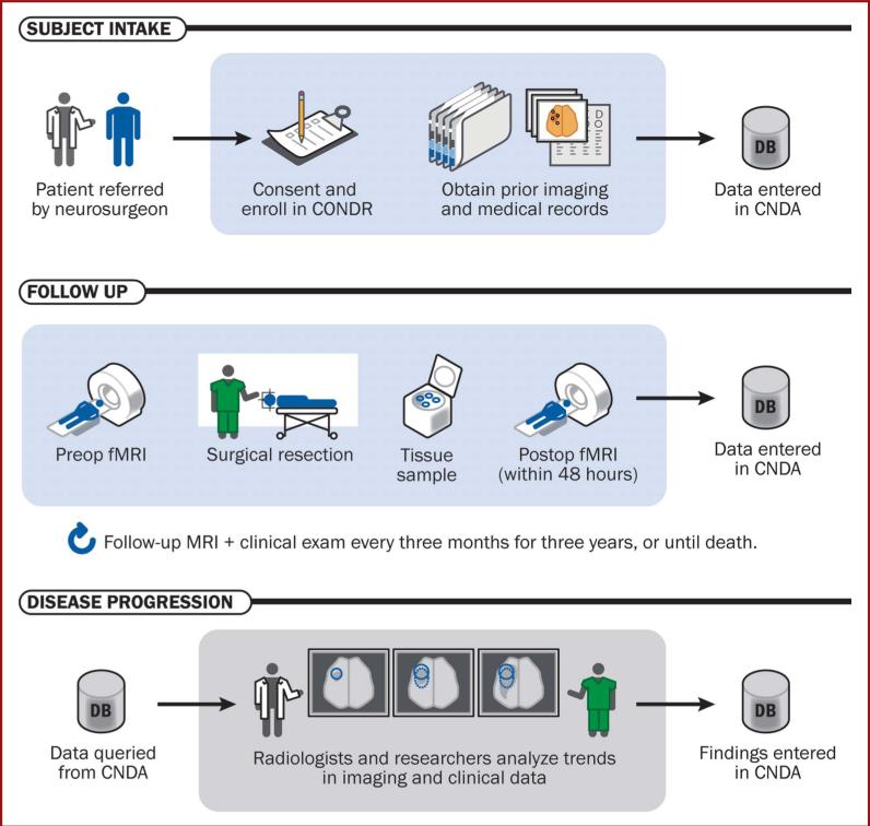
Subjects are selected from patients seen by members of the Neurosurgical, Medical, and Radiation Oncology services at Swedish Hospital and Washington University School of Medicine. The principal investigator or a collaborating physician or representative nurse coordinator at either site contacts patients who have a planned surgery for a presumed primary brain tumor. Patients must be willing to have tissue taken and archived at the time of surgery and meet the study inclusion and exclusion criteria. Patients are enrolled in the study before or within 1 week of undergoing surgery for a malignant brain tumor. The overall schematic for patient registration and subsequent data collection in the study is described in Figure 1.
FIGURE 1.
Subject data collection. Schematic timeline of subject enrollment in the CONDR workflow, including clinical, imaging, and surgical encounters. CONDR, Comprehensive Neuro-Oncology Data Repository.
Image Collection
Imaging sequences are obtained according to the clinical standard of care agreed to at each institution at the following intervals: preoperatively, postoperatively before the initiation of adjunctive chemotherapyand radiation, following completion of radiation, and every 2 months until death or 3 years following enrollment. The CONDR standard imaging protocol follows ACRIN Protocol 668647 with the addition of a precontrast high-resolution T1-weighted sequence. It includes magnetization-prepared rapid acquisition with gradient echo (MP-RAGE) / Spoiled Gradient Echo (SPGR) (SPGR) pre- and post-Gd-enhanced volumetric acquisition, diffusion acquired as diffusion tensor imaging (25 direction, bmax = 1400 s/mm2),13 and dynamic susceptibility contrast48,49 to generate relative cerebral blood flow and cerebral blood volume parameter maps. These sequences are standardized across GE and Siemens systems at each site. As the imaging data are collected for clinical purposes, deviations from the standard protocol were allowed. Imaging data are de-identified and transferred to the CONDR central database.
Clinical Data Collection
Patients have clinical data collected preoperatively, postoperatively, and serially every 2 months after enrollment. A standard data-collection tool is used to identify data points of interest including but not limited to patient age, sex, Karnofsky Performance Status score, tumor location, extent of resection, pre-treatment medications and dosage, chemotherapeutic regimen and radiation regimen, seizure status, pathological status, steroid usage, need for further surgery or changes in treatment regimens, treatment-associated complications, and overall survival and time to progression as defined by the Macdonald and Revised Assessment in Neuro-Oncology criteria.31,34 This information, along with data related to the imaging and surgical encounters, is archived on tools within the CONDR archive that are standardized and readily allow subsequent data extraction for the purposes of subsequent analyses (Table 1).
TABLE 1.
Data Obtained at Each Clinical Encountera
| Encounter Type | Data Acquired | Relative Timeline |
|---|---|---|
| Enrollment | Medical history, demographics, medications and other treatments | At diagnosis |
| Preop imaging | Standard CONDR imaging, additional imaging, radiological assessment | At enrollment/diagnosis |
| Surgery | Tissue samples, surgical details, intraop imaging | At enrollment/diagnosis |
| Postop imaging | Standard CONDR imaging, additional imaging | <48 h after surgery. |
| Clinical follow-up | KPS score, chemotherapeutic regimen and radiation regimen, seizure status, pathological status, steroid usage, need for further surgery or changes in treatment regimens, treatment-associated complications, and overall survival and time to progression as defined by the Macdonald and RANO criteria | ~ every 2 mo postsurgery |
CONDR, Comprehensive Neuro-Oncology Data Repository; KPS, Karnofsky Performance Status; preop, preoperative; intraop, intraoperative; postop, postoperative; RANO, Revised Assessment in Neuro-Oncology.
Tissue-Based Data Collection
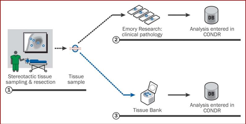
For each subject enrolled in CONDR, surgical specimens are sent for pathological analysis, as is part of the clinical standard of care. The clinical operative protocol for patients with malignant brain tumors includes surgery guided by intraoperative navigation (Stealth/Medtronic). In CONDR cases, each surgical case also includes the removal of specific surgical specimens/biopsies (tissue that would be taken as a part of the intended surgical resection) for research purposes. Specimen/biopsy collection includes removal of an approximately 5-mm tissue sample with immediate recording by using the intraoperative navigation system, of the biopsy location. This location is captured in axial, sagittal, and coronal planes, and is also recorded in 3-dimensional coordinate patient space, which can be subsequently transformed and recorded on the patient's preoperative imaging studies. The biopsies are removed before significant removal of tumor tissue to minimize effects of brain shift. Biopsy specimens are divided and sent for tissue banking for genetic analysis and for advanced histopathological analysis. This process is described schematically in Figure 2. Tissue is removed from the enhancing tumor, surrounding tissue with abnormal T2 or fluid attenuated inversion recovery signal, and when clinically indicated, from surrounding normal tissue (in instances when normal tissue must be removed to access a deeper tumor).
FIGURE 2.
Tissue sample procedure. As part of the CONDR workflow, at the time of tumor resection, specimens are taken within and at the margin of the tumor resection. When a biopsy specimen is removed, its location is recorded on the intraoperative navigation system. The specimen is split in half for subsequent archival and analysis. CNDA, Central Neuroimaging Data Archive; CONDR, Comprehensive Neuro-Oncology Data Repository; fMRI, functional magnetic resonance imaging; preop, preoperative; postop, postoperative.
Image Processing
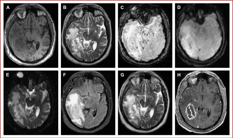
The CONDR image-processing framework includes 3 major components: (1) computation of perfusion and diffusion parameter maps, including cerebral blood flow, cerebral blood volume, fractional anisotropy, and mean diffusivity; (2) spatial coregistration of all structural and derived images; and (3) generation of regions of interest (ROIs) for the biopsy sites. For cross-modal spatial registration, a post-gadolinium T1 image with a standard voxel size 1 × 1 × 2 mm is used as the reference target. The registration is performed by optimization of joint intensity gradient target function50 for the 12-parameter affine transform. Perfusion and diffusion parameter maps are computed by 3 different sets of tools: first, the US Food and Drug Administration-approved nordicICE Neurolab image-processing workstation; second, the Siemens Leonardo workstation; and third, a suite of in-house algorithms based on references.51,52 All diffusion and perfusion parameter maps are transformed to the target space by using a precomputed transform generated by transformation of the original echo planar image (EPI) to the T1w target. The quality of registration is validated both automatically by comparing image similarity metrics with those of good registrations, and manually for echo planar imaging sequences, which are susceptible to distortions that lead to increased registration error. The resulting 4-dimensional data set (the fourth dimension being an MRI modality) is used to quantitatively analyze the contribution of the available modalities to prognosis and treatment planning. To generate the ROIs, the spatial coordinates of the biopsy sites that are recorded from the intraoperative navigation system are registered to the images through manual alignment by a radiologist. Automated approaches for alignment are currently being developed. An example subject's processed image data are illustrated in Figure 3.
FIGURE 3.
Common tumor protocol sequences in a single CONDR study: A, precontrast MP-RAGE; B, high-resolution T2; C, SWI; D, T1 GRE* (this scan was taken from a different patient); E, coronal 5-mm T1; F, T2 FLAIR; G, T2 BLADE; H, high-resolution post-gadolinium T1. The same patient slice in the common space is shown except for D. CONDR, Comprehensive Neuro-Oncology Data Repository; MP-RAGE, magnetization-prepared rapid acquisition gradient echo; FLAIR, fluid attenuated inversion recovery; SWI, susceptibility weighted imaging; GRE, gradient echo sequences.
Tissue Analysis
Biopsied tissue obtained from the neurosurgical tumor bed and corresponding to specific spatial coordinates from neuroimaging studies is separated into fragments designated for molecular and genetic studies, which are snap frozen in liquid nitrogen and for histopathological assessment. The latter is fixed in formalin, processed,and embedded atSwedish Medical Center or Washington University.Five consecutive unstained tissue sections arecut at 5 μm for each biopsy. Sections are packaged and shipped to Dr Brat at the Winship Cancer Institute at Emory University, where they are stained, whole-slide scanned for digital pathology, and analyzed for specific pathological correlates, as detailed below.
Unstained slides for each biopsy are stained with hematoxylin and eosin (H & E), the proliferation marker MIB-1 (Dako), and the vascular marker CD 31 (Abcam). Two unstained slides are filed for future investigation. Slides are scanned at ×40 in an Olympus Nanozoomer Whole Slide Scanner.
Images are accessed through Aperio Spectrum and annotated by using Aperio Imagescope, which allows for the creation of multiple layers of mark-up and annotation using a human-machine interface, with Dr Brat as the neuropathologist. For H & E-stained images, the following layers are created, marked and annotated: (1) tumor tissue; (2) normal/reactive tissue; (3) necrosis; and (4) total tissue area. Each markup creates x, y coordinates that are saved in an XML file to the corresponding image. This allows calculation of the percentage of the tissue occupied by tumor tissue, necrosis, and normal/reactive tissue.
Computer-based algorithms are used to segment and characterize selected structures in digitized H & E and immunohistochemical slides. Within the layer of neoplastic tissue on H & E images, normal nuclei are segmented to obtain a cell density (ratio of total nuclear area to total tissue area). On images of MIB-1-stained slides, a threshold-based algorithm is used to identify positively stained nuclei and total nuclei (stained lightly with hematoxylin) and to obtain a proliferation index as the ratio of MIB-1-stained nuclei to total nuclei. Similarly, a vascular density is calculated within the neoplastic tissue as a percentage of CD31 staining to total surface area.
In addition to these automated metrics, a detailed manual review form is completed for each tissue sample. The form, modified from the protocol used by the Cancer Genome Atlas Neuropathology Review, captures cellular and tissue elements that may have correlates with imaging and molecular studies, including cellular differentiation of glioma cells (fibrillary, gemistocytic, giant cell, sarcomatoid, small cell, or oligodendroglial); qualitative vascular changes (endothelial hypertrophy, hyperplasia, complex hyperplasia, thrombosis); qualitative necrosis (zonal vs pseudopalisading); inflammation (macrophage/monocyte, lymphocytes); and analysis of microcalcification, microcysts and edema. These features are graded as absent (0%), minimal (in <5% of tissue), present (in >5% and <50% of tissue), and abundant (>50% of tissue).
We anticipate conducting future genomic assays of the obtained tissue samples. To enable these assays, the samples are maintained within the tumor bank at the Washington University School of Medicine.
Longitudinal Follow-up
The subjects are followed by their neurosurgeon, as well as by their radiation and medical oncology teams. Ongoing medical histories are recorded in the CONDR database by extracting information from patient records and through communication with their health care providers. The standard of care at both institutions is to obtain a clinical MRI (using the CONDR tumor protocol as described above) 4 weeks following the completion of radiation and chemotherapy, and then approximately every 2 months unless the patient has interim evidence of neurological deterioration. At each follow-up time point, clinical and imaging data are entered into the CONDR database. Figure 1 again illustrates these longitudinal procedures.
Analyses
The CONDR informatics platform will answer initial questions regarding standardization of image data collection, processing, and analysis. After the collection of data in a large cohort of subjects, we plan to use the repository to facilitate preliminary correlations of clinical, imaging, and tissue-based markers, with the objective of determining whether imaging markers can serve the same role in prognosis that clinical and histopathological markers have come to play in recent decades. In addition, the role of anatomic and advanced imaging markers in definitions of progression will be considered. Representative markers that can be studied are listed in Table 2.
TABLE 2.
Representative Markers that Will be Considered for Preliminary Analyses Within the Initial CONDR Data Seta,b
| Clinical | Imaging | Tissue |
|---|---|---|
| Age | rCBV (perfusion) | MGMT methylation |
| Sex | Ktrans (perfusion) | IDH-1 |
| KPS | ADC (diffusion) | IDH-2 |
| Extent of resection | Functional diffusion map (diffusion) | Cellularity (MIB-1) |
| Time to progression | Permeability (DCE) | Blood vessel density and area |
| Overall survival | Volumetric measures |
CONDR, Comprehensive Neuro-Oncology Data Repository; KPS, Karnofsky Performance Status; MGMT, O6-methylguanylmethyltransferase; rCBV, cerebral blood volume; ADC, apparent diffusion coefficient; IDH, isocitrate dehydrogenase; DCE, dynamic contrast-enhanced; Ktrans, volume transfer constant.
Key markers from each data domain will be considered for preliminary correlative analyses within the scope of the current project.
DISCUSSION
Research into clinical, imaging, and tissue-based biomarkers and their correlation with clinical outcomes in patients with malignant brain tumors is ongoing at multiple sites across the country. However, few sites have bridged the gap that prevents sites from sharing and comparing data, particularly when imaging data are acquired on different imaging platforms, using different imaging sequences, and without the technology available to facilitate data sharing. Our goal within this study is to not just continue and expand ongoing research projects focused on imaging biomarkers and clinical outcomes, but also to facilitate the centralized collection, processing, and analysis of the data by using a novel data management resource. This will provide a unique tool for the creation of a much larger data set than is currently available with the use of existing standards at even the strongest individual sites. The resource will also record data including clinical and tissue-based data along with both raw and processed imaging data. The availability of this informatics platform has the potential to allow imaging-based studies to move beyond small series obtained at individual institutions or secondary cohorts of larger studies, but to facilitate prospective studies that allow imaging markers to be considered as key end points in a clinical trial, and clinical correlative studies powered to validate the use of these markers in neuro-oncology research and practice.
TRIAL STATUS
One hundred thirty-two subjects have been enrolled in CONDR. CONDR is actively enrolling additional subjects at both sites and laying the groundwork for expansion to additional sites.
SAFETY CONSIDERATIONS
As all treatments included in the clinical care for subjects enrolled in this study are standard of care; no adverse events are anticipated as a result of study participation. Study protocols allow for the inclusion of additional imaging sequences in selected patient cohorts, but additional contrast administration or exposure is not expected.
Research records are handled as carefully as possible to maintain patient confidentiality according to applicable state and federal law, including the Health Insurance Portability and Accountability Act.
Should patient confidentiality be breached and allow identification of a participant by someone other than an investigator in this study, the IRB is notified and the participant contacted. Further data collection would be halted for all participants until procedures are identified to prevent recurrence of the breach.
Because archival of subject's tissue for the potential purposes of genetic analysis is a part of this study, the particular risks of genetic studies, even in the setting of deidentified data, are reviewed with subjects before obtaining informed consent.
FOLLOW-UP
Research subjects are followed for at least 3 years or until death as described above in the Methodology section.
DATA MANAGEMENT AND STATISTICAL ANALYSIS
Data Management
The CONDR study has developed a comprehensive data management system to capture all study data in a single integrated resource. The system is built on the Central Neuroimaging Data Archive (CNDA) at Washington University. The CNDA is an instance of the XNAT imaging informatics platform53 and serves as the University's primary research image database. XNAT consists of an image repository to store raw and postprocessed images, a database to store metadata and nonimaging measures, and user interface tools for accessing, querying, and exploring data. The CNDA currently hosts over 500 separate clinical and basic research studies and includes data obtained from a number of modalities including PET, MRI, and CT.
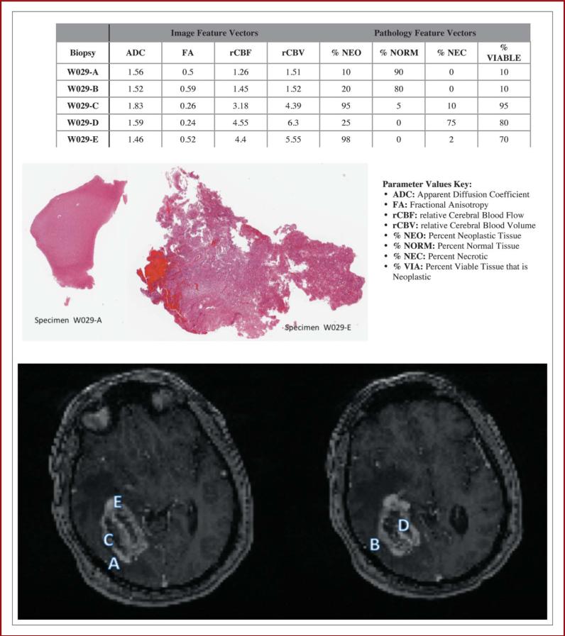
The CNDA has been customized in a variety of ways specifically to host CONDR. Electronic case report forms have been generated for entry of the various clinical and experimental data obtained in CONDR, including medical histories, surgical encounters, pathology, and chemotherapy records. Also, a system was added to the CNDA for capturing digitized histopathology slides and quantitative analyses (Figure 4). Finally, postprocessed diffusion, perfusion, and functional MRI images have been incorporated into the database for subsequent quantitative analysis. The database is currently being extended to enable automated execution of image-processing streams and storage of ROI-based quantitative measures. The CONDR platform was designed to be easily adopted by future neuro-oncology trials. All components are open source and available to other investigators to support imaging-based neuro-oncology studies.
FIGURE 4.
A representative CONDR subject. In this case, a right temporal-occipital glioblastoma was removed via craniotomy. Biopsy specimens(A-E) were taken from the overlying cortical tissue (A, B), the marginal enhancing tissue (C, E), and tissue at the center of the tumor (D). The locations of these biopsies are represented on the MRI shown above. Image and histopathology parameters are defined for each biopsy sample. Images of the whole-mount pathology slide for specimens A and E are shown. The distinction between relatively normal cortex and tumor tissue is visible even at low magnification. For each of these specimens, parallel tissue specimens have been archived for genomic analysis. CONDR, Comprehensive Neuro-Oncology Data Repository.
Statistical Analysis
The total number of available measures within CONDR is quite large and will require complex multivariate statistical methods to fully explore them. In addition, during the early stages of the project, the number of available subjects is relatively small, limiting the power to use such methods. In recognition of these complexities, we have developed a phased approach to analysis of the CONDR database. Initially, we are conducting hypothesis-based analyses on select clinical, genetic, and imaging markers. Secondarily, we will conduct analyses using advanced multivariate statistical methods to mine the full richness of the database. In constraining the scope of the analyses completed within the initial phases of this project, we hope to ensure that significant scientific findings will be delivered with the available resources. This is aligned with the primary objective of this proposal, which is to build the CONDR platform. Then, through our own follow-on projects, we will broaden the scope of our analyses to more deeply mine the data, and we expect that, by making CONDR openly accessible to the research community, external investigators will bring a wide variety of novel approaches to bear on the CONDR data.
Within the scope of the current project, we plan to analyze a select subset of high-interest markers as described in Table 2. This choice highlights key markers that have already been identified as high interest based on previous clinical, imaging and tissue-based studies.3,8,9,11,12,19,37-44
In the later years of this project, we will expand our analyses to include more advanced application of the CONDR tools. By contrast, in hypothesis generation, we will screen an extremely large number of possible hypotheses. Corrections for multiple comparisons are more important here, and even after such formal statistical adjustments, we tend not to strongly believe the results from such explorations until they are independently validated in a replication data set. Although the hypothesis generation aspect of the proposal is by definition an exploratory technique, it has potentially great scientific impact, because it may lead to new insights as to which features of brain images are important for clinical outcome, and which are strongly governed by genetics.
For the more exploratory, hypothesis generation aspect of the project, we will make use of a variety of high-dimensional data reduction techniques. Although there are many agnostic machine learning type methods for discovering relationships in large data sets,54,55 one very promising class of techniques that leverage known biological information with statistical information from the data itself are network models. In this paradigm, complex clinical traits are the “outputs” of pathways that are extremely intricate, yet composed of relatively simple subpaths, each involving relatively few exposures, genes, and intermediate measured factors, including various brain morphology characteristics.
QUALITY ASSURANCE
This study is a prospective registry archiving clinical, imaging, and tissue-based data. Standards of clinical research practice as mandated by the IRBs at each participating institution are followed rigorously, with particular attention to the maintenance of subject confidentiality.
EXPECTED OUTCOMES
The CONDR infrastructure and patient database will be the primary deliverables of the study. The infrastructure is open source and available to other research groups who are developing imaging-based oncology protocols. The CONDR patient database will be made openly available to the research community for further analysis and exploration. In addition, within our own team, we are conducting baseline analyses of multimodal imaging predictors of tumor response to therapy, markers to distinguish recurrence from treatment-associated change, and predictors of patient outcomes/prognosis. We are also considering correlations between imaging and tissue-based markers and both imaging and tissue-based markers of tumor heterogeneity. We anticipate findings from these initial analyses to motivate further prospective multicenter trials founded on imaging markers in these same sorts of studies.
DURATION OF PROJECT
The CONDR protocol will continue enrolling subjects until at least June 2014 and will continue to perform analyses until June 2017; however, so long as the study continues to accrue subjects at both sites, the protocol will continue. It is expected that expansion to additional sites and consideration of additional disease types (eg, metastatic brain tumors) can be undertaken beginning in 2014.
PROJECT MANAGEMENT
Washington University School of Medicine
Daniel Marcus, PhD—Principal Investigator, Informatics Database Development.
Tammie Benzinger, MD, PhD—Neuroradiology—image acquisition and analysis.
Josh S Shimony, MD, PhD—Neuroradiology—image analysis.
Dhana Rajderkar, MD—Neuroradiology—image analysis.
Keith Rich, MD—Neurological Surgery—subject recruitment and tissue acquisition; transition to additional tumor types (metastases).
Michael Chicoine, MD—Neurological Surgery—subject recruitment and tissue acquisition; intraoperative MR imaging component.
Albert Kim, MD—Neurological Surgery—subject recruitment and tissue acquisition; tissue-based studies.
Misha Milchenko, PhD—Computer Science—protocols for image registration, processing, and analysis.
Abraham Snyder, MD, PhD—Radiology—Image processing and algorithms.
Pamela LaMontagne, PhD—Research Coordinator and image data analysis.
Swedish Medical Center
Sarah Jost Fouke, MD—Principal Investigator—Neurosurgery—subject recruitment, clinical and image analysis.
Bart Keogh, MD—Neuroradiology—image acquisition, processing, and analysis.
Xu Feng, PhD—Neuroradiology—image processing and analysis.
John Henson, MD—Neuroradiology—image acquisition and analysis.
Amanda Brown, BA—Research Coordinator.
Colleen Ottinger, BA—Research Coordinator.
Emory University
Daniel Brat, MD, PhD—Pathology—tissue analysis.
David Gutman, MD, PhD—Pathology—tissue analysis.
Merete Williams—Pathology—tissue analysis.
ETHICS
This study is conducted according to US and international standards of Good Clinical Practice and applicable government regulations. This protocol will be submitted to the Swedish Hospital IRB for a formal approval of the study conduct. All participants in this study will be provided an IRB-approved informed consent form describing the study and providing sufficient information for participants to make informed decisions about their participation in this study. The study participant MUST be consented with the IRB-approved informed consent form before the participant is subjected to any investigational study procedures and before data are uploaded to the CONDR registry. The approved consent form MUST be signed and dated by the study participant or legally acceptable representative and the investigator-designated research staff obtaining the consent.
Acknowledgments
This study was supported by the NIH/National Institute of Neurological Disorders and Stroke (R01NS066905).
ABBREVIATIONS
- CNDA
Central Neuroimaging Data Archive
- CONDR
Comprehensive Neurooncology Data Repository
- H & E
hematoxylin and eosin
- IRB
institutional review board
- ROI
region of interest
Footnotes
Disclosures
The authors have no personal, financial, or institutional interest in any of the drugs, materials, or devices described in this article.
REFERENCES
- 1.Davis FG, Freels S, Grutsch J, Barlas S, Brem S. Survival rates in patients with primary malignant brain tumors stratified by patient age and tumor histological type: an analysis based on Surveillance, Epidemiology, and End Results (SEER) data, 1973-1991. J Neurosurg. 1998;88(1):1–10. doi: 10.3171/jns.1998.88.1.0001. [DOI] [PubMed] [Google Scholar]
- 2.Tooth HH. Presidential Address: Some observations on the growth and survival-period of intracranial tumours, based on the records of 500 cases, with special reference to the pathology of the gliomata. Brain. 1912;6(Neurol Sect):61–108. doi: 10.1177/003591571300600801. [DOI] [PMC free article] [PubMed] [Google Scholar]
- 3.Stupp R, Mason WP, van den Bent MJ, et al. Radiotherapy plus concomitant and adjuvant temozolomide for glioblastoma. N Engl J Med. 2005;352(10):987–996. doi: 10.1056/NEJMoa043330. doi: 10.1056/NEJMoa043330. [DOI] [PubMed] [Google Scholar]
- 4.Kelly PJ, Daumas-Duport C, Kispert DB, Kall BA, Scheithauer BW, Illig JJ. Imaging-based stereotaxic serial biopsies in untreated intracranial glial neoplasms. J Neurosurg. 1987;66(6):865–874. doi: 10.3171/jns.1987.66.6.0865. doi: 10.3171/jns.1987.66.6.0865. [DOI] [PubMed] [Google Scholar]
- 5.Surawicz TS, Davis F, Freels S, Laws ER, Jr, Menck HR. Brain tumor survival: results from the National Cancer Data Base. J Neurooncol. 1998;40(2):151–160. doi: 10.1023/a:1006091608586. [DOI] [PubMed] [Google Scholar]
- 6.Prados MD, Levin V. Biology and treatment of malignant glioma. Semin Oncol. 2000;27(3 suppl 6):1–10. [PubMed] [Google Scholar]
- 7.Fewer D, Wilson CB, Boldrey EB, Enot KJ, Powell MR. The chemotherapy of brain tumors. Clinical experience with carmustine (BCNU) and vincristine. JAMA. 1972;222(5):549–552. [PubMed] [Google Scholar]
- 8.Hegi ME, Diserens AC, Gorlia T, et al. MGMT gene silencing and benefit from temozolomide in glioblastoma. N Engl J Med. 2005;352(10):997–1003. doi: 10.1056/NEJMoa043331. [DOI] [PubMed] [Google Scholar]
- 9.Stratton MR, Campbell PJ, Futreal PA. The cancer genome. Nature. 2009;458(7239):719–724. doi: 10.1038/nature07943. doi: 10.1038/nature07943. [DOI] [PMC free article] [PubMed] [Google Scholar]
- 10.Wakimoto H, Aoyagi M, Nakayama T, et al. Prognostic significance of Ki-67 labeling indices obtained using MIB-1 monoclonal antibody in patients with supratentorial astrocytomas. Cancer. 1996;77(2):373–380. doi: 10.1002/(SICI)1097-0142(19960115)77:2<373::AID-CNCR21>3.0.CO;2-Y. [DOI] [PubMed] [Google Scholar]
- 11.Yan H, Parsons DW, Jin G, et al. IDH1 and IDH2 mutations in gliomas. N Engl J Med. 2009;360(8):765–773. doi: 10.1056/NEJMoa0808710. doi: 10.1056/NEJMoa0808710. [DOI] [PMC free article] [PubMed] [Google Scholar]
- 12.Zhao S, Lin Y, Xu W, et al. Glioma-derived mutations in IDH1 dominantly inhibit IDH1 catalytic activity and induce HIF-1alpha. Science. 2009;324(5924):261–265. doi: 10.1126/science.1170944. doi: 10.1126/science.1170944. [DOI] [PMC free article] [PubMed] [Google Scholar]
- 13.Basser PJ. Inferring microstructural features and the physiological state of tissues from diffusion-weighted images. NMR Biomed. 1995;8(7-8):333–344. doi: 10.1002/nbm.1940080707. [DOI] [PubMed] [Google Scholar]
- 14.Barboriak DP. Imaging of brain tumors with diffusion-weighted and diffusion tensor MR imaging. Magn Reson Imaging Clin N Am. 2003;11(3):379–401. doi: 10.1016/s1064-9689(03)00065-5. [DOI] [PubMed] [Google Scholar]
- 15.Provenzale JM, Mukundan S, Barboriak DP. Diffusion-weighted and perfusion MR imaging for brain tumor characterization and assessment of treatment response. Radiology. 2006;239(3):632–649. doi: 10.1148/radiol.2393042031. doi: 10.1148/radiol.2393042031. [DOI] [PubMed] [Google Scholar]
- 16.Khayal IS, Polley MY, Jalbert L, et al. Evaluation of diffusion parameters as early biomarkers of disease progression in glioblastoma multiforme. Neuro Oncology. 2010;12(9):908–916. doi: 10.1093/neuonc/noq049. [DOI] [PMC free article] [PubMed] [Google Scholar]
- 17.Choyke PL, Dwyer AJ, Knopp MV. Functional tumor imaging with dynamic contrast-enhanced magnetic resonance imaging. J Magn Reson Imaging. 2003;17(5):509–520. doi: 10.1002/jmri.10304. doi: 10.1002/jmri.10304. [DOI] [PubMed] [Google Scholar]
- 18.Hu LS, Eschbacher JM, Dueck AC, et al. Correlations between perfusion MR imaging cerebral blood volume, microvessel quantification, and clinical outcome using stereotactic analysis in recurrent high-grade glioma. AJNR Am J Neuroradiol. 2012;33(1):69–76. doi: 10.3174/ajnr.A2743. [DOI] [PMC free article] [PubMed] [Google Scholar]
- 19.Law M, Oh S, Babb JS, et al. Low-grade gliomas: dynamic susceptibility-weighted contrast-enhanced perfusion MR imaging–prediction of patient clinical response. Radiology. 2006;238(2):658–667. doi: 10.1148/radiol.2382042180. doi: 10.1148/radiol.2382042180. [DOI] [PubMed] [Google Scholar]
- 20.Maia AC, Jr, Malheiros SM, da Rocha AJ, et al. MR cerebral blood volume maps correlated with vascular endothelial growth factor expression and tumor grade in nonenhancing gliomas. AJNR Am J Neuroradiol. 2005;26(4):777–783. [PMC free article] [PubMed] [Google Scholar]
- 21.Burtscher IM, Holtås S. Proton magnetic resonance spectroscopy in brain tumours: clinical applications. Neuroradiology. 2001;43(5):345–352. doi: 10.1007/s002340000427. [DOI] [PubMed] [Google Scholar]
- 22.Callot V, Galanaud D, Le Fur Y, Confort-Gouny S, Ranjeva JP, Cozzone PJ. (1)H MR spectroscopy of human brain tumours: a practical approach. Eur J Radiol. 2008;67(2):268–274. doi: 10.1016/j.ejrad.2008.02.036. doi: 10.1016/j.ejrad.2008.02.036. [DOI] [PubMed] [Google Scholar]
- 23.Alavi JB, Alavi A, Chawluk J, et al. Positron emission tomography in patients with glioma. A predictor of prognosis. Cancer. 1988;62(6):1074–1078. doi: 10.1002/1097-0142(19880915)62:6<1074::aid-cncr2820620609>3.0.co;2-h. [DOI] [PubMed] [Google Scholar]
- 24.Barker FG II, Chang SM, Valk PE, Pounds TR, Prados MD. 18-Fluorodeoxyglucose uptake and survival of patients with suspected recurrent malignant glioma. Cancer. 1997;79(1):115–126. [PubMed] [Google Scholar]
- 25.Chen W, Cloughesy T, Kamdar N, et al. Imaging proliferation in brain tumors with 18F-FLT PET: comparison with 18F-FDG. J Nucl Med. 2005;46(6):945–952. [PubMed] [Google Scholar]
- 26.De Witte O, Levivier M, Violon P, et al. Prognostic value positron emission tomography with [18F]fluoro-2-deoxy-D-glucose in the low-grade glioma. Neurosurgery. 1996;39(3):470–476. doi: 10.1097/00006123-199609000-00007. discussion 476-477. [DOI] [PubMed] [Google Scholar]
- 27.Glantz MJ, Hoffman JM, Coleman RE, et al. Identification of early recurrence of primary central nervous system tumors by [18F]fluorodeoxyglucose positron emission tomography. Ann Neurol. 1991;29(4):347–355. doi: 10.1002/ana.410290403. doi: 10.1002/ana.410290403. [DOI] [PubMed] [Google Scholar]
- 28.Hoekstra CJ, Paglianiti I, Hoekstra OS, et al. Monitoring response to therapy in cancer using [18F]-2-fluoro-2-deoxy-D-glucose and positron emission tomography: an overview of different analytical methods. Eur J Nucl Med. 2000;27(6):731–743. doi: 10.1007/s002590050570. [DOI] [PubMed] [Google Scholar]
- 29.Keogh BP, Henson JW. Clinical manifestations and diagnostic imaging of brain tumors. Hematol Oncol Clin North Am. 2012;26(4):733–755. doi: 10.1016/j.hoc.2012.05.002. doi: 10.1016/j.hoc.2012.05.002. [DOI] [PubMed] [Google Scholar]
- 30.Geer CP, Simonds J, Anvery A, et al. Does MR perfusion imaging impact management decisions for patients with brain tumors? A prospective study. AJNR Am J Neuroradiol. 2012;33(3):556–562. doi: 10.3174/ajnr.A2811. doi: 10.3174/ajnr.A2811. [DOI] [PMC free article] [PubMed] [Google Scholar]
- 31.Macdonald DR, Cascino TL, Schold SC, Jr, Cairncross JG. Response criteria for phase II studies of supratentorial malignant glioma. J Clin Oncol. 1990;8(7):1277–1280. doi: 10.1200/JCO.1990.8.7.1277. [DOI] [PubMed] [Google Scholar]
- 32.Quant EC, Wen PY. Response assessment in neuro-oncology. Curr Oncol Rep. 2011;13(1):50–56. doi: 10.1007/s11912-010-0143-y. doi: 10.1007/s11912-010-0143-y. [DOI] [PubMed] [Google Scholar]
- 33.Vogelbaum MA, Jost S, Aghi MK, et al. Application of novel response/progression measures for surgically delivered therapies for gliomas: Response Assessment in Neuro-Oncology (RANO) Working Group. Neurosurgery. 2012;70(1):234–243. doi: 10.1227/NEU.0b013e318223f5a7. discussion 243-244. doi: 10.1227/NEU.0b013e318223f5a7. [DOI] [PubMed] [Google Scholar]
- 34.Wen PY, Macdonald DR, Reardon DA, et al. Updated response assessment criteria for high-grade gliomas: response assessment in neuro-oncology working group. J Clin Oncol. 2010;28(11):1963–1972. doi: 10.1200/JCO.2009.26.3541. doi: 10.1200/JCO.2009.26.3541. [DOI] [PubMed] [Google Scholar]
- 35.Vidiri A, Carapella CM, Pace A, et al. Early post-operative MRI: correlation with progression-free survival and overall survival time in malignant gliomas. J Exp Clin Cancer Res. 2006;25(2):177–182. [PubMed] [Google Scholar]
- 36.Sorensen AG, Emblem KE, Polaskova P, et al. Increased survival of glioblastoma patients who respond to antiangiogenic therapy with elevated blood perfusion. Cancer Res. 2012;72(2):402–407. doi: 10.1158/0008-5472.CAN-11-2464. doi: 10.1158/0008-5472.CAN-11-2464. [DOI] [PMC free article] [PubMed] [Google Scholar]
- 37.Ellingson BM, Cloughesy TF, Zaw T, et al. Functional diffusion maps (fDMs) evaluated before and after radiochemotherapy predict progression-free and overall survival in newly diagnosed glioblastoma. Neuro Oncol. 2012;14(3):333–343. doi: 10.1093/neuonc/nor220. doi: 10.1093/neuonc/nor220. [DOI] [PMC free article] [PubMed] [Google Scholar]
- 38.Ellingson BM, Malkin MG, Rand SD, et al. Volumetric analysis of functional diffusion maps is a predictive imaging biomarker for cytotoxic and anti-angiogenic treatments in malignant gliomas. J Neurooncol. 2011;102(1):95–103. doi: 10.1007/s11060-010-0293-7. doi: 10. 1007/s11060-010-0293-7. [DOI] [PMC free article] [PubMed] [Google Scholar]
- 39.Chenevert TL, Stegman LD, Taylor JM, et al. Diffusion magnetic resonance imaging: an early surrogate marker of therapeutic efficacy in brain tumors. J Natl Cancer Inst. 2000;92(24):2029–2036. doi: 10.1093/jnci/92.24.2029. [DOI] [PubMed] [Google Scholar]
- 40.Mardor Y, Pfeffer R, Spiegelmann R, et al. Early detection of response to radiation therapy in patients with brain malignancies using conventional and high b-value diffusion-weighted magnetic resonance imaging. J Clin Oncol. 2003;21(6):1094–1100. doi: 10.1200/JCO.2003.05.069. [DOI] [PubMed] [Google Scholar]
- 41.Moffat BA, Chenevert TL, Lawrence TS, et al. Functional diffusion map: a noninvasive MRI biomarker for early stratification of clinical brain tumor response. Proc Natl Acad Sci U S A. 2005;102(15):5524–5529. doi: 10.1073/pnas.0501532102. doi: 10.1073/pnas.0501532102. [DOI] [PMC free article] [PubMed] [Google Scholar]
- 42.Gao X, Zhang XN, Zhang YT, Yu CS, Xu DS. Magnetic resonance imaging in assessment of treatment response of gamma knife for brain tumors. Chin Med J (Engl) 2011;124(12):1906–1910. [PubMed] [Google Scholar]
- 43.Nelson SJ. Assessment of therapeutic response and treatment planning for brain tumors using metabolic and physiological MRI. NMR Biomed. 2011;24(6):734–749. doi: 10.1002/nbm.1669. doi: 10.1002/nbm.1669. [DOI] [PMC free article] [PubMed] [Google Scholar]
- 44.Galbán CJ, Chenevert TL, Meyer CR, et al. Prospective analysis of parametric response map-derived MRI biomarkers: identification of early and distinct glioma response patterns not predicted by standard radiographic assessment. Clin Cancer Res. 2011;17(14):4751–4760. doi: 10.1158/1078-0432.CCR-10-2098. doi: 10.1158/1078-0432.CCR-10-2098. [DOI] [PMC free article] [PubMed] [Google Scholar]
- 45.Maia AC, Jr, Guedes BV, Lucas A, Jr, da Rocha AJ. Diffusion MR imaging for monitoring treatment response. Neuroimaging Clin N Am. 2011;21(1):153–178, viii-ix. doi: 10.1016/j.nic.2011.02.004. doi: 10.1016/j.nic.2011.02.004. [DOI] [PubMed] [Google Scholar]
- 46.Henson JW, Ulmer S, Harris GJ. Brain tumor imaging in clinical trials. AJNR Am J Neuroradiol. 2008;29(3):419–424. doi: 10.3174/ajnr.A0963. doi: 10.3174/ajnr.A0963. [DOI] [PMC free article] [PubMed] [Google Scholar]
- 47. [November 21, 2013];ACRIN. ACRIN 6686 (RTOG 0825) advanced MRI imaging manual. 2009 Available at: http://www.acrin.org/Portals/0/Protocols/6686/6686%20MRmanual%2010012009.pdf.
- 48.Kwong KK, Chesler DA, Weisskoff RM, et al. MR perfusion studies with T1-weighted echo planar imaging. Magn Reson Med. 1995;34(6):878–887. doi: 10.1002/mrm.1910340613. [DOI] [PubMed] [Google Scholar]
- 49.Wong EC, Buxton RB, Frank LR. Quantitative imaging of perfusion using a single subtraction (QUIPSS and QUIPSS II). Magn Reson Med. 1998;39(5):702–708. doi: 10.1002/mrm.1910390506. [DOI] [PubMed] [Google Scholar]
- 50.Rowland DJ, Garbow JR, Laforest R, Snyder AZ. Registration of [18F]FDG microPET and small-animal MRI. Nucl Med Biol. 2005;32(6):567–572. doi: 10.1016/j.nucmedbio.2005.05.002. doi: 10.1016/j.nucmedbio.2005.05.002. [DOI] [PubMed] [Google Scholar]
- 51.Lee JJ, Bretthorst GL, Derdeyn CP, et al. Dynamic susceptibility contrast MRI with localized arterial input functions. Magn Reson Med. 2010;63(5):1305–1314. doi: 10.1002/mrm.22338. doi: 10.1002/mrm.22338. [DOI] [PMC free article] [PubMed] [Google Scholar]
- 52.Jones DK, Horsfield MA, Simmons A. Optimal strategies for measuring diffusion in anisotropic systems by magnetic resonance imaging. Magn Reson Med. 1999;42(3):515–525. [PubMed] [Google Scholar]
- 53.Marcus DS, Olsen TR, Ramaratnam M, Buckner RL. The Extensible Neuro-imaging Archive Toolkit: an informatics platform for managing, exploring, and sharing neuroimaging data. Neuroinformatics. 2007;5(1):11–34. doi: 10.1385/ni:5:1:11. [DOI] [PubMed] [Google Scholar]
- 54.Gu CC, Rao DC, Stormo G, Hicks C, Province MA. Role of gene expression microarray analysis in finding complex disease genes. Genet Epidemiol. 2002;23(1):37–56. doi: 10.1002/gepi.220. doi: 10.1002/gepi.220. [DOI] [PubMed] [Google Scholar]
- 55.Province MA, Shannon WD, Rao DC. Classification methods for confronting heterogeneity. Adv Genet. 2001;42:273–286. doi: 10.1016/s0065-2660(01)42028-1. [DOI] [PubMed] [Google Scholar]


